Standing MRI: How Upright Open Scanners Work, What They Cost, and When to Choose One
Standing MRI explained: 🆕 how upright open scanners work, what they cost, who benefits, accuracy compared to recumbent MRI, and how to prepare for a...
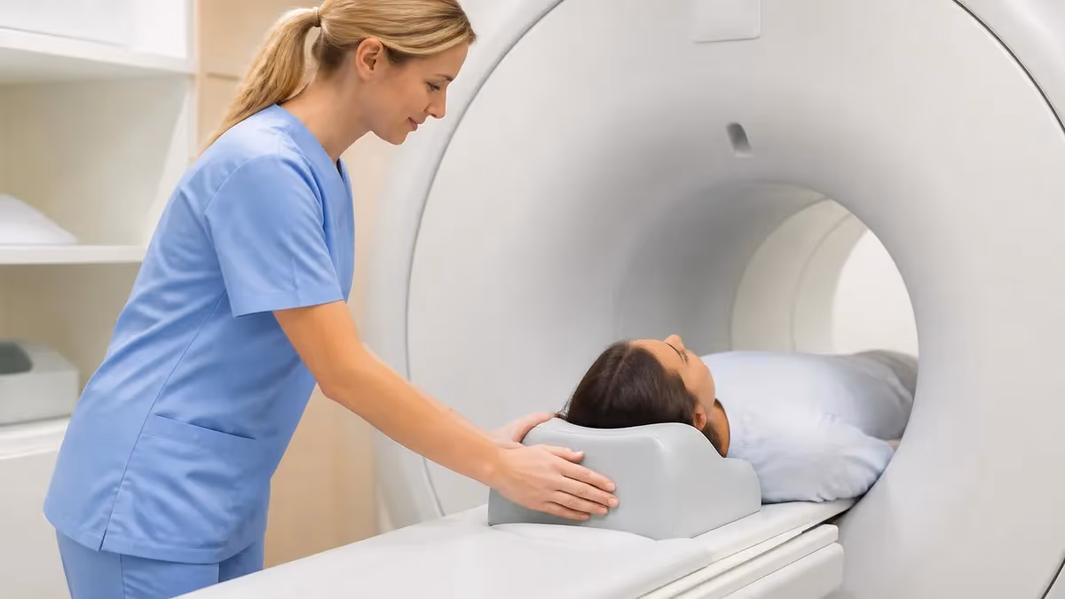
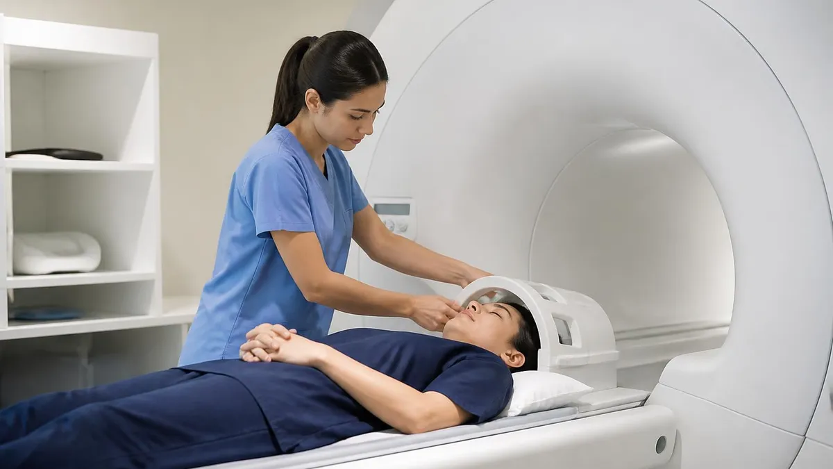
A standing MRI is an upright, weight-bearing magnetic resonance imaging exam that scans the spine, joints, and soft tissues while the patient is sitting, standing, bending, or flexing rather than lying flat on a sliding table. Unlike a conventional closed-bore scanner that forces the body into a horizontal, gravity-neutral position, a standing MRI uses a vertically opened magnet design that allows the spine to bear load just as it does in everyday life. That single change in posture can reveal disc bulges, spondylolisthesis, ligament laxity, and joint instabilities that recumbent imaging routinely misses.
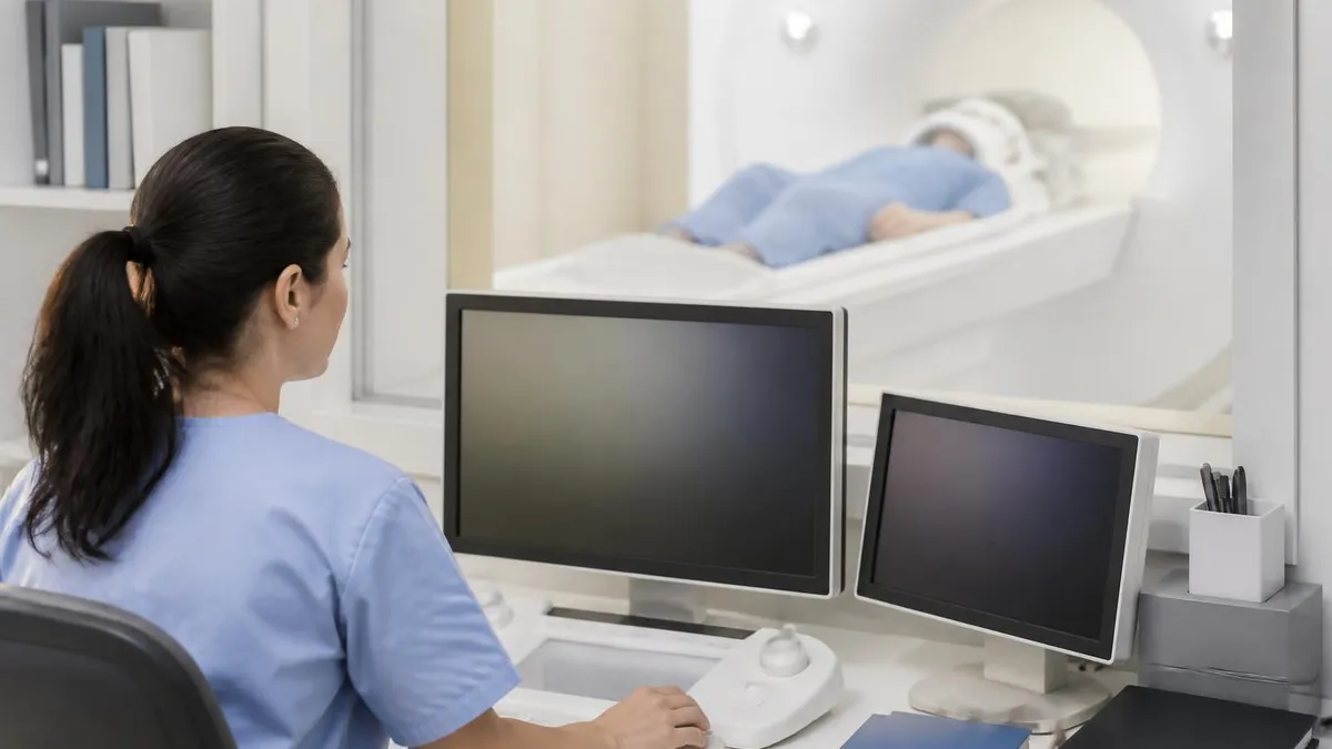
The technology was pioneered in the late 1990s with the FONAR Upright Multi-Position MRI, the first commercial system capable of imaging the spine under physiologic load. Today, several manufacturers including FONAR, Esaote, and Paramed market positional and weight-bearing MRI scanners across roughly 150 to 200 outpatient sites in the United States. Most operate at 0.5 to 0.6 Tesla, far below the 1.5T or 3T field strength of a closed bore, which means longer scan times but dramatically better access for claustrophobic, obese, pediatric, and post-surgical patients who cannot tolerate a tunnel.
The clinical case for standing MRI is strongest in cervical and lumbar spine evaluations where symptoms are positional. A patient whose sciatica only fires up after twenty minutes of walking or whose neck pain disappears the moment they lie down often produces a completely unremarkable supine scan. Image them upright with the same gravitational loading the disc experiences in real life, and a hidden L5-S1 disc herniation, foraminal narrowing, or unstable spondylolisthesis frequently becomes obvious. Orthopedists, chiropractors, and personal-injury attorneys lean heavily on this dynamic capability.
Cost is a frequent surprise. A self-pay standing MRI in the United States typically runs $400 to $1,200 per body region, sometimes higher in major metros, which is comparable to or modestly above a recumbent scan at the same facility. Most major insurers, Medicare, and workers compensation programs cover positional MRI when prior authorization establishes medical necessity, especially when prior supine imaging was non-diagnostic. Cash-pay discounts and bundled multi-region pricing are common at independent imaging centers, and HSA and FSA funds apply.
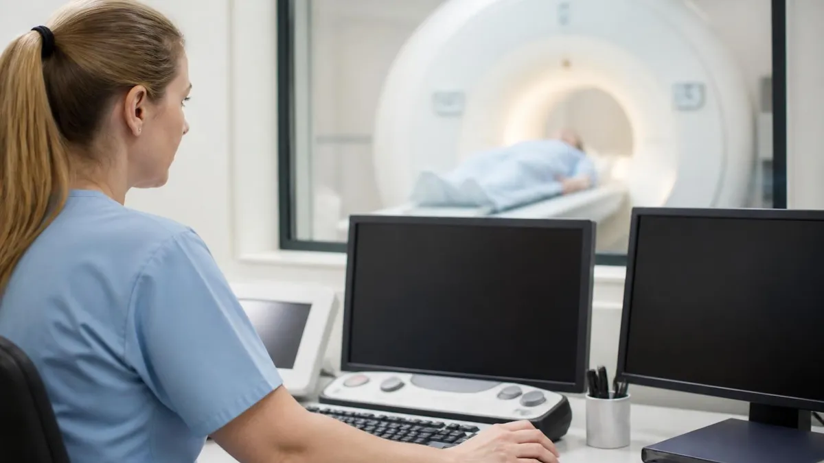
For radiologic technologists preparing for the ARRT MRI registry or working at an upright facility, standing MRI introduces a small but important set of physics and safety considerations. Field strength, gradient performance, RF coil design, patient positioning protocols, and motion artifact mitigation all differ meaningfully from a Siemens or GE closed bore. Understanding these differences is essential for producing diagnostic images, protecting patients with implants, and answering the increasingly common registry questions about open, low-field, and weight-bearing systems.
This guide walks through how a standing MRI works, what it shows that conventional MRI cannot, who is a strong candidate, what costs and insurance to expect, how accuracy compares to high-field systems, and what to do before and during your scan. Whether you are a patient referred for an upright study after a non-diagnostic supine MRI, a clinician deciding when to order one, or a technologist studying for board exams, the sections below cover the clinical, technical, and practical ground you need.
By the end you should be able to answer the three questions that drive most upright MRI decisions: is the patient's complaint truly positional, will the available 0.5T resolution be diagnostic for the suspected pathology, and is the facility, coil, and protocol appropriate for the body region of interest. Get those three right and standing MRI becomes one of the most powerful tools in musculoskeletal and spinal imaging.
Standing MRI by the Numbers
How a Standing MRI Scanner Is Built
Two flat magnet poles sit above and below the patient with open space on all four sides. This C-shaped or H-shaped geometry lets patients stand, sit, or be tilted while maintaining a usable imaging volume between the poles.
A motorized platform tilts from fully upright at 90° down through any intermediate angle to fully supine at 0°. The same patient can be scanned in multiple positions during one appointment to capture true positional pathology.
Flexible cervical, lumbar, knee, and shoulder coils wrap around the body region of interest. Coil selection is critical at low field because signal-to-noise margins are tighter than on a 1.5T closed bore scanner.
Most upright systems use 0.5–0.6T permanent or resistive magnets rather than superconducting magnets. This eliminates cryogen costs and quench risk but reduces SNR, requiring longer acquisition times and thicker slices.
Because patients are upright and may experience orthostatic symptoms, technologists maintain direct line-of-sight, two-way audio, and a panic ball throughout the scan. Patients can be lowered to supine within seconds if they feel faint.
The strongest clinical case for a standing MRI is in patients whose symptoms are clearly positional. Lumbar disc disease, cervical radiculopathy, spondylolisthesis, and ligamentous instability all respond to gravity, and many findings that drive surgical decision-making only become visible when the spine carries load. A recumbent scan unloads the discs, opens the foramina, and reduces the slip of a degenerative spondylolisthesis, often producing a falsely reassuring image that contradicts the patient's actual experience.
Upright imaging excels at quantifying anterolisthesis and retrolisthesis. A grade I slip seen on a supine MRI may convert to grade II or even grade III under load, which directly changes the management conversation between conservative care and surgical fusion. Flexion and extension views captured in the same session allow radiologists to measure dynamic instability in millimeters of translation, a measurement that simply cannot be made when the patient is lying motionless on a closed-bore table.
Disc herniations behave similarly. A central or paracentral protrusion that is barely contacting the thecal sac in supine position can balloon outward by two to four millimeters under axial load, definitively compressing a nerve root and explaining symptoms a referring physician was struggling to localize. The same is true of cervical foraminal stenosis aggravated by extension and of thoracic outlet pathology that only manifests when the arms are abducted and the head is rotated. For more background on the modality, see our history of MRI overview.
Beyond the spine, upright MRI has carved out a niche in knee, shoulder, ankle, and temporomandibular joint imaging. A weight-bearing knee MRI can demonstrate meniscal extrusion, patellofemoral maltracking, and tibiofemoral joint space narrowing that disappears when the knee is unloaded. TMJ studies benefit enormously from imaging the joint in open-mouth and closed-mouth positions during the same exam, which is awkward and cramped inside a closed bore but trivial in an upright scanner.
The other major patient population is those who simply cannot tolerate a closed bore. Severe claustrophobia, panic disorder, post-traumatic stress, autism spectrum disorder, and a fear of confined spaces all make conventional MRI impossible without sedation. An upright open scanner with the patient seated in a chair-like position, facing outward toward family and staff, removes the tunnel entirely and converts a sedation case into a routine awake scan. Bariatric patients and patients with severe kyphosis or contractures also benefit.
Pediatric imaging is another underappreciated use case. Young children who would otherwise require general anesthesia in a closed bore can sometimes complete an upright scan awake with a parent in direct view. The trade-off is the longer acquisition time of low-field imaging, which can offset the anesthesia advantage if the child cannot stay still. Careful candidate selection, age-appropriate explanation, and the use of headphones and movies projected onto the open-bore wall meaningfully improve pediatric success rates.
Finally, standing MRI plays a documented role in personal-injury, workers compensation, and disability evaluation. Plaintiff and defense experts both use upright imaging to objectively demonstrate or refute positional spinal pathology in litigation. While a positional finding alone does not establish causation, a clear difference between supine and upright images is far more persuasive than a verbal description of pain that worsens with activity.
MRI Practice Test Questions
Prepare for the MRI - Magnetic Resonance Imaging exam with our free practice test modules. Each quiz covers key topics to help you pass on your first try.
MRI Knowledge
MRI Exam Questions covering Knowledge. Master MRI Test concepts for certification prep.
MRI Physics
Free MRI Practice Test featuring Physics. Improve your MRI Exam score with mock test prep.
MRI Anatomy and Pathology
MRI Test Prep for MRI Anatomy and Pathology. Practice MRI Quiz questions and boost your score.
MRI Anatomy and Positioning
MRI Questions and Answers on MRI Anatomy and Positioning. Free MRI practice for exam readiness.
MRI Contrast Agents
Free MRI Quiz on MRI Contrast Agents. MRI Exam prep questions with detailed explanations.
MRI Patient Care and Positioning
MRI Practice Questions for MRI Patient Care and Positioning. Build confidence for your MRI certification exam.
Standing MRI vs Conventional MRI
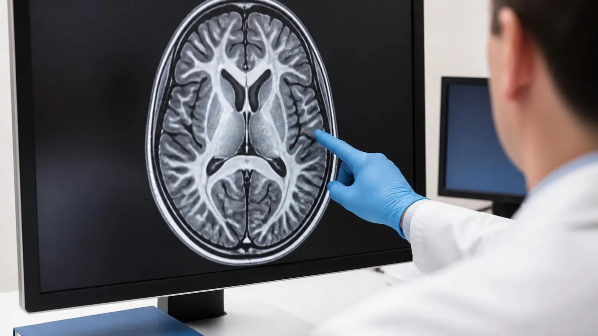
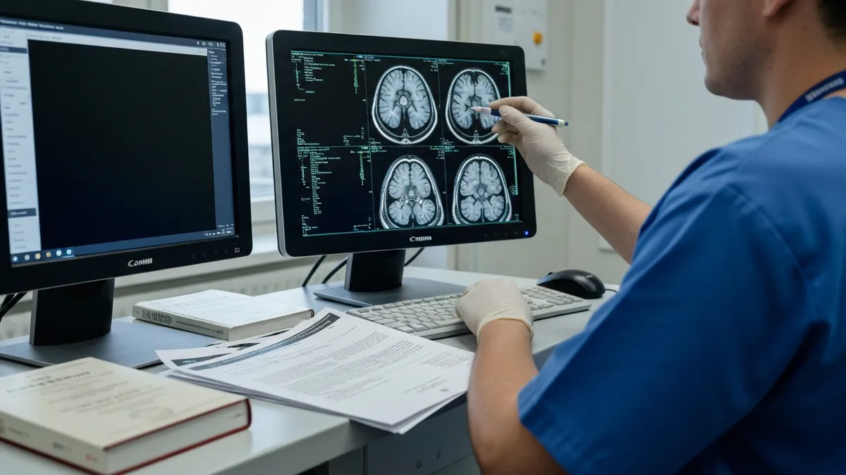
Standing MRI scanners almost universally operate at 0.5T to 0.6T, while modern closed-bore systems run at 1.5T or 3T. Because signal-to-noise ratio scales roughly linearly with field strength, a 0.5T image inherently carries far less signal per voxel than a 3T image. Technologists compensate with longer scan times, thicker slices, lower in-plane resolution, and optimized coil selection. For gross spinal anatomy this trade is acceptable, but small lesions, subtle marrow edema, and tiny labral tears can be missed.
Field homogeneity also matters. Permanent-magnet open systems achieve respectable uniformity inside a small imaging volume, but rapid drop-off near the magnet edges limits coverage. For a single lumbar segment or knee joint this is rarely an issue, but full thoracic spine imaging or wide field-of-view body scans are not practical on most upright units. Patients needing comprehensive whole-spine or contrast-enhanced oncologic imaging are nearly always better served by a high-field closed bore.
Standing MRI: Advantages and Trade-Offs
- +Demonstrates positional pathology invisible on supine MRI
- +Open design eliminates claustrophobia for nearly all patients
- +Accommodates bariatric, kyphotic, and post-surgical patients
- +Allows flexion, extension, rotation, and dynamic imaging
- +Pediatric patients often avoid general anesthesia
- +Family member can remain in direct view during scan
- +Useful objective evidence in workers comp and personal injury cases
- −Lower field strength reduces signal-to-noise ratio
- −Scan times are typically longer than 1.5T or 3T systems
- −Limited availability with only ~200 US centers
- −Standing immobility is difficult for severe pain patients
- −Not ideal for tumor, MS, cardiac, or vascular imaging
- −Some insurers require prior authorization or denial of supine MRI first
- −Smaller imaging volume limits whole-spine or body coverage
Patient Checklist Before Your Standing MRI
- ✓Confirm prior authorization with your insurer at least one week before the scan
- ✓Bring any previous MRI, CT, or X-ray images and reports on disc or via portal
- ✓List all implants including pacemakers, neurostimulators, cochlear devices, and surgical hardware
- ✓Disclose pregnancy, possible pregnancy, or current breastfeeding to scheduling staff
- ✓Remove jewelry, hairpins, underwire bras, and metal-threaded clothing before arrival
- ✓Eat a normal meal and stay well hydrated to reduce dizziness while upright
- ✓Take routine medications including pain medication unless told otherwise
- ✓Wear loose, two-piece clothing without zippers, snaps, or metallic prints
- ✓Arrive 30 minutes early to complete safety screening and consent forms
- ✓Tell the technologist immediately if you feel faint, dizzy, or numb during the scan
Order upright MRI when symptoms are clearly positional
If a patient's pain reliably worsens with standing, walking, or specific postures and improves with lying down, a recumbent MRI may underestimate the true pathology. In these cases, an upright weight-bearing scan in flexion, extension, and neutral can convert a non-diagnostic study into a surgical decision tool. Reserve the modality for genuine positional complaints rather than ordering it routinely.
Pricing for a standing MRI in the United States falls into a relatively narrow band. Self-pay rates at independent imaging centers typically range from $400 to $1,200 per body region, with most facilities clustered between $600 and $900 for a single lumbar or cervical spine study. Multi-region bundles are common: lumbar plus cervical, or bilateral knee studies, are often discounted 20 to 30 percent below the sum of individual prices. Hospital outpatient departments charge meaningfully more, sometimes two to three times the independent center rate.
Insurance coverage has improved substantially over the past decade. Medicare reimburses upright MRI under the same CPT codes used for conventional MRI of the spine, knee, or shoulder when medical necessity is documented. Most commercial insurers including Aetna, Cigna, UnitedHealthcare, and Blue Cross Blue Shield plans cover positional MRI with prior authorization, especially when a prior supine MRI was inconclusive or contradicted clinical findings. Workers compensation carriers routinely approve upright imaging for positional spine complaints.
Authorization denials are most common when the referring provider has not first documented a trial of conservative therapy or a prior non-diagnostic supine scan. The best practice is for the ordering clinician to include a clear positional history, prior imaging dates, and a statement that supine imaging was inadequate to explain symptoms. Peer-to-peer reviews succeed at high rates when these elements are present. Independent MRI imaging centers usually have authorization specialists who handle this paperwork directly.
Geographic access remains the largest practical limitation. The roughly 150 to 200 upright MRI centers in the United States are concentrated in major metropolitan areas, with significant gaps across the Mountain West, rural South, and northern New England. Patients in underserved areas often drive two to four hours to reach the nearest facility. Some employers and workers compensation carriers cover travel and lodging when upright imaging is required for case adjudication.
Cash pricing transparency has improved markedly with federal price disclosure rules. Most upright centers now publish self-pay prices on their websites or provide written estimates within 24 hours of request. HSA and FSA funds apply to upright MRI in full when the scan is ordered by a licensed clinician. Several centers offer payment plans for uninsured patients, and a few participate in direct-pay healthcare networks that reduce costs further.
For uninsured patients with clearly positional symptoms, the cost-benefit calculation usually favors the upright scan if surgery is on the table. A single weight-bearing study that either confirms operative pathology or rules it out is far cheaper than weeks of additional conservative therapy followed by a surgical decision made on incomplete imaging. Conversely, for vague mechanical back pain without red flags or positional features, the marginal benefit over a high-quality supine MRI is small.
One final cost consideration is image quality versus location. Patients sometimes choose a more distant high-quality 0.6T upright center over a closer 0.3T low-field open scanner. The difference in diagnostic yield can be substantial. Before scheduling, ask the facility specifically about field strength, coil availability for your body region, scan time, and whether positional sequences in flexion and extension are routinely included.
Although standing MRI scanners run at lower field strengths than closed-bore systems, they are still powerful magnets and pose identical risks for ferromagnetic implants, pacemakers, aneurysm clips, cochlear devices, and metallic foreign bodies. Complete the full MRI safety questionnaire at every visit, even if you have been scanned before, and disclose any new implants, surgeries, or occupational metal exposure since your last study.
Diagnostic accuracy is the question every referring clinician should ask before ordering an upright MRI. Peer-reviewed comparisons of 0.6T weight-bearing scans against 1.5T supine scans consistently show two findings: closed-bore systems produce higher overall image quality, and upright systems detect positional pathology that supine systems miss. The clinically important question is therefore not which scanner is better in the abstract, but which is better for the specific question being asked.
For lumbar disc herniations, sensitivity of upright MRI for compressive disc disease is broadly comparable to 1.5T supine imaging when the patient is symptomatic at the time of the scan. For spondylolisthesis grading and dynamic instability, upright imaging is meaningfully superior because the spine is loaded. For multiple sclerosis plaque burden, oncologic staging, or detailed cartilage mapping, closed-bore high-field imaging is clearly preferred and upright scans should not be substituted.
Resolution limits matter most in small joint and pediatric imaging. A 0.5T scan of a wrist or pediatric elbow may not resolve a subtle scaphoid stress fracture or a small osteochondral lesion that 3T imaging would catch. For these cases, referring physicians should weigh patient cooperation against image quality and frequently choose closed-bore imaging with mild sedation over an upright scan. Conversely, for a knee meniscal extrusion or patellofemoral maltracking question, weight-bearing imaging adds information that even 3T supine cannot provide.
Motion artifact is the dominant practical limitation. Holding a load-bearing posture for forty-five minutes is difficult even for healthy patients, and minor sway translates directly into image blur on a long acquisition. Skilled technologists mitigate this with posture supports, frequent micro-breaks, sequence ordering that captures the most important images first, and selective lowering of the platform to supine for sequences where positioning is not clinically critical. mri with and without contrast cpt is rarely performed on upright systems because gadolinium-enhanced sequences usually demand higher field strength.
Coil selection drives much of the difference between a good upright scan and a great one. A flexible cervical coil that wraps closely around the neck dramatically improves SNR compared to a generic body coil. The same is true for dedicated knee, shoulder, and lumbar coils. Patients and referring clinicians can and should ask whether the facility has the appropriate dedicated coil for the body region being studied. A facility that scans every patient with the same general-purpose coil is signaling lower-quality imaging.
Radiologist subspecialty experience also matters more than at a high-field center. Reading a 0.5T upright spine study requires familiarity with the appearance of positional pathology, common artifacts of the modality, and the contrast and signal characteristics of low-field imaging. Centers with fellowship-trained musculoskeletal or neuroradiology readers consistently produce more clinically useful reports than centers that rely on general radiologists unfamiliar with the modality.
The bottom line is straightforward. Standing MRI is a focused tool for positional spine and joint pathology, claustrophobic patients, and selected pediatric and bariatric populations. Used for the right indications, it produces information no other modality can. Used as a generic substitute for closed-bore imaging, it disappoints. Match the modality to the clinical question and the outcomes are excellent.
Preparing well for an upright MRI makes the difference between a diagnostic study and a wasted appointment. Start the day with a normal breakfast or lunch and good hydration, because the most common complication of standing MRI is orthostatic dizziness from prolonged upright posture. Patients who arrive fasted, dehydrated, or sleep-deprived are far more likely to need a position change mid-scan, which can compromise the very weight-bearing sequences the study was ordered to obtain.
Bring a complete imaging history. Prior supine MRI, CT, X-ray, and any operative reports give the upright radiologist a comparison baseline and dramatically improve report quality. Most centers accept images on CD, USB drive, or through electronic image-sharing portals. If you have had previous spine surgery, bring the operative note or at minimum the date, level, and type of hardware placed. Disclose every implant and foreign body during safety screening, including dental work, body piercings, and tattoos with metallic pigment.
Wear comfortable, loose, two-piece clothing free of zippers, metal snaps, underwire, and metallic prints. Many facilities provide gowns or scrub pants, but starting in MRI-appropriate clothing saves time. Leave jewelry, watches, hair clips, and credit cards at home or in a locker. If you wear glasses, contact lenses, hearing aids, or removable dentures, ask the technologist before the scan whether each item can stay in place during your specific protocol.
Manage pain proactively. Take your routine pain medication on schedule before the appointment unless your physician has told you otherwise. There is no benefit to arriving in maximum pain, and movement during the scan from discomfort is a major cause of non-diagnostic images. Patients with severe radiculopathy or post-surgical pain should specifically ask whether the protocol can be shortened, broken into segments, or modified to limit time in the most provocative position.
During the scan, communicate. The technologist can see and hear you continuously and can pause the sequence within seconds if you feel faint, numb, or unable to hold position. Many patients tolerate the scan better when they know breaks are available on request. Use the panic ball or call button without hesitation. Pushing through severe symptoms to avoid restarting a sequence almost always backfires when the resulting images are too blurred to interpret.
After the scan, ask when and how results will be delivered. Most upright centers produce a preliminary report within 24 to 48 hours and a final report within three to five business days. Request that images and the report be sent simultaneously to your referring clinician and to your patient portal. Bring printed copies to your follow-up appointment if your provider is outside the imaging network, and keep the original CD or digital file for future comparison studies.
For technologists studying for the ARRT MRI registry, treat every upright case as a teaching encounter. Pay attention to coil selection rationale, the effect of patient positioning on image quality, motion artifact patterns specific to standing acquisition, and the safety considerations unique to a vertically oriented magnet. These details show up on board exams under questions about low-field imaging, open systems, and patient management in non-traditional positions.
MRI Questions and Answers

The History of MRI: From Discovery to Modern Medicine

MRI With and Without Contrast: How It Works, What to Expect

MRI Medical Abbreviation: What MRI Stands For and Why It Matters
MRI Imaging Centers: Complete Guide to Independent Outpatient MRI Facilities
St Jude Pacemaker MRI Compatibility List: A Complete Safety and Scanning Guide
About the Author
Medical Laboratory Scientist & Clinical Certification Expert
Johns Hopkins UniversityDr. Sandra Kim holds a PhD in Clinical Laboratory Science from Johns Hopkins University and is certified as a Medical Technologist (MT) and Medical Laboratory Scientist (MLS) through ASCP. With 16 years of clinical laboratory experience spanning hematology, microbiology, and molecular diagnostics, she prepares candidates for ASCP board exams, MLT, MLS, and specialist certification tests.
Join the Discussion
Connect with other students preparing for this exam. Share tips, ask questions, and get advice from people who have been there.
View discussion (6 replies)