MRI MRCP: How Magnetic Resonance Cholangiopancreatography Works, What It Shows, and What to Expect
MRI MRCP explained — 📗 how magnetic resonance cholangiopancreatography images bile and pancreatic ducts, prep tips, results, costs, and what to expect.
An MRI MRCP, short for magnetic resonance cholangiopancreatography, is a specialized magnetic resonance imaging study that produces remarkably detailed, non-invasive pictures of the biliary tree, gallbladder, and pancreatic ducts. Unlike older diagnostic methods that required an endoscope to be threaded through the upper digestive tract, an MRI MRCP uses powerful magnets and radio waves to generate images while the patient simply lies still on the scanner table. Radiologists rely on these images to detect gallstones, strictures, tumors, congenital variants, and inflammation that would otherwise require an invasive procedure to visualize.
The technology behind MRCP works because bile and pancreatic juice are static, fluid-filled structures that appear extremely bright on heavily T2-weighted sequences. Surrounding solid tissues appear dark, creating a natural contrast that highlights the ductal anatomy without intravenous contrast in most cases. Patients receive a comprehensive look at the common bile duct, intrahepatic ducts, cystic duct, and pancreatic duct in a single session — typically lasting between thirty and forty-five minutes from start to finish.
Because MRCP is non-ionizing, it avoids the radiation exposure associated with CT scans and the procedural risks of endoscopic retrograde cholangiopancreatography (ERCP). That makes it an attractive first-line option for pregnant patients, younger patients with recurrent abdominal pain, and anyone who needs repeat imaging over time. Many imaging providers, including most major hospital systems and outpatient MRI imaging centers, now offer MRCP as a standard protocol on 1.5T and 3.0T scanners.
Referring physicians order MRCP when they suspect a problem in the hepatobiliary system but want to avoid the complications associated with ERCP, which carries a small but real risk of pancreatitis, bleeding, and perforation. Common indications include unexplained jaundice, abnormal liver function tests, suspected choledocholithiasis, recurrent pancreatitis, and pre-operative mapping before liver or pancreatic surgery. The exam can also evaluate post-surgical complications such as bile leaks or anastomotic strictures following liver transplantation.
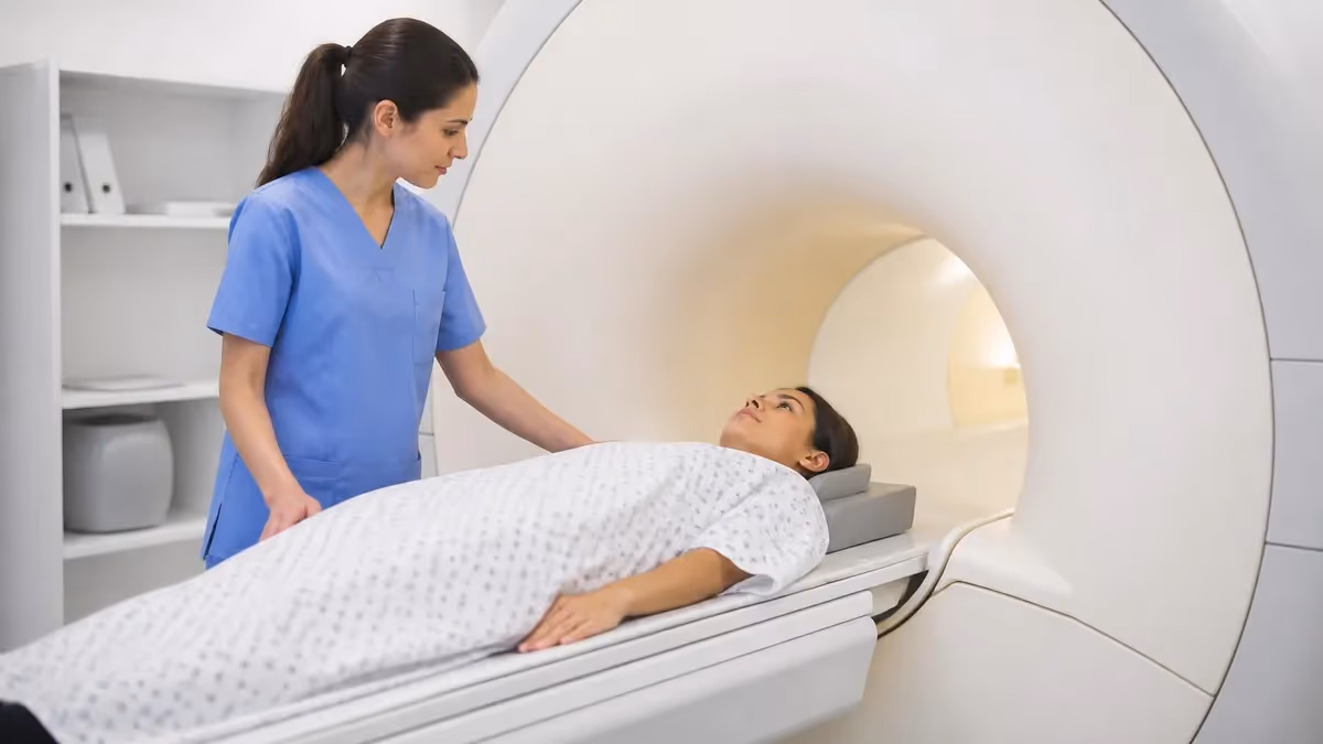
One of the most attractive features of MRCP is that it requires very little mri of cervical spine. There are no incisions, no sedation in most cases, and no intravenous contrast for the basic study. Patients are typically asked to fast for four to six hours beforehand to reduce duodenal fluid and improve gallbladder distension, but otherwise the experience is identical to a routine abdominal MRI. For patients who are claustrophobic, anxious, or unable to hold their breath, technologists can adjust sequences or offer mild oral sedation when medically appropriate.
Modern MRCP protocols often pair the heavily T2-weighted ductal images with conventional T1 and T2 abdominal sequences, plus optional contrast-enhanced phases using gadoxetate disodium (Eovist) to assess hepatic function and bile excretion. This combined approach gives radiologists a complete picture of the liver, biliary tree, pancreas, and surrounding organs in one visit. Throughout this article we will walk through how the exam is performed, what it shows, how to prepare, how much it costs, and how to interpret the report you receive.
Whether you have been scheduled for your first MRCP or you are a technologist or student studying for a board exam, understanding the physics, anatomy, and clinical role of this imaging study will help you feel confident going into the scanner and discussing the findings afterward. The remainder of this guide breaks the topic down into clear sections covering protocol, mri appointment abnormalities look like on the images, comparison with ERCP and ultrasound, and answers to the questions patients ask most often.
MRI MRCP by the Numbers

MRCP Protocol and Sequences
A respiratory-triggered, fat-saturated three-dimensional sequence with very long echo times that makes static fluid extremely bright. This is the workhorse sequence that produces the maximum intensity projection (MIP) images radiologists use to map the ductal tree.
Rapid breath-hold thick-slab acquisitions, typically 40–70 mm, give a quick overview of the biliary tree in coronal and oblique planes. These images are useful for orientation and patient triage, especially when 3D sequences fail due to motion.
Half-Fourier single-shot turbo spin echo sequences provide background anatomy of the liver, pancreas, and surrounding structures. They help radiologists localize ductal abnormalities and identify masses, cysts, or fluid collections that explain the symptoms.
Dual-echo T1 imaging detects fatty liver, hepatic iron overload, and adrenal lesions that are often incidental on MRCP. The opposed-phase signal drop also helps characterize adenomas and other lipid-containing masses.
For functional biliary imaging or evaluation of bile leaks, gadoxetate disodium is administered intravenously. Delayed hepatobiliary-phase images at 20 minutes show bile excretion into the ducts, helping identify leaks, strictures, and biliary anatomy variants.
What MRI MRCP actually shows on the final images is a striking three-dimensional roadmap of every fluid-filled channel in the upper abdomen. The intrahepatic bile ducts appear as branching white lines threading through the dark liver parenchyma, joining together to form the right and left hepatic ducts, then the common hepatic duct, the cystic duct from the gallbladder, and finally the common bile duct that empties into the duodenum at the ampulla of Vater. The pancreatic duct runs horizontally through the gland and joins the common bile duct just before the ampulla.
Gallstones, the most common reason MRCP is ordered, appear as round or faceted dark filling defects against the bright bile background. Stones as small as three or four millimeters can be detected with high-resolution 3D sequences, although the sensitivity drops considerably for stones under five millimeters. Radiologists also look for stones lodged in the cystic duct, which can produce Mirizzi syndrome by compressing the adjacent common hepatic duct. The shape of the obstruction and the degree of upstream ductal dilation help differentiate stones from tumors.
Strictures of the bile duct, whether benign or malignant, show up as focal narrowings with proximal dilation. A short, smooth, tapered stricture often indicates a benign cause such as chronic pancreatitis or a post-surgical scar. A longer, irregular, shouldered stricture is more concerning for cholangiocarcinoma. Primary sclerosing cholangitis produces a characteristic beaded appearance with multifocal strictures and saccular dilations affecting both intrahepatic and extrahepatic ducts. Reviewing prior MRI with and without contrast studies side-by-side often clarifies whether a finding is new or stable.
The pancreatic duct is a critical structure that MRCP visualizes exceptionally well. A normal duct measures up to three millimeters in the head, two millimeters in the body, and one and a half millimeters in the tail. Dilation, abrupt cutoffs, side branch dilation, or filling defects all raise concern for pancreatic pathology including chronic pancreatitis, intraductal papillary mucinous neoplasm (IPMN), or pancreatic adenocarcinoma. The double duct sign — simultaneous dilation of both the pancreatic and common bile ducts — is a classic indicator of a periampullary mass.
MRCP also detects congenital anatomic variants that may have clinical significance. Pancreas divisum, in which the dorsal and ventral pancreatic ducts fail to fuse, is identified by tracing the dorsal duct as it crosses anterior to the common bile duct and drains into the minor papilla. Choledochal cysts, classified by the Todani system into five types, appear as focal or diffuse cystic dilations of the biliary tree. These variants are important to recognize because they affect surgical planning and increase the lifetime risk of cholangiocarcinoma.
Beyond the ductal anatomy, the supporting sequences on an MRCP exam evaluate the liver parenchyma, pancreas, gallbladder wall, and adjacent organs. Radiologists often identify incidental findings such as hepatic cysts, hemangiomas, focal nodular hyperplasia, renal cysts, and adrenal nodules. While most of these are benign, some require further characterization with dedicated contrast-enhanced MRI. Reporting templates typically address each organ system in turn so the referring physician receives a complete abdominal assessment, not just a ductal study.
One challenge with MRCP interpretation is differentiating real pathology from artifact. Pneumobilia after sphincterotomy, surgical clips, breathing motion, and pulsatile flow can all create dark spots that mimic stones. Experienced radiologists cross-check 3D images against 2D thick-slab images and source data to confirm any finding. Knowing the patient's surgical history, recent ERCP, or stent placement is essential for accurate interpretation, so technologists are trained to gather a focused clinical history before scanning begins.
MRI Practice Test Questions
Prepare for the MRI - Magnetic Resonance Imaging exam with our free practice test modules. Each quiz covers key topics to help you pass on your first try.
MRI Knowledge
MRI Exam Questions covering Knowledge. Master MRI Test concepts for certification prep.
MRI Physics
Free MRI Practice Test featuring Physics. Improve your MRI Exam score with mock test prep.
MRI Anatomy and Pathology
MRI Test Prep for MRI Anatomy and Pathology. Practice MRI Quiz questions and boost your score.
MRI Anatomy and Positioning
MRI Questions and Answers on MRI Anatomy and Positioning. Free MRI practice for exam readiness.
MRI Contrast Agents
Free MRI Quiz on MRI Contrast Agents. MRI Exam prep questions with detailed explanations.
MRI Patient Care and Positioning
MRI Practice Questions for MRI Patient Care and Positioning. Build confidence for your MRI certification exam.
MRCP vs ERCP vs Ultrasound
MRCP is entirely non-invasive, uses no ionizing radiation, and provides excellent visualization of the biliary tree and pancreatic duct in a single thirty-minute exam. It is the preferred test when there is moderate clinical suspicion of biliary disease but no immediate need for therapeutic intervention. Sensitivity for common bile duct stones larger than six millimeters approaches 95 percent, and specificity is generally above 90 percent in experienced hands.
The main limitations of MRCP are spatial resolution compared to direct cholangiography, difficulty detecting very small stones, and contraindications related to implanted devices, severe claustrophobia, or inability to lie still for the required time. MRCP is also purely diagnostic — it cannot remove stones, place stents, or take biopsies. When intervention is anticipated, ERCP remains the appropriate next step after MRCP findings guide planning.
Is MRCP the Right Test for You?
- +No ionizing radiation makes it safe for pregnant patients and repeat imaging
- +Non-invasive with no incisions, sedation, or recovery time required
- +High sensitivity for common bile duct stones larger than six millimeters
- +Visualizes the entire biliary and pancreatic ductal anatomy in one exam
- +Detects congenital variants like pancreas divisum and choledochal cysts
- +Avoids the 3–10% risk of post-ERCP pancreatitis associated with endoscopy
- +Provides comprehensive abdominal organ assessment alongside ductal imaging
- −Cannot perform therapeutic intervention like stone removal or stent placement
- −Reduced sensitivity for stones smaller than five millimeters
- −Contraindicated in patients with certain implanted devices or severe claustrophobia
- −Requires 30–45 minutes of lying still inside the scanner bore
- −More expensive than ultrasound as a first-line screening test
- −Image quality degrades with breathing motion or uncooperative patients
- −Limited availability after hours compared to CT or ultrasound
MRI MRCP Preparation Checklist
- ✓Fast from solid food and fluids for four to six hours before your appointment
- ✓Continue taking essential medications with small sips of water unless told otherwise
- ✓Remove all metal jewelry, piercings, hair accessories, and dental appliances
- ✓Wear loose clothing without metal zippers, buttons, or wire underwire bras
- ✓Complete the MRI safety screening form covering implants, surgeries, and pregnancy status
- ✓Notify staff if you have pacemakers, aneurysm clips, cochlear implants, or metal fragments
- ✓Inform the technologist about claustrophobia so mild oral sedation can be arranged
- ✓Drink a small amount of pineapple or blueberry juice if provided to suppress bowel signal
- ✓Empty your bladder immediately before entering the scanner room for comfort
- ✓Bring a list of prior abdominal surgeries and recent imaging studies for the radiologist
MRCP relies on naturally bright fluid, not injected contrast
The reason MRCP works without intravenous contrast in most cases is that bile and pancreatic juice are static fluids that produce extremely bright signal on heavily T2-weighted sequences with long echo times. Surrounding solid tissue produces almost no signal at TE values above 600 ms, so the ductal anatomy stands out automatically. This physics-based contrast is what makes MRCP uniquely non-invasive and reproducible.
The cost of an MRI MRCP varies dramatically depending on where you have the exam performed, what insurance you carry, and whether contrast is added to the protocol. Hospital-based imaging departments typically charge between two thousand and five thousand dollars in total billed charges, while freestanding outpatient imaging centers often charge between five hundred and fifteen hundred dollars for the same study. Insured patients with deductibles still unmet may pay several hundred dollars out of pocket, while patients who have met their deductible often pay only a small co-insurance percentage.
Insurance coverage for MRCP is generally straightforward when the exam is ordered for a clearly documented indication such as suspected choledocholithiasis, abnormal liver function tests, recurrent pancreatitis, or pre-operative planning. Most major insurers, including Medicare and Medicaid, cover MRCP when the appropriate ICD-10 diagnosis codes accompany the order. Pre-authorization is commonly required, and your ordering physician's office typically handles that process. If pre-authorization is denied, a peer-to-peer review with the insurance medical director often reverses the decision when the clinical justification is strong.
Scheduling an MRCP usually takes one to two weeks at most outpatient centers, although urgent cases can often be accommodated within twenty-four to forty-eight hours when ordered as STAT. Saturday and evening appointments are increasingly available as imaging centers compete on convenience. When choosing a facility, ask whether they use a 1.5T or 3.0T scanner — the higher field strength generally produces better quality MRCP images — and whether subspecialty-trained abdominal radiologists interpret the studies.
Patients with high-deductible insurance plans or no insurance should ask about cash-pay or self-pay rates, which are often substantially lower than the billed charges. Many imaging centers offer transparent self-pay pricing on their websites or by phone, and some negotiate further discounts for payment at the time of service. Comparing prices across two or three facilities in your area can easily save a thousand dollars or more for the same study performed on similar equipment.
Health savings accounts (HSAs) and flexible spending accounts (FSAs) can be used to pay for MRCP costs, including any portion not covered by insurance. Keep the itemized bill and the order from your physician in case the tax-advantaged account administrator requests documentation. If you are referred for MRCP as part of an evaluation that ultimately leads to surgery, the imaging costs are part of the overall episode of care and may count toward an out-of-pocket maximum that limits your total annual spending.
For patients who need repeat MRCP imaging — such as those with primary sclerosing cholangitis, IPMN surveillance, or post-transplant monitoring — establishing a long-term relationship with a single imaging facility offers important advantages. Prior studies can be compared directly, the radiologists become familiar with your anatomy, and the protocols can be standardized across exams. Consistency in technique improves the radiologist's ability to detect subtle interval changes that might otherwise be attributed to differences in image acquisition.
If your physician orders MRCP, ask whether contrast will be administered and whether a same-day diagnostic abdominal MRI is also planned. Combining studies can save time and money, but it also extends the appointment to sixty or even ninety minutes. Patients who are uncomfortable with long scan times should discuss splitting the exams across two visits, or pre-medicating with anxiolytics, before the day of the appointment.
Tell the MRI technologist about any implanted devices including pacemakers, defibrillators, cochlear implants, aneurysm clips, neurostimulators, and insulin pumps before entering the scanner room. Some older devices are absolute contraindications to MRI, while others are MR-conditional and require specific scanning parameters. Failure to disclose implants can result in device malfunction, tissue heating, or serious injury during the exam.
Reading the report you receive after an MRI MRCP becomes much easier when you understand the standard reporting structure radiologists use. The report typically begins with a clinical history summary, lists the technique used (sequences and contrast administration), and then walks through each organ system in turn: liver, biliary tree, gallbladder, pancreas, spleen, kidneys, adrenals, bowel, and vasculature. Each section notes findings and provides measurements where relevant. The impression section at the bottom synthesizes the key findings and provides recommendations for follow-up.
Common phrases in MRCP reports include descriptors of duct caliber, signal characteristics, and filling defects. A normal common bile duct measures up to six or seven millimeters, increasing slightly with age and after cholecystectomy. A duct that measures eight millimeters or more in a younger patient with intact gallbladder is considered dilated and warrants explanation. Filling defects are described by size, shape, location, and whether they are mobile on different sequences — features that help distinguish stones from sludge, tumor, or artifact.
When the report mentions the pancreatic duct, pay attention to its caliber and contour. A normal duct is smooth, tapering, and measures up to three millimeters in the head. Irregular contour, focal dilation, or side-branch ectasia raises concern for chronic pancreatitis or branch-duct IPMN. The radiologist will often recommend interval surveillance MRI for small IPMNs that meet criteria for low-risk lesions, with the surveillance interval depending on cyst size and growth rate. Discussing prior shoulder mri without contrast cpt or other unrelated studies is rarely useful, but prior abdominal imaging always provides important context.
If your report mentions cholangiocarcinoma, the description usually includes location (intrahepatic, perihilar/Klatskin, or distal), length of involvement, vascular invasion, and lymph node assessment. Klatskin tumors are further classified by the Bismuth-Corlette system based on which hepatic ducts are involved. This information is critical for surgical planning, and patients with these findings will typically be referred to a hepatobiliary surgical oncology team for multidisciplinary review.
Choledocholithiasis findings will include the size, number, and location of stones within the common bile duct. Radiologists will note whether the stones are obstructing flow, whether upstream ducts are dilated, and whether there is any evidence of cholangitis such as duct wall thickening or enhancement. These findings drive the decision between same-admission ERCP, elective ERCP, or surgical common bile duct exploration during cholecystectomy.
If the report notes a normal MRCP with no biliary or pancreatic abnormality, the reader should still review the rest of the abdominal findings carefully. Incidental hepatic lesions, renal cysts, adrenal nodules, and ovarian cysts are common and may require characterization with additional imaging or simply documentation for the medical record. Your physician will integrate the MRCP findings with your symptoms, lab results, and physical exam to decide what comes next.
Patients often want to discuss the MRCP report directly with the radiologist, and many practices now offer patient consultation services to support this. While the ordering physician remains the primary point of communication, a radiologist consultation can be helpful when the findings are complex or when a second opinion on a specific finding is desired. Bringing the report and prior imaging to the consultation ensures the most productive conversation possible.
Preparing for your MRI MRCP appointment with a few practical steps can make the experience significantly more comfortable and produce better diagnostic images. Start by confirming the fasting requirement directly with the imaging center the day before — most ask for four to six hours of fasting, but some prefer eight hours for optimal gallbladder distension. Avoid carbonated beverages in the hours before the exam because gas in the duodenum can degrade image quality and create artifacts that obscure the distal common bile duct.
Wear comfortable, metal-free clothing to the appointment. Most facilities provide a gown, but arriving in soft pants and a simple shirt without zippers, snaps, or wire underwire shortens the check-in process. Leave jewelry, watches, and accessories at home or in a secure locker. Hair clips, bobby pins, and certain hair extensions can also cause artifacts or pose safety issues, so address those before you arrive. If you wear hearing aids, they must be removed before entering the scanner room.
Plan for the exam to take longer than the scan itself. Allow about ninety minutes total: check-in and safety screening (15 minutes), changing and IV placement if contrast is planned (10 minutes), scanning (30–45 minutes), and post-exam observation if contrast was given (15 minutes). Arriving fifteen minutes early helps ensure you start on time and reduces stress that can make it harder to lie still in the scanner.
If you have a history of claustrophobia or anxiety in enclosed spaces, talk with your referring physician before the appointment about a small dose of oral lorazepam or a similar short-acting anxiolytic. The medication should be taken about thirty to sixty minutes before the scan, and you must bring a driver because you cannot operate a vehicle for several hours afterward. Wide-bore 70 cm scanners are now standard at most modern facilities and significantly reduce claustrophobia compared to older 60 cm bores.
During the scan itself, the technologist will give you breath-hold instructions for some sequences and ask you to breathe normally for others. Listen carefully and follow the instructions exactly — breath-holds typically last fifteen to twenty seconds, and inconsistent timing degrades image quality. Many centers now use respiratory triggering or navigator gating that synchronizes acquisition with your natural breathing cycle, eliminating the need for prolonged breath-holds and producing sharper images.
After the exam, you can resume normal activities immediately unless you received intravenous contrast or oral sedation. If gadoxetate or another gadolinium agent was administered, drink extra water for the next twenty-four hours to support renal clearance. Patients with significant kidney disease should have had their renal function checked beforehand to ensure safe contrast administration. side effects of mri from gadolinium are uncommon and usually mild — a brief metallic taste, transient nausea, or a feeling of warmth at the injection site.
Results are typically available within twenty-four to seventy-two hours, although STAT studies can be read within an hour. Your referring physician will receive the report electronically and will contact you to discuss the findings and next steps. Many patient portals now provide direct access to imaging reports and even the images themselves, although interpreting them without medical training can lead to unnecessary worry. Save your questions for the follow-up appointment when you can review the report with a clinician who knows your full history.
MRI Questions and Answers

The History of MRI: From Discovery to Modern Medicine

MRI Medical Abbreviation: What MRI Stands For and Why It Matters

MRI With and Without Contrast: How It Works, What to Expect
Shoulder MRI: What It Shows, Procedure, and Reading the Report
MRI Imaging Centers: Complete Guide to Independent Outpatient MRI Facilities
About the Author
Medical Laboratory Scientist & Clinical Certification Expert
Johns Hopkins UniversityDr. Sandra Kim holds a PhD in Clinical Laboratory Science from Johns Hopkins University and is certified as a Medical Technologist (MT) and Medical Laboratory Scientist (MLS) through ASCP. With 16 years of clinical laboratory experience spanning hematology, microbiology, and molecular diagnostics, she prepares candidates for ASCP board exams, MLT, MLS, and specialist certification tests.
Join the Discussion
Connect with other students preparing for this exam. Share tips, ask questions, and get advice from people who have been there.
View discussion (6 replies)