Boy Killed During MRI Scan: What Happened and How MRI Safety Failures Cause Tragic Incidents
✏️ Boy killed during MRI scan: learn how the 2001 Michael Colombini tragedy and other MRI safety incidents shaped modern scanner safety protocols.
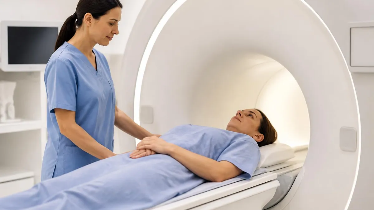
The phrase boy killed during MRI scan most often refers to the 2001 death of six-year-old Michael Colombini at Westchester Medical Center in Valhalla, New York. A ferromagnetic oxygen tank was carried into Zone IV of the MRI suite while Michael was being scanned. The tank became a projectile, accelerating toward the magnet bore and striking the child in the head. Michael died two days later. The case became the most widely cited MRI safety tragedy in modern radiology and reshaped how facilities zone, screen, and supervise every magnetic resonance scan performed in the United States.
MRI safety incidents are rare relative to the tens of millions of scans performed each year, but when they happen they are catastrophic. The static magnetic field of a clinical 1.5T or 3T scanner is always on, even when the technologist is not actively imaging. Any iron, nickel, or cobalt object carried into the scan room can become a high-velocity projectile. Oxygen tanks, IV poles, scissors, hairpins, hospital beds, and floor buffers have all been pulled into magnet bores, sometimes injuring or killing patients and staff who happened to be in the path.
This article walks through what happened to Michael Colombini, the chain of failures that allowed an oxygen tank into Zone IV, and the other documented MRI fatalities and serious injuries that have followed. We explain the four-zone screening model the American College of Radiology now recommends, the role of ferromagnetic detectors, and the way burns, hearing damage, and contrast reactions also contribute to the broader MRI safety record. The goal is not sensationalism — it is to make clear how preventable most of these tragedies actually were.
If you are a patient, a parent, or a student preparing for a registry exam, understanding MRI safety incidents helps you ask better questions before a scan and recognize the warning signs of a poorly run facility. Technologists carry the heaviest responsibility, but everyone who enters the controlled area of an MRI suite shares a piece of it. The fundamental rule is unchanged since 2001: nothing ferromagnetic crosses the Zone III line without explicit clearance from the MR-trained personnel on duty.
We will also cover the regulatory response. The Joint Commission issued Sentinel Event Alert #38 in 2008 to address MRI safety after Michael's death and a series of follow-up incidents. The ACR's Manual on MR Safety, now in its current 2024 edition, codified the zoning system and personnel categories that most hospitals follow. The FDA tracks adverse events through its MAUDE database, and reports show that despite improved protocols, projectile events, thermal injuries, and quench-related incidents still occur every year in the United States.
Finally, we will look at what has changed and what has not. Ferromagnetic detection systems at the door of Zone IV are now common but not universal. Screening forms are more thorough, but verbal screening remains inconsistent. Sedated and unconscious patients depend entirely on the staff around them, and most fatalities involve some combination of communication breakdown, fatigue, and assumed familiarity. Reading these case studies is uncomfortable, but it is the most direct way to understand why MRI safety culture matters as much as the physics behind the images.
Throughout the article you will find links to related topics such as mri with and without contrast cpt, quiz tiles for registry prep, and a detailed FAQ. By the end you should be able to explain the Colombini case, name the four ACR zones, list the most common projectile objects, and describe the screening steps a properly run MRI department uses on every single patient, every single day.
MRI Safety Incidents by the Numbers
Timeline of the Michael Colombini Case
July 25, 2001 — Scan Begins
Oxygen Tank Carried In
Projectile Strike
July 27, 2001 — Death
2008 — Sentinel Event Alert #38
2024 — ACR Manual Updated
To understand why a boy was killed during an MRI scan, you have to understand what happens when ferromagnetic material enters a strong static magnetic field. Clinical MRI scanners operate at field strengths of 1.5 Tesla or 3 Tesla, which is roughly 30,000 to 60,000 times the strength of Earth's natural magnetic field. The field is always on. There is no switch the technologist can flip to make the scanner safe to enter, short of a quench — a controlled venting of the superconducting magnet's liquid helium that is essentially a one-time emergency action.
When a ferromagnetic object enters this field, it experiences a force pulling it toward the magnet's center, where field strength is highest. Small objects like hairpins, paper clips, and pens move quickly. Larger objects like oxygen tanks, IV poles, scissors, and floor buffers can accelerate to dangerous speeds in fractions of a second. The closer the object gets to the bore, the stronger the pull. This nonlinear behavior is why staff often describe the projectile effect as feeling like the object was "ripped" from their hands without warning.
The classic MRI projectile incidents involve oxygen cylinders. Standard medical D and E cylinders are made of steel and weigh between 5 and 15 pounds when full. At magnet bore field strengths, that weight is effectively multiplied by the magnetic attraction, and the cylinder behaves like a missile. Aluminum cylinders, which are non-ferromagnetic, were available before 2001 but were not universally deployed. After Michael Colombini's death, aluminum became standard in hospital MRI suites, often color-coded so staff can distinguish them at a glance from older steel tanks.
Other objects that have caused documented injuries include stethoscopes, hemostats, defibrillator paddles, scissors, mop buckets, vacuum cleaners, wheelchairs, and even police firearms carried by officers escorting patients. Any of these can become projectiles. The danger is not limited to objects clearly identifiable as metal — many modern devices contain hidden ferromagnetic components in batteries, motors, or springs. This is why ferromagnetic detection systems and rigorous screening matter, and why "I thought it was safe" is the most common phrase appearing in adverse event reports filed with the FDA.
Patients with implants face a different but related risk. Older aneurysm clips, certain pacemakers, cochlear implants, and some orthopedic hardware can heat, move, or malfunction in the magnetic field. The MR safety labeling system — MR Safe, MR Conditional, and MR Unsafe — was introduced in part to help technologists make rapid decisions about implanted devices. Conditional implants can usually be scanned, but only under specific field strength, gradient, and SAR limits documented by the manufacturer. Skipping that documentation step has caused serious patient injuries, including burns and pacemaker reprogramming events.
Burns are another frequently reported MRI safety incident. They occur when conductive loops form against the patient's skin — for example, when an EKG lead, a wet washcloth, or even crossed arms create a closed electrical loop. Radiofrequency energy from the scanner heats the loop, sometimes producing second-degree burns within minutes. Tattoo pigments with iron oxide content have caused similar focal burns, though most modern inks are not ferromagnetic enough to be a problem. Padding, careful patient positioning, and avoidance of skin-to-skin contact loops are the standard countermeasures.
Hearing damage is the most common MRI-related injury overall. Gradient coil noise during fast sequences can exceed 110 decibels, comparable to a chainsaw or rock concert. Every patient should be offered ear protection — foam plugs, padded headphones, or both — and pediatric patients require especially careful coverage. While not a fatality risk, untreated noise exposure can cause permanent hearing loss after a single scan, particularly in infants and children whose ear canals provide less natural attenuation than adult ears do.
MRI Practice Test Questions
Prepare for the MRI - Magnetic Resonance Imaging exam with our free practice test modules. Each quiz covers key topics to help you pass on your first try.
MRI Knowledge
MRI Exam Questions covering Knowledge. Master MRI Test concepts for certification prep.
MRI Physics
Free MRI Practice Test featuring Physics. Improve your MRI Exam score with mock test prep.
MRI Anatomy and Pathology
MRI Test Prep for MRI Anatomy and Pathology. Practice MRI Quiz questions and boost your score.
MRI Anatomy and Positioning
MRI Questions and Answers on MRI Anatomy and Positioning. Free MRI practice for exam readiness.
MRI Contrast Agents
Free MRI Quiz on MRI Contrast Agents. MRI Exam prep questions with detailed explanations.
MRI Patient Care and Positioning
MRI Practice Questions for MRI Patient Care and Positioning. Build confidence for your MRI certification exam.
Documented MRI Safety Incidents
The Michael Colombini case remains the defining MRI fatality in the United States. A steel oxygen cylinder was brought into the scan room by an anesthesiologist responding to falling oxygen saturation in the sedated six-year-old patient. The cylinder was pulled into the bore and struck the child in the head, causing fatal injuries. The hospital later settled a wrongful death suit with the family in 2010.
The case became the centerpiece of MRI safety training nationwide. It is cited in nearly every ACR safety document, every residency lecture on MR safety, and every registry exam question about projectile risk. The legacy of the Colombini case is the universal adoption of aluminum cylinders in MRI areas, mandatory screening of every item before Zone IV entry, and visible signage warning of the always-on magnetic field.
Ferromagnetic Detection Systems: Are They Worth It?
- +Detect hidden ferromagnetic objects before they reach the magnet bore
- +Alert staff to items missed by verbal and visual screening
- +Provide an objective second layer of safety beyond human judgment
- +Recommended by the ACR Manual on MR Safety as best practice
- +Reduce projectile incidents and near-misses in published studies
- +Help protect sedated or unconscious patients who cannot self-report
- −Initial purchase and installation costs can exceed $20,000 per unit
- −Require regular calibration and staff training to function correctly
- −Generate false positives that can slow patient throughput
- −Cannot detect implants already inside the patient's body
- −Some legacy MRI suites lack space for proper threshold installation
- −Not yet mandated by federal regulation, only recommended by professional bodies
Essential MRI Safety Checklist
- ✓Confirm patient identity and the specific exam ordered before entering Zone III.
- ✓Complete a written and verbal MR safety screening form for every patient.
- ✓Ask about implants, surgical hardware, pacemakers, and metal fragments in eyes.
- ✓Have the patient change into MR-safe scrubs to eliminate hidden metallic items.
- ✓Pass the patient and all accompanying staff through a ferromagnetic detector.
- ✓Verify all ancillary equipment entering Zone IV is labeled MR Safe or MR Conditional.
- ✓Use only aluminum oxygen cylinders in MRI suites and never carry steel cylinders in.
- ✓Provide hearing protection and confirm it is properly placed before scanning begins.
- ✓Maintain continuous visual and audio contact with sedated or anxious patients.
- ✓Document any near-miss or adverse event through the facility's incident reporting system.
Nothing ferromagnetic crosses Zone IV — ever, under any circumstance.
Every documented MRI fatality involves a violation of this rule. There is no acceptable exception, no "just this once," and no situation in which carrying an unscreened metal object into the scan room is safer than pausing to swap it for an MR-safe alternative. If wall oxygen fails, switch to an aluminum cylinder. If the patient is crashing, move them out of Zone IV before resuscitating with standard equipment. The magnet does not care about urgency.
The American College of Radiology defines four zones inside an MRI facility, and understanding them is the foundation of every safety policy in modern radiology. Zone I is the freely accessible public area outside the controlled MRI environment — the waiting room, the front desk, the corridor. Anyone can be there. Zone II is the transitional area where patients are greeted, screened, and changed into scrubs. Staff supervise Zone II, but it is not yet considered restricted. Most facility entry points sit somewhere between Zone I and Zone II.
Zone III is the controlled area immediately surrounding the magnet room. Only screened patients and MR personnel may enter. Zone III is where ferromagnetic detection systems are typically installed, and where the screening forms and final verbal confirmation take place. Doors leading from Zone II to Zone III must be locked, supervised, or both. This is the line that the steel oxygen tank in the Colombini case should never have crossed. The Joint Commission specifically calls out Zone III access control in Sentinel Event Alert #38.
Zone IV is the MRI scanner room itself. The static magnetic field is always present, and only fully screened individuals carrying only fully screened equipment may enter. Zone IV is sometimes marked with a red line on the floor and prominent signage warning of the magnet. The ACR recommends that Zone IV be locked when not in use and that no one enter alone outside of an emergency. Patients exiting Zone IV pass back through Zone III to Zone II to Zone I as they leave.
The ACR also defines two categories of MR personnel. Level 1 MR personnel have completed basic MR safety training and can work within Zone III under supervision. Level 2 MR personnel — typically MR technologists, MR radiologists, and MR safety officers — have completed advanced training and can supervise patients and ancillary staff in Zone IV. The distinction matters because non-MR personnel like janitors, transport staff, and even ER physicians count as patients for safety purposes and must be escorted and screened just like a patient would be.
Screening is the heart of zone enforcement. A properly designed MRI screening form asks about pacemakers, implanted defibrillators, neurostimulators, cochlear implants, aneurysm clips, heart valves, stents, drug pumps, port-a-caths, joint replacements, surgical hardware, foreign metal fragments, tattoos, pregnancy, and previous reactions to gadolinium contrast. It also asks about recent surgeries, occupation (welders and machinists may have metal fragments in their eyes), and any history of MRI scans. Patients are asked these questions twice — once on paper and once verbally.
Some facilities use orbital radiographs to screen patients with potential metal fragments in the eyes before scanning. The risk is that intraocular metal can move under magnetic force and damage the retina. The ACR recommends screening anyone with a history of metal-on-metal grinding, welding without eye protection, or penetrating eye trauma. Plain film radiography is inexpensive and reliable, and the small radiation dose is acceptable when weighed against the consequences of intraocular metal motion inside a 1.5T or 3T field.
Sedated patients, pediatric patients, and unconscious patients receive extra attention because they cannot self-report. The screening process shifts to medical records, parental interview, and physical examination — sometimes including portable radiographs if implant history is unclear. The Colombini case is a constant reminder that sedated children depend entirely on the adults around them, and that those adults must communicate before, during, and after the scan about every piece of equipment crossing the Zone IV threshold.
A quench is the controlled or uncontrolled venting of liquid helium from the superconducting magnet, used to rapidly eliminate the magnetic field in an emergency. Quenching costs tens of thousands of dollars and takes days or weeks to recover from. It is a last resort, used only when a life is at immediate risk inside Zone IV and the projectile or pinning hazard cannot be resolved any other way.
For patients, the lessons of MRI safety incidents are mostly about asking questions. Before your scan, ask whether the facility uses a ferromagnetic detection system. Ask how they screen for implants, especially if you have any hardware in your body. Ask whether the technologist is MR-safety certified and whether a radiologist is available during your scan. If you are bringing a child, ask whether they use aluminum oxygen cylinders and how they monitor sedated pediatric patients during the exam. A facility that answers confidently is probably doing things right.
Empty your pockets fully before changing into scrubs. Hairpins, coins, keys, phones, and credit cards all behave unpredictably in a 1.5T field. If you have a tattoo, especially an older one made with iron oxide pigments, tell the technologist — they may want to monitor that area or position a cold compress nearby. If you have ever worked with metal, especially welding or grinding, mention it during screening even if you think the chance of an eye fragment is low. Orbital radiographs take minutes and resolve the question completely.
For technologists and trainees, the lesson is humility. Most MRI projectile incidents involve experienced staff who knew better and assumed they could make a one-time exception. The anesthesiologist in the Colombini case was responding to a real medical emergency. The fact that the rule felt like an obstacle in the moment is exactly why the rule exists. Use checklists every time. Use ferromagnetic detection every time. Verify implant status every time. The cost of slowing down by ninety seconds is zero compared to the cost of a projectile event.
For facility administrators and MR safety officers, the lessons are structural. Adequate staffing matters. Training matters. Posting a safety officer who can be paged at any hour matters. Auditing screening forms quarterly matters. So does enforcing a culture where any staff member — including the most junior tech — can stop a scan if something feels wrong, without fear of retaliation. Many adverse events trace back to a tech who sensed a problem but didn't speak up because they didn't think they had authority. Authority must be explicit and protected.
For the broader medical community, the lesson is communication. ER physicians, anesthesiologists, surgeons, and floor nurses often interact with MRI patients without realizing how different MR safety is from CT or X-ray safety. A piece of equipment that is fine in the ICU can be lethal in Zone IV. Every hospital should ensure that all clinical staff receive at least basic MR safety orientation, and that everyone — including new residents and rotating staff — knows the four zones and what they mean. Familiarity prevents tragedies.
Reading about cases like Michael Colombini, Rajesh Maru, and the Swedish firearm incident is not pleasant, but it is the most direct way to internalize why these protocols exist. For more on safe contrast protocols, including how gadolinium reactions are screened and managed, see our guide to MRI with and without contrast. Understanding MRI safety holistically — magnet, contrast, noise, sedation, screening — gives you a full picture of what your radiology team is doing on your behalf.
Finally, consider that MRI is still one of the safest imaging modalities in medicine. It uses no ionizing radiation, the vast majority of scans are uneventful, and the diagnostic value is enormous. The point of studying MRI safety incidents is not to scare patients away from a scan their doctor has ordered. The point is to make sure the small fraction of preventable tragedies becomes even smaller, and that every patient, family, and technologist walks out of Zone IV the same way they walked in.
If you are preparing for an MRI registry exam or studying MR safety as part of clinical training, focus your review on the specific elements that appear in adverse event reports. Memorize the four ACR zones and know the boundary between Zone III and Zone IV cold. Know the two MR personnel levels and what each is allowed to do. Know the difference between MR Safe, MR Conditional, and MR Unsafe labels. These three concepts — zones, personnel, and labels — drive most MR safety registry questions and most real-world incident investigations.
Study the most common projectile objects. Steel oxygen cylinders, scissors, hemostats, IV poles, stethoscopes, wheelchairs, mop buckets, and floor buffers are the classics. Know that aluminum cylinders are MR safe and steel cylinders are not, and be ready to explain why color-coded equipment matters. Be ready to describe the screening process in detail — the form, the verbal confirmation, the ferromagnetic detector — and explain the special considerations for sedated, pediatric, and unconscious patients who cannot self-report.
Understand the basics of RF burns. Know that conductive loops against the skin can heat to second-degree burn levels in minutes. Be able to explain why patients should not cross their arms or legs and why wet clothing should be removed before scanning. Recognize that older tattoo pigments containing iron oxide can warm during scans, even if modern inks rarely cause problems. Know the role of padding between skin contacts, and the importance of monitoring sedated patients who cannot report discomfort during the scan.
Hearing protection is another reliable exam topic. Gradient noise in fast sequences can exceed 110 decibels, and ear protection is required for every patient regardless of age. Pediatric patients, especially infants, need extra care because their ear canals attenuate less noise than adult ears. Be ready to describe the choice between foam plugs and padded headphones, and to explain that hearing protection should be verified before the scan begins, not assumed based on the patient's word alone.
For contrast safety, study acute gadolinium reactions and the much rarer but serious nephrogenic systemic fibrosis (NSF) associated with older linear gadolinium agents in patients with severe kidney disease. Modern macrocyclic agents have largely eliminated NSF risk, but eGFR screening before contrast is still standard. Know the rough thresholds — below 30 mL/min/1.73m² typically requires consultation with a radiologist — and know that hydration, premedication, and alternative imaging are options for high-risk patients.
Practice quenching scenarios. Know that a quench is initiated by pressing the magnet rundown unit (MRU) button, that liquid helium boils off rapidly, and that the room should be evacuated and the door propped open if helium vents into the scan room. Asphyxiation and frostbite are real risks. Quenches are reserved for situations where a person is pinned or about to be struck and there is no other way to remove them from the magnetic field. Routine emergencies do not require a quench.
Finally, build a mental checklist that mirrors the one you would use in real practice. Confirm the patient. Confirm the order. Complete the screening. Empty the pockets. Change into scrubs. Pass through the ferromagnetic detector. Verify implants. Place hearing protection. Position the patient with no skin loops. Confirm sedation status. Enter Zone IV with only MR-safe equipment. Maintain communication during the scan. Document any near-miss event. If your routine includes these steps every time, you are operating at the standard the Colombini family fought to establish twenty-five years ago.
MRI Questions and Answers

The History of MRI: From Discovery to Modern Medicine

MRI Medical Abbreviation: What MRI Stands For and Why It Matters

MRI With and Without Contrast: How It Works, What to Expect
Shoulder MRI: What It Shows, Procedure, and Reading the Report
MRI Imaging Centers: Complete Guide to Independent Outpatient MRI Facilities
About the Author
Medical Laboratory Scientist & Clinical Certification Expert
Johns Hopkins UniversityDr. Sandra Kim holds a PhD in Clinical Laboratory Science from Johns Hopkins University and is certified as a Medical Technologist (MT) and Medical Laboratory Scientist (MLS) through ASCP. With 16 years of clinical laboratory experience spanning hematology, microbiology, and molecular diagnostics, she prepares candidates for ASCP board exams, MLT, MLS, and specialist certification tests.
Join the Discussion
Connect with other students preparing for this exam. Share tips, ask questions, and get advice from people who have been there.
View discussion (6 replies)