MRI Specialty Topics: SI Joint, Pregnancy, Dental Implants 2026 July
MRI specialty topics: 🧠 SI joint, SLAP, lumbar, disc herniation stages, stress MRI, pregnancy, dental implants, mobile trailers & noise explained.
Magnetic resonance imaging covers a much wider range of clinical questions than most patients realize, and the specialty topics around it tend to generate the most questions before a scan. From an MRI SI joint study used to investigate sacroiliac pain, to MRI when pregnant, dental implant compatibility, mobile MRI trailers, and the puzzle of MRI white spots brain findings, each scenario has its own protocol, prep, and safety profile.
Ask three different radiologists about the same study and you'll get three slightly different opinions on sequence choice and read priorities, which is part of why specialty MRI questions show up so often on registry exams.
This guide walks through the most commonly searched specialty MRI topics in a structured way. You'll see how SI joint, SLAP lesion, lumbar, and disc herniation MRIs differ in protocol and indication, what an MRI stress test actually looks like, why MRI machines are so loud, and how dental hardware affects image quality on the jaw and brain. We'll also cover mobile MRI trailers, Spanish-language patient instructions, and the always-anxious topic of white-matter spots seen on a brain scan.
It's written for students prepping for the ARRT MRI registry, technologists building experience in specialty exams, and patients trying to make sense of a referral that came back with an MRI order. Use it as a fast reference before exams, before a shift, or before your appointment. By the end you should understand why your scan is taking 45 minutes, why the technologist keeps asking about implants, and what the most common interpretive language on the report actually means in clinical practice.
Specialty MRI by the Numbers
Most specialty MRI exams fall somewhere in a 30-to-60-minute window. Short studies like an MRI tailbone (coccyx) protocol can finish in under 25 minutes when limited to a focused sagittal stack, while a detailed MRI spinal lumbar with contrast may stretch past an hour, especially if the radiologist requests post-contrast axials and dynamic sequences. The acoustic noise from gradient coils sits between 100 and 120 decibels, which is why ear protection isn't optional under any modern safety policy. Continuous exposure at those levels without protection would exceed OSHA permissible limits in minutes.
Around a third of clinical MRI orders include intravenous gadolinium contrast, used most often for tumor characterization, infection, post-operative scar evaluation, and vascular imaging. The contrast agent shortens T1 relaxation in tissues that take it up, producing bright signal on T1-weighted sequences. Different agents (macrocyclic vs linear) carry different stability profiles, and macrocyclic agents are now strongly preferred because they release fewer free gadolinium ions in tissue.
Pregnancy MRI is generally accepted as the safest cross-sectional option because it uses no ionizing radiation. The American College of Radiology supports non-contrast MRI at any trimester when the study will change clinical management. Gadolinium itself crosses the placenta, so contrast is avoided in pregnancy unless the benefit is clearly greater than any theoretical fetal risk.
Specialty topics like SI joint imaging, stress MRI, and the rising use of mobile MRI trailers all sit inside that same safety framework. Each adds its own checklist on top of the standard ferromagnetic screen — implants, sedation needs, contrast hold times, and patient mobility.
Why MRI Beats CT for Many Specialty Studies
MRI delivers superior soft-tissue contrast, which is why it dominates joint, spine, pelvic, and brain imaging. There's no ionizing radiation, so it's the preferred tool for pregnancy, pediatric follow-up, and any patient who needs repeat scans every few months. Functional sequences like diffusion-weighted imaging, MR spectroscopy, and dynamic contrast-enhanced perfusion let radiologists evaluate cartilage, ligament integrity, marrow edema, and even tiny labral tears like a SLAP lesion — details a CT scan simply cannot resolve at any dose.
The trade-off is time and patient cooperation. MRI requires the patient to stay still for long stacks of sequences, often inside a tight 60-cm bore, with the constant percussive noise we'll cover later. For joints in particular — shoulder SLAP lesions, sacroiliac inflammation, lumbar disc herniation — the diagnostic payoff justifies the discomfort. A 45-minute sacroiliac protocol can map active sacroiliitis years before plain films show any structural damage, which is exactly why rheumatologists order it so often for suspected ankylosing spondylitis and other spondyloarthropathies. Catching disease while it's still pre-radiographic changes the treatment ladder.
Claustrophobic patients sometimes need an open or wide-bore scanner, which can be a 70-cm bore at 1.5T or 3T, or a true open low-field magnet at 0.3T to 0.7T. Image quality is lower at low field strength, so wide-bore high-field is usually the better compromise for specialty studies. Some centers offer mild oral anxiolytics like a single low-dose benzodiazepine to help patients tolerate the experience without losing the ability to hold position.
The next sections break the topic into four practical buckets: spine and joint imaging, cardiac stress MRI, specialty cases like pregnancy and dental implants, and mobile MRI services. Each one comes up regularly on board exams and on Spanish-language patient education sheets — many facilities now offer MRI test in Spanish instructions to keep informed consent clear. Translated forms covering contrast, pregnancy disclosure, and ferromagnetic items are increasingly part of the standard intake packet at any imaging center that sees a meaningful number of bilingual patients.
Four Buckets of Specialty MRI
Covers SI joint (mri si / mri sij), SLAP lesion of the shoulder, lumbar spine, and the stages of disc herniation. High soft-tissue resolution makes MRI the gold standard for orthopedic and rheumatologic workups, with no radiation.
An MRI stress test combines pharmacologic stress (adenosine or regadenoson) with perfusion imaging to detect myocardial ischemia. Useful when nuclear imaging is contraindicated, inconclusive, or when scar burden also needs assessment.
Includes MRI when pregnant (non-contrast preferred), MRI with dental implant or implants (titanium usually safe, artifact possible), and pediatric protocols. Each carries unique screening checks and consent language.
An MRI trailer brings imaging to community hospitals, prisons, and rural clinics. Same field strength as fixed units, but with extra logistical screening, weight loads, and weather considerations to manage.
Joint and spine MRIs lead the specialty request list. The MRI sij protocol — sometimes written as MRI SI for sacroiliac — uses oblique coronal STIR and T1 sequences along the long axis of the joint to catch bone marrow edema, erosions, and sclerosis. Active inflammation lights up on fluid-sensitive sequences like STIR or fat-suppressed T2, while chronic changes show on T1 as low signal erosions or high signal fatty marrow conversion. Reading these studies takes practice, because subtle bone marrow edema can be missed if the slice angle is off.
For shoulders, an MRI SLAP lesion study often pairs with an arthrogram, injecting dilute gadolinium into the glenohumeral joint so the labrum stands out against the contrast. The ABER (abducted external rotation) position helps unfurl superior labral tears that hide on standard axials. Without arthrography, subtle SLAP tears are easy to miss, which is why orthopedic surgeons often request the MR arthrogram protocol from the outset.
Lumbar imaging is the most common spine study globally. An MRI spinal lumbar protocol covers sagittal T1 and T2 plus axials through the lower three discs, with optional STIR if marrow pathology is suspected. Radiologists describe findings using the MRI stages of disc herniation — bulge, protrusion, extrusion, and sequestration — terms that show up constantly on registry exams and surgical planning notes.
MRI soft tissue neck is a related study used for masses, abscess, retropharyngeal space evaluation, and lymphadenopathy assessment in the cervical region. Each protocol has standard sequences candidates need to memorize for boards, along with the typical pathology each sequence highlights.
Specialty MRI Topics in Depth
Sacroiliac joint MRI uses small field-of-view oblique coronals through both joints, with STIR or fat-suppressed T2 to highlight active inflammation. SLAP shoulder studies need ABER positioning or MR arthrography for subtle superior labral tears. Lumbar MRI grades disc pathology by herniation stage: bulge (broad-based ≤3mm), protrusion (focal, neck wider than dome), extrusion (dome wider than neck), and sequestration (free fragment). The lumbar protocol also evaluates facet arthropathy, ligamentum flavum thickening, central canal stenosis, and neural foraminal narrowing on every slice.
Cardiac stress MRI deserves its own attention. An MRI stress test uses a vasodilator drug — typically regadenoson or adenosine — to mimic exercise stress while perfusion images are acquired during a gadolinium first-pass injection. Compared to nuclear stress tests, cardiac MRI offers higher spatial resolution and no radiation, plus simultaneous assessment of wall motion, late gadolinium enhancement for scar, and overall ventricular function in a single study.
It's especially useful for patients with poor echo windows, contraindications to nuclear imaging, or known cardiomyopathy where tissue characterization matters as much as ischemia detection. Stress MRI also offers excellent assessment of viability, helping cardiologists decide whether revascularization will actually recover function.
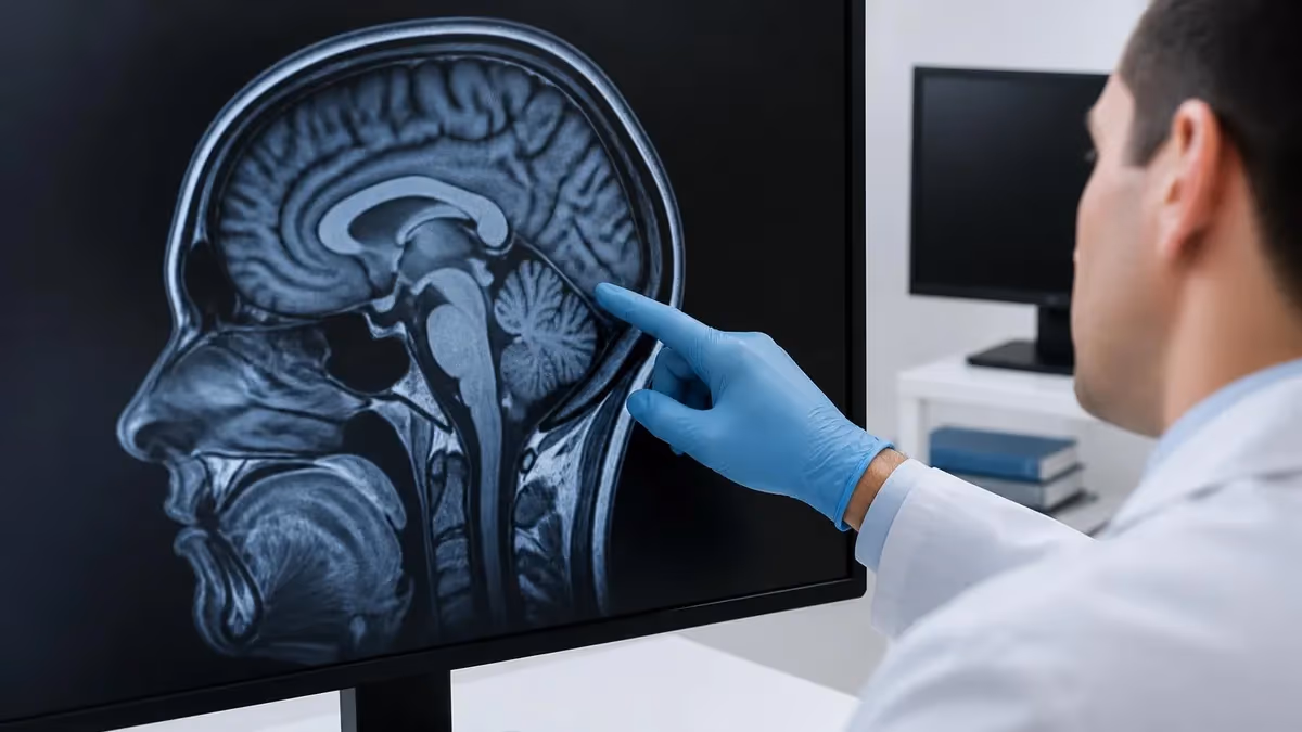
Beyond cardiac, the MRI white spots brain question is one of the most common patient concerns after a brain scan. These T2/FLAIR hyperintensities can represent small vessel ischemic disease, demyelination, migraine-related lesions, post-traumatic gliosis, perivascular spaces, or simply normal age-related white matter changes. Location, distribution, shape, size, and patient age all guide interpretation. A 70-year-old with periventricular cap-shaped hyperintensities is a very different clinical story from a 30-year-old with ovoid Dawson's-finger lesions running perpendicular to the ventricles on sagittal FLAIR.
Radiologists never make a definitive diagnosis from the image alone — clinical history, neurologic exam, lab work like vitamin B12 and Lyme titers, and often follow-up imaging close the case. Multiple sclerosis criteria require dissemination in both space and time, which sometimes means waiting six months for a repeat scan with new lesions to confirm.
This is also why a careful Spanish-language MRI test explanation matters: patients deserve clarity before worrying about a single phrase like "nonspecific white matter changes" on a printed report. Many imaging centers have moved to layered patient-friendly reports that translate the radiology language into something a non-medical reader can actually parse without panicking.
An MRI trailer is a fully shielded MRI suite on wheels — same magnet strength, same safety zones. Patients still need full ferromagnetic screening before approaching the trailer, and the 5-gauss line is marked outside the unit. Cold weather, weight limits, and limited space for emergencies are the main operational differences. Never assume a mobile unit is somehow safer or weaker than a fixed scanner — the magnet inside is identical to the hospital version.
Preparation for specialty MRI studies follows a predictable pattern, but each subtype has its own quirks worth memorizing in advance. Cardiac stress MRI requires caffeine restriction for at least 12 hours so the vasodilator drug works correctly; even decaf coffee can blunt the response enough to ruin the study and force a reschedule.
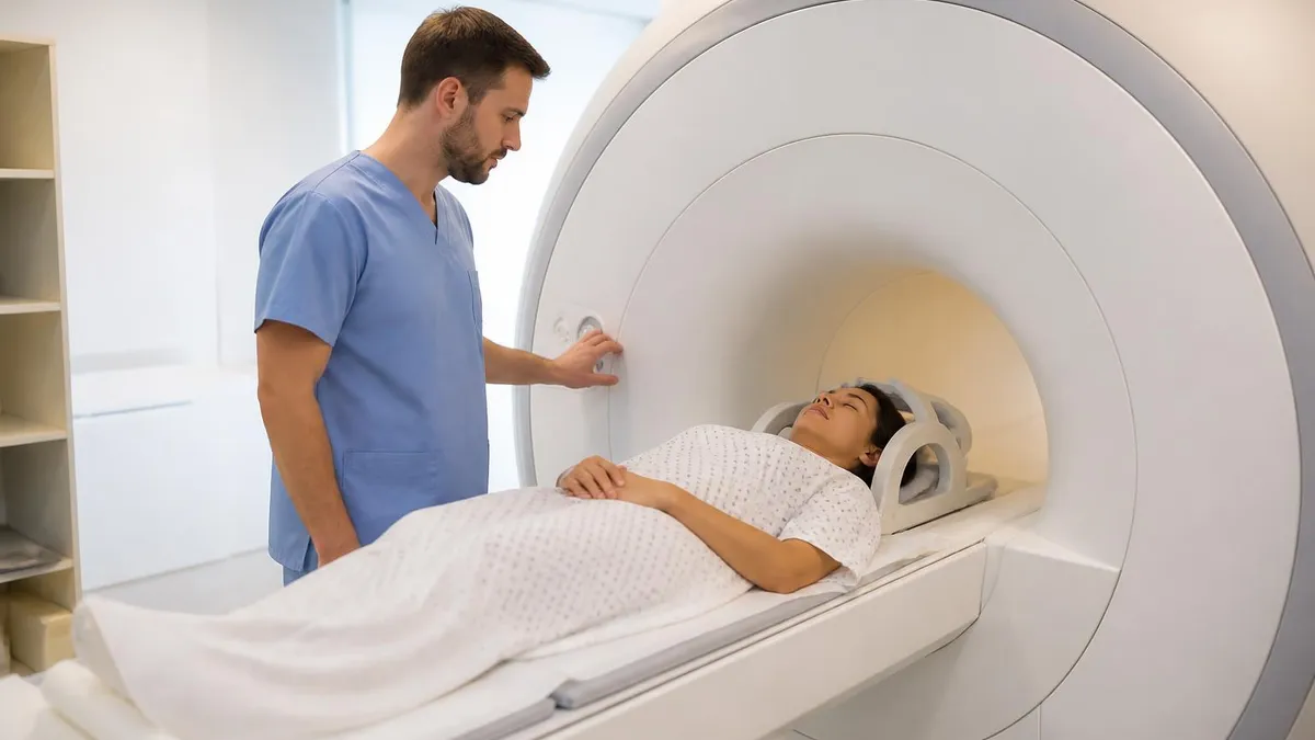
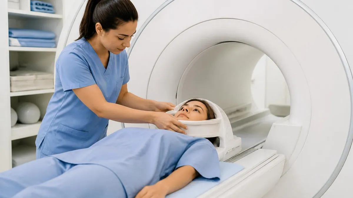
Pelvic and SI joint studies often ask patients to arrive with a partially full bladder, especially when fluid distension helps separate adjacent organs and reduce motion artifact from bowel peristalsis. Pregnancy MRI typically skips contrast unless medically necessary, and many centers schedule pregnant patients into morning slots when nausea is least disruptive to remaining still in the bore for 30 to 45 minutes.
Dental implant patients should bring documentation of the implant brand and material if available — the screening tech can check it against the MRI safety database if there's any doubt. Most titanium hardware is fine for both 1.5T and 3T scans, but the screening record still needs to be in the chart and signed off by the supervising radiologist on duty. For tailbone, lumbar, and SI joint imaging, patients should mention any recent steroid injections, since those can change marrow signal and affect interpretation of active inflammation.
For any specialty MRI — from a coccyx study to a mobile trailer visit — the checklist below covers the items techs and patients most often forget. Treat it as a pre-flight before the scan rather than a list to review in the parking lot. The smoother the screening goes, the faster the scan can start, the better the image quality you get from a still cooperative patient, and the less likely a patient is to have to come back another day for a repeat.
Specialty MRI Prep Checklist
- ✓List every implanted device: pacemaker, neurostimulator, cochlear implant, aneurysm clip, dental implant, IUD, port.
- ✓Avoid caffeine for 12 hours before a cardiac MRI stress test so the vasodilator drug works correctly.
- ✓Wear metal-free clothing; remove jewelry, hairpins, magnetic eyelashes, and transdermal patches.
- ✓For pregnancy MRI, confirm the indication is urgent enough to justify the study and that contrast is truly needed.
- ✓Bring a list of past surgeries and ask the tech if any retained metal could distort SI joint, lumbar, or shoulder images.
- ✓Request earplugs AND headphones — single hearing protection is not enough at 110+ dB.
- ✓For mobile MRI trailer visits, check the weather; severe cold or heat may delay scans because the quench pipe and HVAC have tighter tolerances.
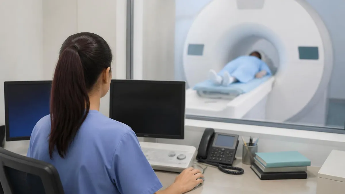
Choosing between a stationary scanner and a mobile MRI trailer comes up more often than patients expect. Rural hospitals, correctional facilities, and even some research studies rely entirely on trailer-based imaging because building a fixed suite costs millions of dollars and requires significant structural shielding work — copper walls, RF doors, and a quench pipe vented to atmosphere. Mobile units offer the same 1.5T or 3T field strengths and the same coil options as their fixed counterparts. What differs is throughput, scheduling flexibility, and the surrounding logistics around emergency response and patient flow.
Stationary MRI suites win on volume and emergency support. If a patient has a contrast reaction or a code event, a hospital crash team is steps away with full advanced cardiac life support equipment. Trailers compensate with predictable rotational schedules, often visiting the same site once a week on a fixed day. For elective specialty MRI — say, a planned MRI sij follow-up to monitor sacroiliitis treatment — either setting works equally well. For an acute neurologic question in a patient with a low GCS or for any contrast-requiring stress study, the fixed in-hospital scanner is the safer call.
The comparison below summarizes the practical trade-offs in plain terms. Use it when counseling patients who have a choice between a community trailer visit and driving an hour to a hospital-based scanner. For most outpatient specialty studies, the mobile option is genuinely equivalent in image quality, and convenience wins. For acute or high-acuity cases, the hospital-based setting wins on safety margin alone.
Stationary vs Mobile MRI
- +Stationary: full hospital crash support and contrast reaction backup
- +Stationary: wider coil inventory for niche studies (breast, MSK, pediatric)
- +Stationary: higher daily throughput, more flexible add-on slots
- +Stationary: typically newer scanners with the quietest sequences available
- −Mobile: brings imaging to rural, correctional, and underserved sites
- −Mobile: same 1.5T/3T field strengths as fixed suites — no quality compromise
- −Mobile: limited add-on slots and tighter scheduling windows
- −Mobile: weather-sensitive operations; cold snaps can delay starts
Before wrapping up, it's worth touching on language access. Many imaging centers in the US now offer their MRI test in Spanish with translated consent forms, in-suite signage, and bilingual technologists. The same applies to written instructions about contrast hold times, breastfeeding pauses, and pre-cardiac stress caffeine restrictions. For patients with limited English proficiency, this isn't a courtesy — it's an informed-consent requirement under most state laws and accreditation standards from the Joint Commission and similar bodies.
If you're studying for the ARRT MRI registry or the AART board, expect questions on safety zones, gadolinium classes, gradient noise, pregnancy protocols, and implant compatibility. The four MRI safety zones (I through IV), the gauss line, gradient slew rates, and the difference between linear and macrocyclic gadolinium agents are all high-yield topics.
The FAQ below covers the topics that show up most often in practice questions and patient discussions. Use it as a final review, and follow it up with the linked practice quiz to lock in the concepts. Specialty MRI knowledge separates a confident tech from one still relying on protocol cards taped to the scanner wall, and registry pass rates reward the candidates who have internalized this layer of detail.
Mastering specialty MRI topics means moving past the basic safety screen and into the protocol-level details — knowing why an SI joint study uses oblique coronals through the joint's long axis, why a SLAP lesion gets an arthrogram with ABER positioning, why pregnancy MRI skips gadolinium contrast in nearly all clinical scenarios, and why dental implants usually pass screening but still cause local susceptibility artifact on jaw imaging.
These are exactly the questions that separate a confident tech or candidate from a hesitant one in the reading room or at the registry exam table. The same set of details also tend to land directly in patient consults and pre-scan phone calls with worried family members.
The FAQ below pulls the most common patient and exam questions into one block so you can scan it quickly before a shift, before an appointment, or before a test session. Pair it with the practice questions linked above for full coverage of the specialty MRI portion of your study plan.
Bookmark this page and return to it whenever a referral comes in for an unfamiliar specialty study — the protocols repeat themselves more than you'd expect, and a few minutes of refresher reading is usually enough to walk into the scanner room with full confidence in your approach, your screening process, and your conversation with the patient on the table.
MRI Questions and Answers
About the Author
Medical Laboratory Scientist & Clinical Certification Expert
Johns Hopkins UniversityDr. Sandra Kim holds a PhD in Clinical Laboratory Science from Johns Hopkins University and is certified as a Medical Technologist (MT) and Medical Laboratory Scientist (MLS) through ASCP. With 16 years of clinical laboratory experience spanning hematology, microbiology, and molecular diagnostics, she prepares candidates for ASCP board exams, MLT, MLS, and specialist certification tests.
Join the Discussion
Connect with other students preparing for this exam. Share tips, ask questions, and get advice from people who have been there.
View discussion (6 replies)