Types of MRI: Complete Guide to Every MRI Scan, Machine, and Imaging Sequence
💡 Explore the main types of MRI scans, machines, and pulse sequences. Learn how each MRI type works, what it shows, and when doctors order it.
When patients hear they need an MRI, most assume there is just one kind of scan. In reality, the many types of MRI form a remarkably diverse family of imaging studies, each tuned to answer a specific clinical question. Some MRIs evaluate the brain, others map cartilage in a knee, and still others trace blood flow through arteries without injecting contrast. Understanding these variations helps patients prepare, helps technologists protocol correctly, and helps clinicians order the right study the first time.
The differences between MRI types fall into three broad buckets: the machine itself, the body region or organ targeted, and the pulse sequences applied during scanning. A 3 Tesla closed-bore scanner is a very different experience from a 0.3 Tesla open unit, even when both are imaging the same lumbar spine. Likewise, a routine brain MRI shares hardware with a functional MRI, yet the data acquired and the information returned look nothing alike. Each variation exists for a reason.
Magnetic resonance imaging was first used clinically in the early 1980s, and you can read more about its origins in our overview of the history of MRI. Since then, manufacturers have pushed field strengths higher, gradient coils stronger, and software smarter. The result is a menu of dozens of distinct exams that radiologists choose from based on symptoms, prior imaging, and patient factors such as implants, claustrophobia, or kidney function.
This guide walks through every major category you are likely to encounter, from the standard structural brain scan to advanced techniques like diffusion tensor imaging, MR spectroscopy, and cardiac MRI. We will also cover the differences between open and closed machines, the role of contrast agents, and how field strength changes image quality. Whether you are a patient, a student preparing for the registry, or a referring provider, this should give you a clear map of the MRI landscape.
You will notice that the term "MRI" is often modified by a prefix or suffix that signals the specific technique: fMRI for functional, MRA for angiography, MRCP for cholangiopancreatography, and so on. These abbreviations are not arbitrary marketing labels. Each represents a distinct pulse sequence design, a particular post-processing pipeline, and a specific diagnostic purpose. Learning the shorthand makes it easier to interpret reports and discuss findings.
Throughout the article, we will reference real clinical scenarios, scan times, and protocols. We will note when intravenous contrast is typically used, when sedation might be considered, and which body parts each MRI type handles best. By the end you should be able to tell a T1-weighted image from a T2, recognize when a doctor might order DWI versus SWI, and understand why a cardiologist might insist on a 1.5T rather than a 3T machine for a particular study.
One last note before we begin: not every facility offers every MRI type. Smaller community hospitals usually provide standard structural and contrast studies, while academic centers and large outpatient networks add functional, spectroscopy, and high-field research scans. Knowing what your local center offers, and what they refer out, can save weeks of delay when a specialized exam is needed.
Types of MRI by the Numbers
Types of MRI Machines
The standard tunnel-shaped scanner operating at 1.5T or 3T. Produces the highest image quality and fastest acquisition times, making it the workhorse of most hospitals and outpatient centers for nearly every body region.
Designed for claustrophobic, larger, or pediatric patients. Field strength is usually 0.3T to 1.2T, with the magnet shaped like two flat plates above and below the patient. Image quality is lower but accessibility is far better.
A compromise between closed and open designs. The bore opening is 70 cm instead of the traditional 60 cm, accommodating heavier patients and reducing claustrophobia while keeping 1.5T or 3T diagnostic quality.
Compact scanners that image only a single limb such as a wrist, elbow, knee, or ankle. The patient sits beside the machine while the joint slides into a small bore. Ideal for orthopedic and sports medicine clinics.
Built directly into a surgical suite so neurosurgeons can scan during a procedure. Used most often in brain tumor resection to confirm complete removal before closing the skull. Requires extensive room shielding and specialized instruments.
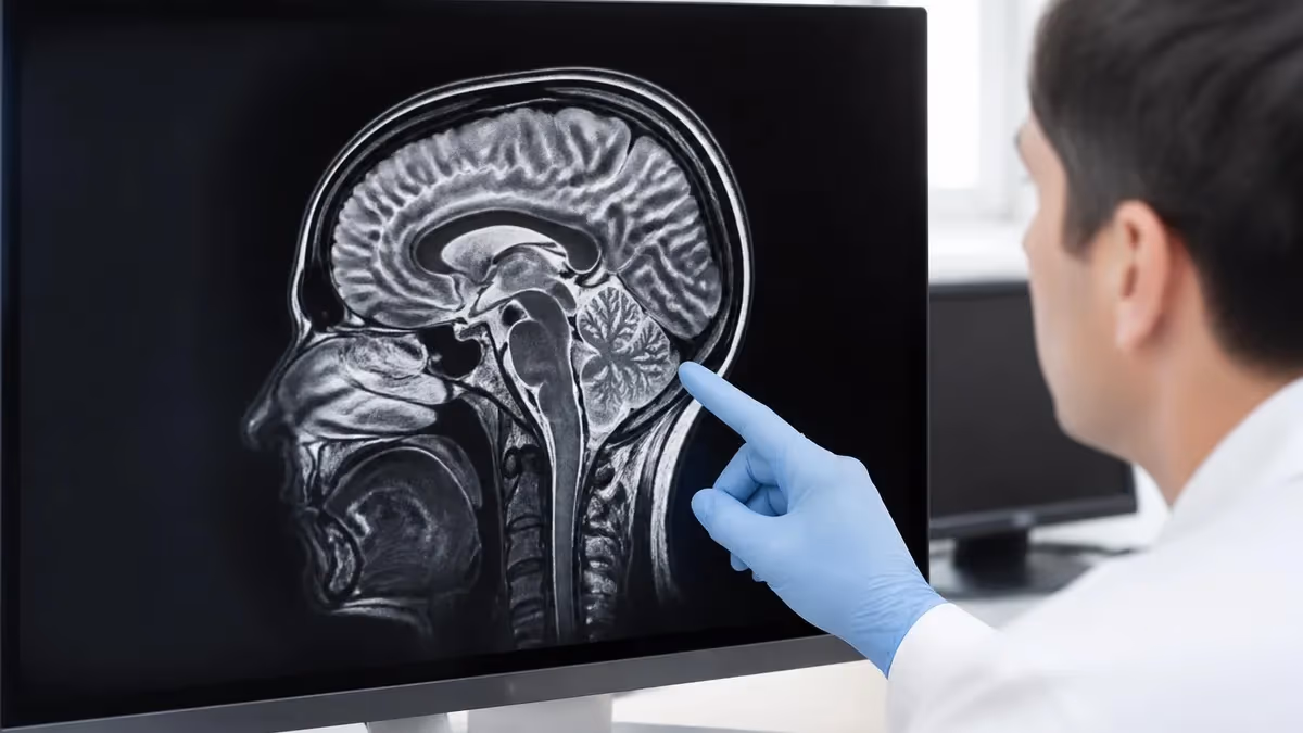
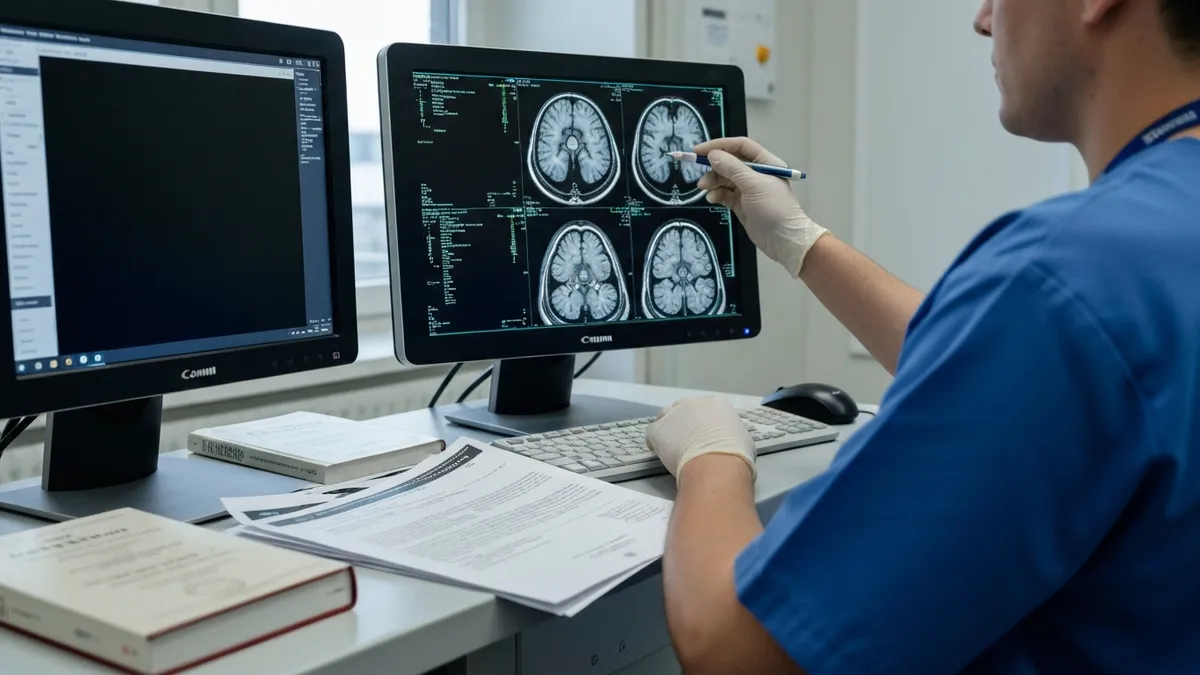
Once the machine is chosen, the next decision is which body region to scan and how to protocol it. Brain MRI is the single most ordered exam in the country, used for headaches, suspected strokes, multiple sclerosis monitoring, tumor surveillance, and dementia workups. A standard brain MRI takes about 30 to 45 minutes and includes T1, T2, FLAIR, diffusion-weighted, and gradient echo sequences. Adding contrast extends the scan by roughly 10 minutes and is essential for tumor, infection, and inflammatory workups.
Spine MRI is the second most common, divided into cervical, thoracic, and lumbar studies. Lumbar spine scans dominate volume because of how often low back pain prompts imaging. Radiologists evaluate disc height, nerve root impingement, facet arthropathy, and any marrow signal abnormalities. Cervical spine MRI is especially important after trauma, when ligamentous injury or cord contusion can hide on CT. thoracic spine mri cpt is the least requested but critical for myelopathy and metastatic workups.
Musculoskeletal MRI covers shoulders, knees, hips, ankles, wrists, and elbows. These studies use dedicated surface coils that wrap around the joint to maximize signal. Sports medicine physicians rely on MRI to grade ligament tears, identify cartilage defects, and detect stress fractures invisible on plain film. A knee MRI typically runs 20 to 30 minutes without contrast, while a shoulder MR arthrogram, which involves injecting dilute gadolinium into the joint, takes longer and requires fluoroscopic guidance.
Abdominal and pelvic MRI evaluate the liver, pancreas, kidneys, adrenal glands, bowel, prostate, uterus, and ovaries. These exams demand breath-holding for many sequences and often include dynamic contrast phases timed to arterial, portal venous, and delayed enhancement. MRCP, a specialized non-contrast study of the biliary tree, has largely replaced diagnostic ERCP for evaluating gallstones and pancreatic duct anatomy. Prostate MRI follows a structured PI-RADS protocol that combines T2, diffusion, and dynamic contrast.
Cardiac MRI deserves its own category because of how technically demanding it is. The heart moves constantly, so sequences must be gated to the ECG and acquired during breath-holds. A complete cardiac MRI evaluates chamber size, wall motion, ejection fraction, valvular flow, myocardial perfusion, and tissue characterization for fibrosis, edema, or iron overload. Many cardiologists now consider cardiac MRI the gold standard for assessing myocarditis, cardiomyopathies, and viability before revascularization.
Vascular imaging through MR angiography and MR venography visualizes arteries and veins anywhere in the body without ionizing radiation. Some MRA techniques use gadolinium contrast, while time-of-flight and phase-contrast methods rely purely on flow physics. This is invaluable for patients with renal disease who cannot safely receive iodinated CT contrast. To understand when contrast is required versus optional, see our deep dive on MRI with and without contrast.
Breast MRI rounds out the most common body-region studies. It is performed prone using a dedicated coil with the breasts hanging into openings. Indications include high-risk screening, evaluating the extent of newly diagnosed cancer, and assessing response to neoadjuvant chemotherapy. The exam always uses gadolinium because enhancement kinetics are central to interpretation. Specialized exams for prostate, fetal anatomy, and small bowel enterography fill out the rest of the body-region menu.
MRI Practice Test Questions
Prepare for the MRI - Magnetic Resonance Imaging exam with our free practice test modules. Each quiz covers key topics to help you pass on your first try.
MRI Knowledge
MRI Exam Questions covering Knowledge. Master MRI Test concepts for certification prep.
MRI Physics
Free MRI Practice Test featuring Physics. Improve your MRI Exam score with mock test prep.
MRI Anatomy and Pathology
MRI Test Prep for MRI Anatomy and Pathology. Practice MRI Quiz questions and boost your score.
MRI Anatomy and Positioning
MRI Questions and Answers on MRI Anatomy and Positioning. Free MRI practice for exam readiness.
MRI Contrast Agents
Free MRI Quiz on MRI Contrast Agents. MRI Exam prep questions with detailed explanations.
MRI Patient Care and Positioning
MRI Practice Questions for MRI Patient Care and Positioning. Build confidence for your MRI certification exam.
Common MRI Pulse Sequences
T1-weighted images make fat appear bright and water appear dark, providing crisp anatomic detail. They are the backbone of nearly every MRI protocol because they show borders between tissues clearly and serve as the comparison image for post-contrast sequences. Radiologists use T1 to evaluate fatty marrow, subacute hemorrhage, and proteinaceous fluid, all of which show as high signal.
T2-weighted images reverse that contrast, making water bright and fat darker. Because most pathology involves edema or fluid accumulation, T2 sequences are excellent for finding disease. A torn meniscus, an inflamed joint, a brain tumor, or a herniated disc all stand out because they contain more water than the surrounding healthy tissue. Together, T1 and T2 form the diagnostic foundation of MRI.
MRI vs Other Imaging: Strengths and Weaknesses
- +No ionizing radiation, making it safe for repeated exams and pregnancy
- +Superior soft tissue contrast compared to CT or ultrasound
- +Multiple sequences can answer different questions in one session
- +Excellent for brain, spine, joints, and pelvic organs
- +Functional and metabolic imaging possible with the same hardware
- +Non-contrast vascular imaging avoids iodinated contrast risks
- −Long scan times ranging from 20 minutes to over an hour
- −Loud acoustic noise that requires hearing protection
- −Closed bore design triggers claustrophobia in many patients
- −Implants and metallic foreign bodies may be absolute contraindications
- −Higher cost than CT or ultrasound at most facilities
- −Gadolinium contrast carries small risks in patients with renal failure
Choosing the Right Type of MRI
- ✓Confirm the clinical question — pain, mass, vascular flow, or function
- ✓Verify whether contrast is needed based on suspected pathology
- ✓Check estimated GFR if gadolinium will be administered
- ✓Screen the patient for implants, pacemakers, and metallic foreign bodies
- ✓Consider open or wide-bore for claustrophobia or body habitus
- ✓Match field strength to the body part — 3T excels for brain and MSK
- ✓Select dedicated coils for breast, shoulder, knee, or wrist exams
- ✓Plan ECG gating and breath-holds for cardiac or abdominal studies
- ✓Order specialized protocols such as MRCP, MRA, or prostate PI-RADS when indicated
- ✓Confirm prior imaging is available so radiologists can compare findings
Not all MRIs are interchangeable.
Ordering a routine brain MRI when you actually need MR angiography, or a 1.5T study when 3T would resolve a small pituitary lesion, can mean a missed diagnosis and a repeat exam. Always match the scan type to the clinical question and verify the protocol with the reading radiologist when in doubt.
Contrast-enhanced and functional MRI sit at the cutting edge of the field, opening up information that no other modality provides. Gadolinium-based contrast agents shorten T1 relaxation time, making blood vessels and pathologic tissue appear bright on post-contrast T1 sequences. The compound is injected intravenously, typically at a dose of 0.1 mmol per kilogram of body weight, and excreted unchanged by the kidneys within 24 hours in patients with normal renal function.
Different gadolinium agents have slightly different molecular structures, ranging from linear to macrocyclic. Macrocyclic agents like gadobutrol and gadoterate are considered safer because they hold the gadolinium ion more tightly, reducing the small risk of nephrogenic systemic fibrosis in patients with advanced kidney disease. The FDA still requires a black-box warning on all gadolinium agents, and screening for renal function before administration is standard practice.
Beyond standard contrast, MRI offers several functional and physiologic techniques. Functional MRI, or fMRI, measures changes in blood oxygen level dependent signal as the patient performs tasks like tapping fingers or reading words. Neurosurgeons use fMRI to map eloquent cortex before resecting nearby tumors, while researchers use it to study cognition, depression, and pain processing. Diffusion tensor imaging, a more advanced cousin of DWI, reconstructs white matter tracts so surgeons can avoid critical pathways.
MR spectroscopy goes a step further by measuring the chemical composition of a small voxel of tissue rather than producing an image. Peaks in the spectrum correspond to metabolites like N-acetylaspartate, choline, creatine, and lactate. The pattern can distinguish certain brain tumors from infections, evaluate metabolic disorders, and monitor response to therapy. Cardiac perfusion MRI similarly uses rapid contrast bolus tracking to identify ischemic myocardium during pharmacologic stress.
Whole-body MRI is gaining popularity for cancer screening, especially in patients with hereditary syndromes like Li-Fraumeni. The scan takes 45 to 75 minutes and produces a head-to-toe survey using a combination of T1, T2, STIR, and diffusion sequences. Skeptics caution about incidental findings that lead to follow-up tests, while proponents point to early cancer detection without radiation exposure. The technique is well established for myeloma surveillance.
Fetal MRI is another specialized application that has matured significantly over the last 15 years. Performed without contrast and without sedation, it provides exquisite detail of fetal brain development, complex congenital anomalies, and placental abnormalities. Most fetal MRIs are performed after 22 weeks gestation when fetal motion has slowed enough to produce diagnostic images using ultrafast single-shot sequences.
Finally, intervention has come to MRI. MR-guided focused ultrasound is being used to treat essential tremor, uterine fibroids, and certain prostate tumors. MR-guided biopsies of the breast and prostate locate lesions invisible on ultrasound. These hybrid procedures require specialized non-magnetic equipment and represent a growing niche at large academic centers, expanding what counts as an MRI exam in modern medicine.
Every type of MRI requires rigorous screening for pacemakers, neurostimulators, cochlear implants, aneurysm clips, and metallic foreign bodies. Even MR-conditional devices have specific scanning parameters that must be followed exactly. Skipping or rushing this step can cause device malfunction, tissue heating, or serious injury.
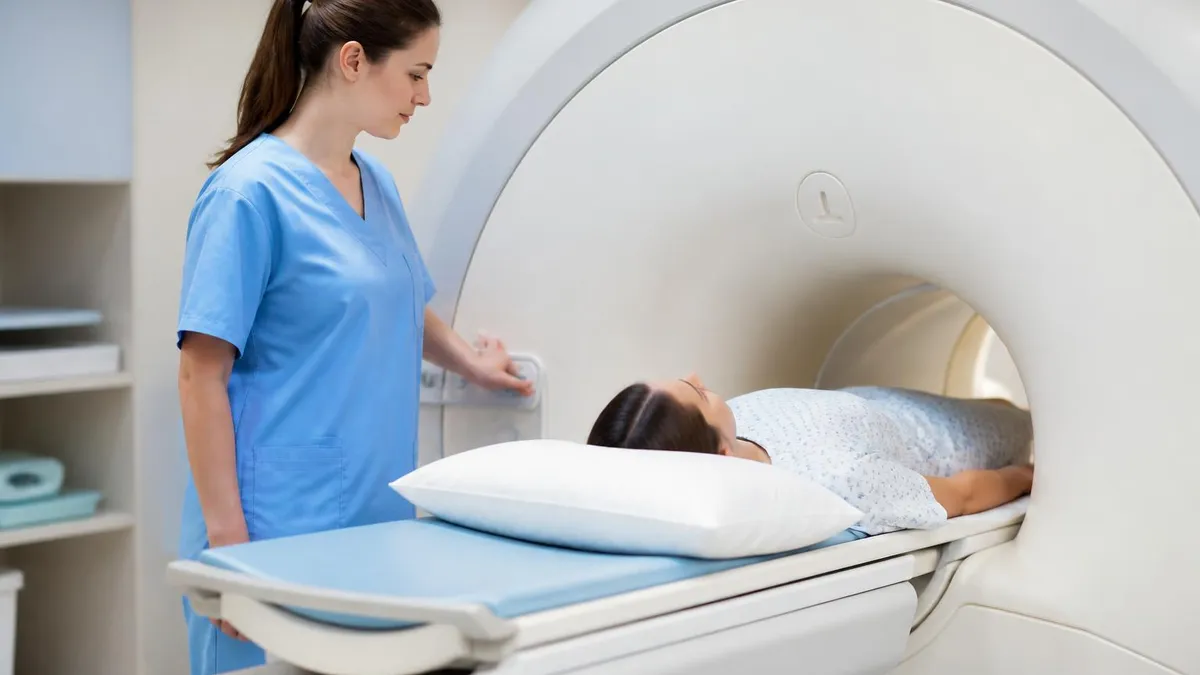
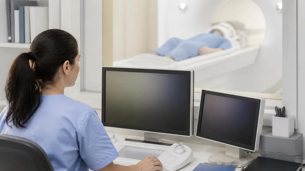
Patient safety and preparation are universal across every type of MRI, even though the specific protocol changes. Before any scan, the patient completes a detailed screening form covering implants, prior surgeries, occupational metal exposure, pregnancy, and allergies. The MRI technologist reviews the form with the patient, looking especially for cardiac devices, cochlear implants, and intracranial aneurysm clips that might be ferromagnetic. When uncertainty exists, prior imaging or the device card is used to confirm MR compatibility.
For most MRI types, the patient changes into scrubs or a hospital gown to remove any metallic snaps, zippers, or underwires. Personal items including phones, watches, jewelry, hearing aids, and credit cards stay outside the scanner room because the magnetic field can damage electronics and erase magnetic strips. Even cosmetic items like eyeliner containing iron oxide pigments and certain hair extensions have caused artifact and discomfort.
Patients with pacemakers, defibrillators, and neurostimulators deserve special attention. Modern MR-conditional devices can be scanned safely when programmed into MRI mode and monitored throughout the exam, but each manufacturer publishes specific scanning conditions. For an example of how complex device-specific safety can be, see our complete mri safety and compatibility. Every implant brand has its own scanning rules, and they update frequently.
Renal function screening matters whenever gadolinium contrast is planned. Patients with an estimated GFR below 30 mL/min face an elevated risk of nephrogenic systemic fibrosis, and the radiologist must weigh diagnostic necessity against potential harm. In acute kidney injury or end-stage renal disease, alternative imaging strategies or use of macrocyclic agents at the lowest effective dose are common. Pregnant patients generally avoid gadolinium unless absolutely necessary in the second or third trimester.
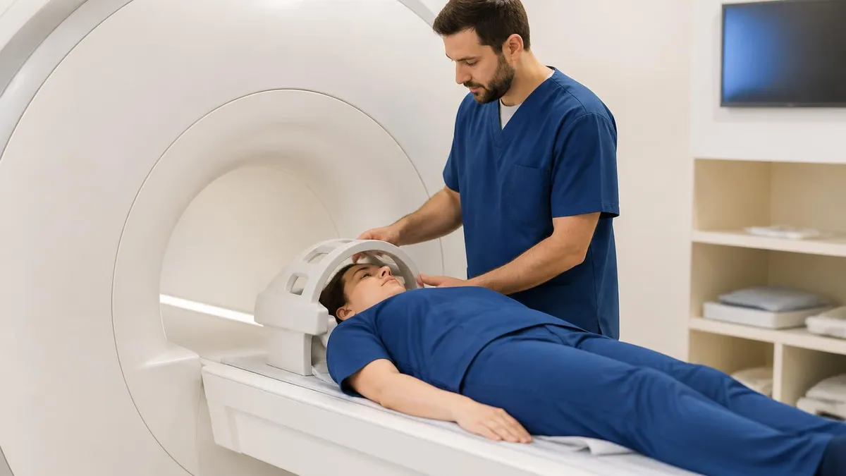
Claustrophobia affects roughly five to ten percent of patients enough to require intervention. Strategies range from listening to music through MR-safe headphones, using a mirror that reflects the room behind the scanner, oral anxiolytics like lorazepam, and finally open-bore or wide-bore alternatives. Pediatric patients under age six often need light sedation or general anesthesia. Many centers now offer comfort coaching sessions before the exam to reduce cancellation rates.
Acoustic noise during MRI can exceed 110 decibels depending on the sequence. Hearing protection in the form of earplugs, headphones, or both is mandatory for every patient and any person who remains in the room. The loudness comes from the gradient coils rapidly changing magnetic field strength, which physically vibrates the coil housing. Newer scanners with quiet technology can reduce noise by 30 to 70 percent on some sequences but cannot eliminate it entirely.
After the scan, most patients can resume normal activity immediately. Those who received sedation need a ride home, and contrast recipients are encouraged to hydrate to help eliminate gadolinium. Images are typically interpreted within 24 hours, although stat exams from the emergency department are read within minutes. Reports are sent to the ordering provider, who reviews findings with the patient and decides on next steps such as additional imaging, biopsy, or treatment.
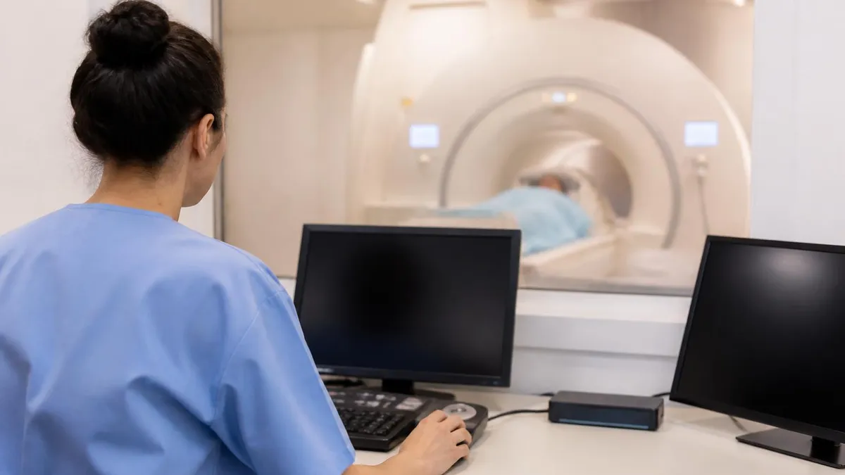
Putting all of this knowledge into practice requires a workflow mindset. When a clinician orders an MRI, they specify the body part, the indication, and whether contrast is required. The scheduler then assigns the appropriate scanner, coil, and time slot. A brain MRI without contrast might be 30 minutes on any 1.5T or 3T scanner with a standard head coil. A cardiac MRI demands a 90-minute slot with an MR-trained technologist, cardiology nurse, and immediate access to a reading cardiologist or radiologist.
Patients can do several things to make their MRI experience smoother. Arrive 30 minutes early to complete paperwork and change. Bring a list of all implants, surgeries, and medications. Eat normally unless told otherwise, since most MRIs do not require fasting except for MRCP, abdominal, and certain pelvic protocols. If you have a history of claustrophobia, request anti-anxiety medication from your ordering provider in advance rather than the day of the exam.
For technologists and students, mastering protocol selection is a career-long skill. Reference materials from the American College of Radiology, manufacturer protocol books, and institutional guidelines should be at every workstation. When a new indication comes through, taking 60 seconds to verify the right sequences saves a callback later. Documenting protocol modifications in the patient record helps the next technologist and the interpreting radiologist understand exactly what was acquired.
Radiologists tailor their reports based on the type of MRI performed. A brain MRI report follows a templated structure covering ventricles, parenchyma, posterior fossa, vasculature, calvarium, and any incidental findings. A musculoskeletal report walks through every ligament, tendon, muscle, bone, and joint surface. Standardized reporting systems like PI-RADS for prostate, LI-RADS for liver, and BI-RADS for breast ensure that referring clinicians receive the information they need in a predictable format.
Quality control is an often invisible but vital part of every MRI department. Daily phantom scans verify that signal-to-noise ratio, geometric accuracy, and slice thickness remain within specification. The American College of Radiology accreditation program requires ongoing documentation, and most insurers will not pay for studies from non-accredited facilities. Behind every type of MRI sits a team of physicists, engineers, and service technicians keeping the magnet, gradients, and software performing reliably.
Emerging technology continues to expand the MRI menu. Artificial intelligence reconstruction can cut scan times by 30 to 50 percent without sacrificing image quality, making it feasible to offer MRI to patients who previously could not tolerate long exams. Portable bedside MRI units operating at very low field strengths are appearing in intensive care units and stroke triage settings. Photon-free quantitative MRI is moving from research into clinical use for liver iron and fat quantification.
The final practical takeaway is this: an MRI is rarely just an MRI. Knowing the type of machine, the sequences acquired, and the clinical context transforms a generic image into actionable medical information. Whether you are scheduling your first scan, preparing for the registry, or refining a department protocol, understanding the full landscape of MRI types makes you a smarter participant in dwi mri. Use the resources at the end of this guide to keep building that knowledge.
MRI Questions and Answers

The History of MRI: From Discovery to Modern Medicine

MRI With and Without Contrast: How It Works, What to Expect

MRI Medical Abbreviation: What MRI Stands For and Why It Matters
MRI Imaging Centers: Complete Guide to Independent Outpatient MRI Facilities
St Jude Pacemaker MRI Compatibility List: A Complete Safety and Scanning Guide
About the Author
Medical Laboratory Scientist & Clinical Certification Expert
Johns Hopkins UniversityDr. Sandra Kim holds a PhD in Clinical Laboratory Science from Johns Hopkins University and is certified as a Medical Technologist (MT) and Medical Laboratory Scientist (MLS) through ASCP. With 16 years of clinical laboratory experience spanning hematology, microbiology, and molecular diagnostics, she prepares candidates for ASCP board exams, MLT, MLS, and specialist certification tests.
Join the Discussion
Connect with other students preparing for this exam. Share tips, ask questions, and get advice from people who have been there.
View discussion (6 replies)