What Does MRI Look Like? Open MRI Pictures and Scan Images Explained 2026 July
💯 See what an open MRI picture looks like, how MRI scans appear, and what different MRI images reveal about brain, spine, and joint anatomy.
If you have ever wondered what an open MRI picture looks like, you are not alone. Patients searching for clarity before their first scan typically want two things at once: a visual of the machine itself and a preview of the grayscale images it produces. An open MRI looks dramatically different from the traditional closed-bore tunnel, and the images it captures reveal soft tissue, bone marrow, blood vessels, and pathology in ways no other imaging tool can match in a single sitting.
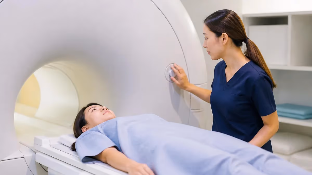
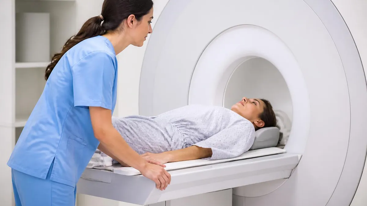
The machine itself resembles two large, flat discs stacked horizontally with a wide gap in between, where the patient lies. Unlike the long tube of a conventional scanner, an open MRI has airflow on three or four sides, no enclosed walls near the face, and often a softer hum during scanning. Many patients describe the experience as lying under a low ceiling rather than inside a tunnel, which is exactly why claustrophobic patients, larger patients, and pediatric patients gravitate toward this design.
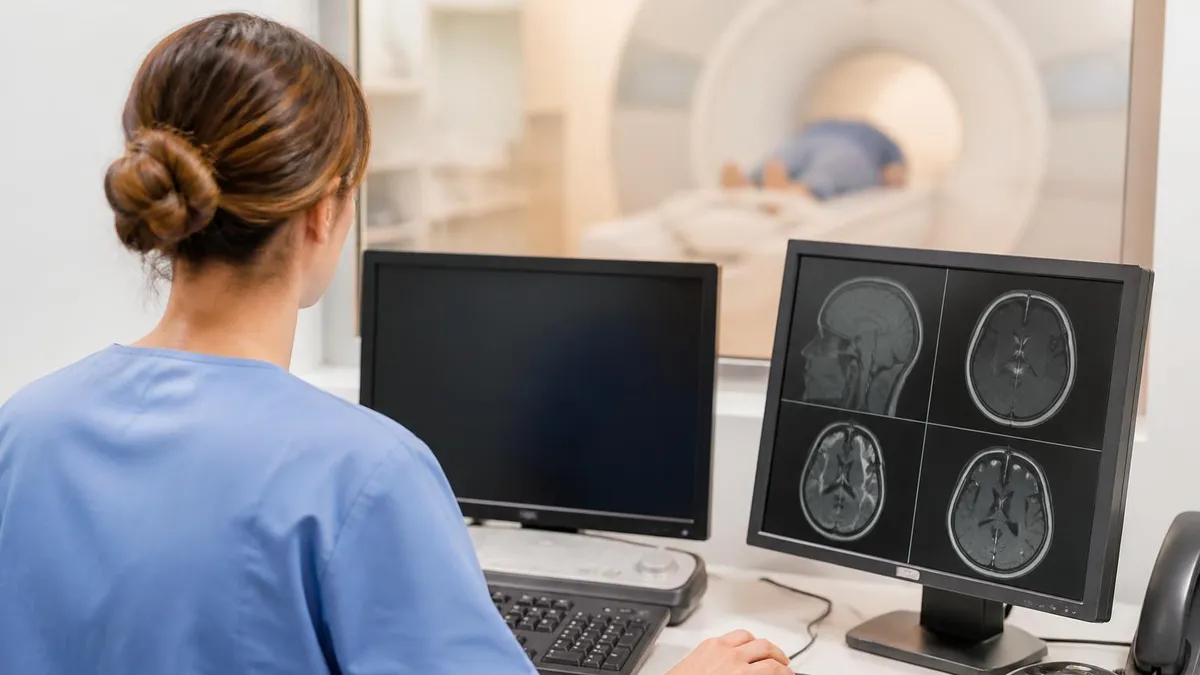
The pictures produced are equally fascinating. MRI images are not photographs in the traditional sense; they are computed maps of how hydrogen protons in your tissues respond to a powerful magnetic field and radiofrequency pulses. A typical brain MRI looks like a series of grayscale cross-sections, each one a thin slice cut through the head from top to bottom, side to side, or front to back. Bright areas, dark areas, and shades of gray each carry diagnostic meaning.
Different sequences produce different looks. A T1-weighted image makes fat appear bright and water appear dark, so brain anatomy is crisp and detailed. A T2-weighted image flips the contrast: fluid lights up, making swelling, edema, and cysts jump off the screen. FLAIR sequences suppress normal cerebrospinal fluid so that abnormal fluid near the ventricles becomes obvious. Diffusion-weighted imaging highlights areas where water movement is restricted, a classic sign of early stroke.
Open MRI pictures of joints and the spine look slightly softer than closed-bore images because most open systems run at lower field strengths, typically 0.3 to 1.0 Tesla compared to 1.5 or 3.0 Tesla in closed scanners. The trade-off is comfort and accessibility for image sharpness. Newer wide-bore and high-field open designs are closing that gap, producing images that rival traditional scanners for many routine indications. For a quick refresher on what the acronym itself stands for, see our guide to the MRI medical abbreviation.
Before your appointment, knowing what to expect visually reduces anxiety more than almost anything else. Patients who have seen photos of the machine, watched a sample scan video, or studied a few example images report shorter perceived scan times and better cooperation during breath-holds. This article walks through both sides of the question: what the equipment looks like, and what the resulting pictures show.
We will compare open and closed designs, explain why bright spots and dark shadows appear where they do, break down brain versus spine versus joint imaging, and finish with practical tips for interpreting your own scan report. By the end, you will be able to look at any MRI image and identify the sequence, the anatomy, and at least the general category of findings the radiologist is evaluating.
MRI Imaging by the Numbers
Open MRI vs Closed MRI: What Each One Looks Like
A long cylindrical tunnel about 60 cm wide and 150-200 cm deep. Patients lie on a sliding table that moves them into the bore. Field strength is typically 1.5T or 3.0T, producing the sharpest images available for clinical diagnosis.
Two flat magnet plates above and below the patient with open sides. No tunnel surrounds the body, which helps claustrophobic, bariatric, and pediatric patients. Field strength is usually 0.3T to 1.0T, producing slightly softer but diagnostically useful images.
A hybrid design with a 70 cm bore opening, shorter tunnel length, and full 1.5T or 3.0T field strength. Looks like a closed MRI from the outside but feels more open inside, accommodating patients up to roughly 550 pounds.
A specialty open system where the patient sits, stands, or bends during imaging. Useful for weight-bearing spine studies, joint kinematics, and patients who cannot lie flat. Less common but valuable for biomechanical assessments.
An MRI picture is fundamentally a grayscale map of proton behavior. When you slide into the scanner, every hydrogen atom in your body briefly aligns with the magnetic field. A radiofrequency pulse knocks them out of alignment, and as they relax back, they release tiny signals that antennas (called coils) detect. A computer reconstructs those signals into an image that resembles a thin slice through your body, as if you had been sectioned with a perfectly even knife.
The first thing most patients notice is that MRI pictures contain no bones in the conventional X-ray sense. Cortical bone has very few mobile hydrogen protons, so it appears dark or signal-void. The bright structures you see are typically fat, bone marrow, muscle, organs, and fluids. This is why MRI excels at imaging soft tissue — ligaments, tendons, brain, cartilage, and disc material show up beautifully where X-rays and CT scans struggle.
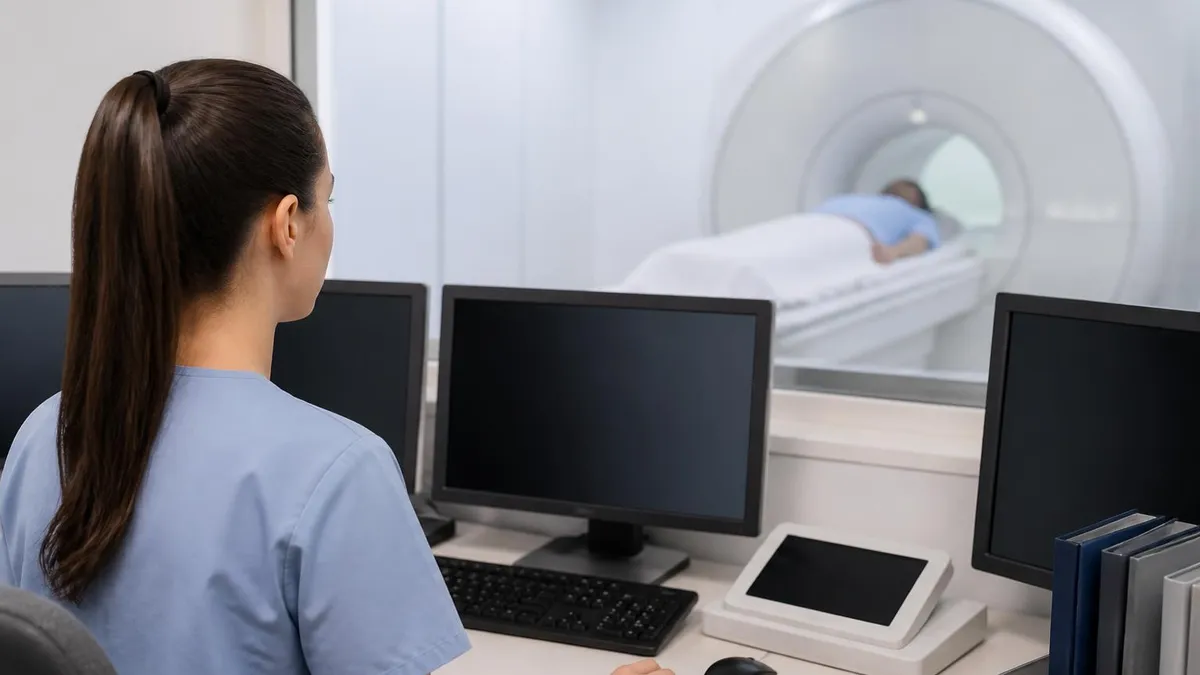
Each MRI study contains multiple series. A series is a stack of images acquired with the same parameters in the same plane. You will typically see axial (horizontal slices), sagittal (side-to-side slices), and coronal (front-to-back slices) orientations. Stacking them mentally gives a three-dimensional understanding of anatomy that a single photograph could never provide. Radiologists scroll through these stacks rather than viewing one image at a time.
The shades of gray are not arbitrary. They encode tissue characteristics that vary by sequence. On a T1-weighted brain image, white matter appears lighter than gray matter because of its fatty myelin content. On a T2-weighted image, the relationship flips, and the cerebrospinal fluid in the ventricles glows white against darker brain tissue. Pathology often disrupts these normal patterns, and that disruption is exactly what the radiologist hunts for.
Resolution depends heavily on the machine and protocol. A 3.0T scanner can resolve structures smaller than a millimeter, while a 0.3T open unit might require thicker slices to maintain signal-to-noise ratio. Both can be diagnostic; they just produce slightly different-looking pictures. If you receive a CD of your images after the appointment, you may notice your scan files are large because each slice is its own high-resolution image and a single study often contains hundreds of them.
Open MRI pictures specifically tend to have a different aesthetic. Lower field strength means the images can appear slightly grainier, with less crisp contrast between similar tissues. However, manufacturers have refined coil design, sequence engineering, and post-processing so dramatically over the past decade that an open MRI of the knee, shoulder, or lumbar spine is often visually indistinguishable from a closed-bore scan for routine diagnoses. For a comparison of dye-enhanced versus standard imaging, see our deep dive on mri with and without contrast cpt.
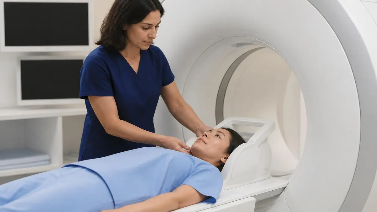
Patient positioning also shapes how the image looks. A poorly centered shoulder or a slightly rotated spine introduces obliquity that makes anatomical landmarks harder to identify. Technologists spend significant time aligning patients precisely so that the resulting pictures are truly axial, truly sagittal, and truly coronal. This is one reason scans take longer than a quick X-ray; the setup is as important as the acquisition itself.
MRI Practice Test Questions
Prepare for the MRI - Magnetic Resonance Imaging exam with our free practice test modules. Each quiz covers key topics to help you pass on your first try.
MRI Knowledge
MRI Exam Questions covering Knowledge. Master MRI Test concepts for certification prep.
MRI Physics
Free MRI Practice Test featuring Physics. Improve your MRI Exam score with mock test prep.
MRI Anatomy and Pathology
MRI Test Prep for MRI Anatomy and Pathology. Practice MRI Quiz questions and boost your score.
MRI Anatomy and Positioning
MRI Questions and Answers on MRI Anatomy and Positioning. Free MRI practice for exam readiness.
MRI Contrast Agents
Free MRI Quiz on MRI Contrast Agents. MRI Exam prep questions with detailed explanations.
MRI Patient Care and Positioning
MRI Practice Questions for MRI Patient Care and Positioning. Build confidence for your MRI certification exam.
What Different MRI Sequences Look Like
T1-weighted images look anatomically crisp. Fat is bright, fluid is dark, and brain white matter appears lighter than gray matter. This sequence is the workhorse for showing structure: cortical folds, ventricle outlines, organ borders, and fat planes between muscles are all clearly defined. Subcutaneous fat lights up as a bright ring around the body, while cerebrospinal fluid in the brain ventricles appears nearly black.
Radiologists rely on T1 images to identify normal anatomy and to detect substances like fat-containing lesions, subacute blood, melanin, and gadolinium contrast enhancement. After contrast injection, T1 post-contrast images show areas of disrupted blood-brain barrier or increased vascularity as bright spots, highlighting tumors, infections, and active inflammation against a darker background.
Open MRI Pictures: Pros and Cons for Patients
- +Far more comfortable for claustrophobic patients with open sides and no enclosing tunnel
- +Accommodates larger patients who exceed traditional bore weight limits
- +Easier for children, elderly patients, and those who need a caregiver beside them
- +Reduces sedation requirements because most patients tolerate the scan without anxiety medication
- +Allows weight-bearing or positional imaging on upright systems for spine biomechanics
- +Lower noise levels on many open systems compared to high-field closed scanners
- +Better visibility and access for technologists monitoring patient comfort
- −Lower field strength (0.3-1.0T) produces slightly softer images than 1.5T or 3.0T scanners
- −Scan times are often longer to compensate for reduced signal-to-noise ratio
- −Not always suitable for advanced applications like cardiac MRI or functional brain imaging
- −Some insurance plans require justification for open MRI when closed-bore is available
- −Specialty coils for very small structures may be limited compared to high-field systems
- −Fewer locations offer open MRI, sometimes requiring longer travel for an appointment
How to Look at Your Open MRI Picture Like a Pro
- ✓Check the corner labels to identify the sequence (T1, T2, FLAIR, STIR, DWI)
- ✓Confirm the imaging plane (axial, sagittal, or coronal) before orienting yourself
- ✓Note the body region and which side is left versus right on the image
- ✓Look at fluid-containing structures first — they reveal the sequence type quickly
- ✓Compare symmetric structures side by side for subtle asymmetries
- ✓Scroll through the entire stack rather than judging one slice in isolation
- ✓Match findings across at least two different sequences for confirmation
- ✓Read the radiologist report alongside the images for guided interpretation
- ✓Identify the bright versus dark pattern of any abnormality you spot
- ✓Ask your doctor to walk you through the specific images mentioned in your report
Water is the Universal Compass
If you are unsure whether you are looking at a T1 or T2 image, glance at any fluid in the picture — the bladder, cerebrospinal fluid, or a joint effusion. If the fluid is bright white, you are on T2. If it is dark, you are on T1. This single trick orients you faster than any other heuristic and works on virtually every MRI image you will ever see.
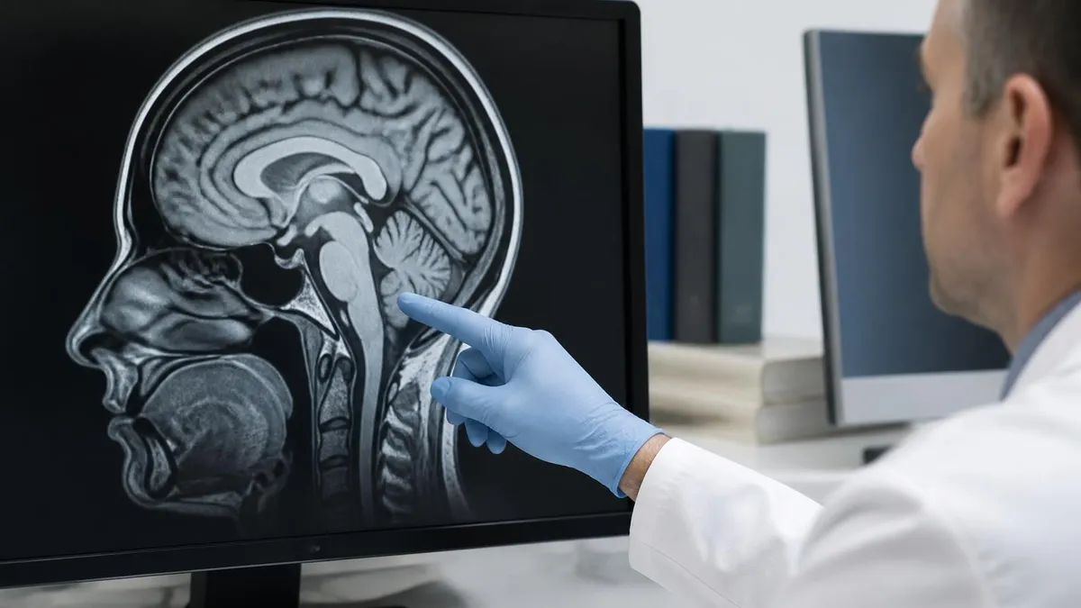
Brain MRI pictures are perhaps the most iconic. An axial brain slice shows a roughly oval outline with the symmetric cerebral hemispheres, the dark butterfly shape of the ventricles, and the textured cortex around the edges. The thalami sit centrally as paired gray ovals, and the brainstem appears as a smaller round structure in lower slices. Sagittal brain images reveal the corpus callosum arching over the ventricles like a comma, with the cerebellum tucked beneath the occipital lobe.
Spine MRI pictures look distinctly different. A sagittal lumbar spine image shows the vertebral bodies stacked vertically with the intervertebral discs between them. On T2, healthy discs glow bright due to their water content while the dark spinal cord runs centrally, surrounded by even brighter cerebrospinal fluid. Disc herniations appear as dark bulges pressing backward into the canal, and any nerve root compression becomes visually obvious in this view.
Knee MRI pictures show the femur and tibia separated by the meniscus and cartilage. The cruciate ligaments cross the joint diagonally on sagittal images, looking like dark bands within the brighter joint fluid. A torn meniscus shows up as a bright line running through the normally dark triangular meniscus shape. Bone bruises appear as patchy bright signal within the marrow on T2 fat-suppressed sequences, often invisible on X-ray but unmistakable on MRI.
Shoulder MRI pictures center on the rotator cuff tendons, the labrum, and the glenohumeral joint. Each tendon appears as a dark band, and any tear shows up as bright fluid signal interrupting that band. The labrum, a fibrocartilaginous rim around the socket, appears as a small dark triangle in cross-section; tears within it look like bright clefts. For patients with orthodontic hardware, our guide on MRI with braces covers what to expect during scans of the head and neck.
abdominal mri cpt code pictures show organs in remarkable detail. The liver appears uniformly mid-gray on T1, the spleen slightly darker, and the kidneys with their distinctive bean shape and corticomedullary differentiation. The biliary tree can be highlighted using a specialized sequence called MRCP that makes bile and pancreatic ducts glow brightly while suppressing surrounding tissue. Pelvic MRI shows the prostate or uterus with exquisite zonal anatomy not visible on any other imaging modality.
Cardiac MRI pictures move. Cine sequences capture the beating heart across the cardiac cycle, displayed as a short loop rather than a single static image. The dark blood pool of the ventricles contrasts with the bright myocardial walls, and any wall motion abnormality becomes visually apparent. Late gadolinium enhancement images, taken minutes after contrast injection, reveal scar tissue as bright patches within the otherwise dark myocardium.
Vascular MRI pictures, called MR angiography, look almost like x-ray contrast studies. Blood vessels appear as bright ribbons against a suppressed background, allowing radiologists to trace the carotids, the circle of Willis, the aorta, and the renal arteries without any catheter procedure. These angiographic views often look strikingly three-dimensional because they are reconstructed from a stack of thin slices into a rotating maximum-intensity projection.
A single MRI study can contain 200 to 500 individual images and consume 100-500 MB on a CD. This is not inefficiency — it reflects the dense diagnostic information packed into each slice. If you receive a disc, install the included DICOM viewer to scroll through all sequences rather than just viewing a few sample JPEGs.
Contrast-enhanced MRI pictures look fundamentally different from non-contrast scans. Gadolinium, the contrast agent used in MRI, shortens the T1 relaxation time of nearby protons, making tissues with disrupted blood-brain barrier or increased vascular permeability light up brilliantly on T1-weighted post-contrast images. A tumor that appeared as a subtle gray mass on a non-contrast scan can suddenly stand out as a vivid white ring against darker normal brain.
Different enhancement patterns reveal different pathologies. A homogeneous enhancing mass typically suggests a meningioma or a highly vascular tumor. A ring-enhancing lesion with a dark center suggests an abscess, necrotic tumor, or demyelinating lesion. A nodular enhancement along a nerve root suggests inflammation or tumor seeding. Radiologists are trained to recognize these patterns the way a botanist recognizes leaf shapes.
Functional MRI pictures look like standard brain images overlaid with brightly colored activation maps. When a patient performs a task — wiggling fingers, listening to words, viewing flashing checkerboards — the corresponding brain regions show increased blood flow, which fMRI detects as a small signal change. Neurosurgeons use these maps before operations to plan around language and motor areas.
Diffusion tensor imaging produces some of the most visually stunning MRI pictures. Each color represents the dominant direction of water diffusion along white matter tracts: red for left-right fibers, green for anterior-posterior fibers, blue for superior-inferior fibers. The resulting image looks like a brightly colored brain wiring diagram, and clinicians use it to track connectivity and detect microstructural injuries.
Spectroscopy, often called MRS, does not produce a picture in the traditional sense. Instead, it generates a graph with peaks representing different metabolites in a small voxel of tissue. Radiologists read these peaks like a chemist reads a mass spectrum, identifying choline, creatine, N-acetylaspartate, and lactate concentrations to characterize brain tumors and metabolic disorders. The image is essentially a chemical fingerprint of the tissue.
Whole-body MRI is increasingly used for cancer screening and staging. The resulting picture is a head-to-toe coronal image that looks like a long, thin scan of the entire body, sometimes accompanied by diffusion-weighted overlays where lesions appear as bright spots against a darkened background. This view can identify metastases, lymphadenopathy, and bone marrow involvement in a single appointment.
Pediatric MRI pictures often look slightly different because growing tissues have different water content. An infant brain shows reversed gray-white matter contrast compared to an adult because myelination is still in progress. Radiologists who interpret pediatric scans use age-specific normal references rather than adult standards. To appreciate how far the technology has evolved, our article on the history of MRI traces the journey from first principles to today's images.
Before your own MRI appointment, ask the scheduling office whether the facility uses an open, wide-bore, or closed-bore system. If you have any history of claustrophobia, weight concerns, or mobility limitations, the open or wide-bore option may save you a great deal of stress. Some imaging centers are happy to send you photos of the specific machine you will be using, and a quick online image search for the model name often yields helpful pictures.
If you receive a CD or USB drive with your images after the appointment, take a moment to install the included DICOM viewer rather than relying on the preview JPEGs. The DICOM software allows you to scroll through every slice, change window and level settings to highlight different tissues, and switch between sequences. This experience makes your radiologist report far more meaningful when you sit down to discuss it with your doctor.
When you review your images for the first time, do not try to make a diagnosis. Even radiologists train for years before reading MRI pictures independently. Instead, use the visual to understand what the report is describing. If the report mentions a herniated disc at L4-L5, scroll to the sagittal lumbar series, find the level, and look at the dark bulge pushing posteriorly. Seeing the finding helps you participate meaningfully in treatment decisions.
Patients often ask whether they should request specific sequences. In general, the radiologist and technologist will choose the appropriate protocol based on your clinical question. However, if you have a specific concern — for example, suspected multiple sclerosis or a small acoustic neuroma — you can ask whether the protocol includes the sequences best suited to detect it (FLAIR for MS, thin-section internal auditory canal sequences for acoustic neuroma).
Repeat MRIs are common for monitoring chronic conditions. When you compare a new scan to an old one, make sure both studies use comparable sequences and slice orientations. A subtle change between two T2-FLAIR images is meaningful; a comparison between a T1 and a T2 is not. Most radiology reports automatically compare to prior studies, but you can ask for this explicitly if your case involves slow-growing tumors or chronic inflammatory disease.
For additional context on common observations in everyday scans, our guide to common MRI findings walks through what radiologists look for in the brain, spine, and joints, along with what each finding typically means clinically. Understanding both the picture and the report together transforms an opaque medical document into a clear conversation with your care team.
Finally, do not be alarmed by incidental findings. Almost every MRI reveals something — a small cyst, a benign lesion, an anatomic variant — that has nothing to do with the original clinical question. Radiologists are trained to mention these findings while clearly distinguishing them from clinically significant pathology. If your report lists several incidental observations, that is normal, and your physician will help you understand which ones matter and which can be safely ignored.
MRI Questions and Answers


About the Author
Medical Laboratory Scientist & Clinical Certification Expert
Johns Hopkins UniversityDr. Sandra Kim holds a PhD in Clinical Laboratory Science from Johns Hopkins University and is certified as a Medical Technologist (MT) and Medical Laboratory Scientist (MLS) through ASCP. With 16 years of clinical laboratory experience spanning hematology, microbiology, and molecular diagnostics, she prepares candidates for ASCP board exams, MLT, MLS, and specialist certification tests.
Join the Discussion
Connect with other students preparing for this exam. Share tips, ask questions, and get advice from people who have been there.
View discussion (6 replies)