
MRI vs fMRI: Key Differences, Clinical Uses, and What Each Scan Reveals 2026
Compare magnetic resonance imaging MRI and functional magnetic resonance imaging fMRI — how each works, clinical uses, and what technologists need to know. 🏆
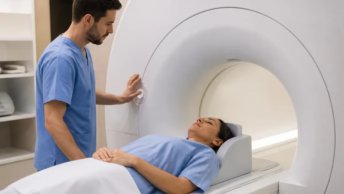
Understanding the distinction between magnetic resonance imaging MRI and functional magnetic resonance imaging fMRI is essential for every imaging professional, radiology student, and clinician who orders or interprets these scans. Both modalities use the same fundamental physics — a powerful magnetic field and radiofrequency pulses to manipulate hydrogen protons — yet their diagnostic purposes, acquisition techniques, and clinical applications differ dramatically. Knowing when each tool is appropriate can make the difference between a definitive diagnosis and an inconclusive workup.
Conventional MRI excels at producing high-resolution, static anatomical images of soft tissue, bone marrow, cartilage, and organ structures. A radiologist reviewing an MRI of the lumbar spine, for example, can precisely measure disc height, identify herniated nucleus pulposus material, and detect nerve root compression with submillimeter detail. This structural clarity makes conventional MRI the gold standard for musculoskeletal pathology, neurological anatomy, oncologic staging, and vascular assessment when combined with contrast agents.
Functional MRI, by contrast, was developed in the early 1990s to map brain activity rather than anatomy. It detects tiny changes in blood oxygenation — a phenomenon called the blood-oxygen-level-dependent, or BOLD, signal — that serve as an indirect proxy for neuronal firing. When a brain region becomes more active, local cerebral blood flow increases, raising the ratio of oxyhemoglobin to deoxyhemoglobin and creating a measurable signal change on a T2*-weighted gradient-echo sequence. This time-series data reveals which cortical and subcortical regions are engaged during a specific cognitive, sensory, or motor task.
For MRI registry candidates and technologists sitting board examinations, the comparison between mri vs fmri appears regularly in physics and clinical applications sections. Understanding BOLD contrast, echo-planar imaging sequences, and the hemodynamic response function are no longer purely academic topics — they increasingly appear in advanced practice settings as fMRI moves from pure research into pre-surgical planning, epilepsy mapping, and psychiatric research protocols at major academic medical centers across the United States.
The equipment used for both modalities is often the same high-field scanner — typically a 1.5T or 3T superconducting magnet — but the pulse sequences, coil configurations, and post-processing pipelines diverge significantly. An fMRI protocol demands faster repetition times, EPI readouts prone to susceptibility artifacts, and sophisticated statistical modeling software that conventional structural protocols simply do not require. Technologists performing fMRI must also manage patient compliance with cognitive paradigms, maintain strict head motion standards, and coordinate with neuropsychologists or researchers administering the tasks.
This article breaks down everything you need to know about the two modalities: how the underlying physics differs, what each scan can and cannot detect, which clinical scenarios call for one versus the other, and how fMRI is reshaping neuroscience and pre-surgical planning in 2026. Whether you are preparing for the MRI registry exam, rotating through a neuroimaging research center, or simply building a deeper conceptual foundation, the comparisons and explanations that follow will give you a comprehensive, evidence-based understanding of both tools.
The content is structured to match the topics most heavily weighted on the ARRT MRI examination blueprint, so you can study with confidence that the time you invest here directly supports your board preparation as well as your day-to-day clinical competence.
MRI vs fMRI by the Numbers
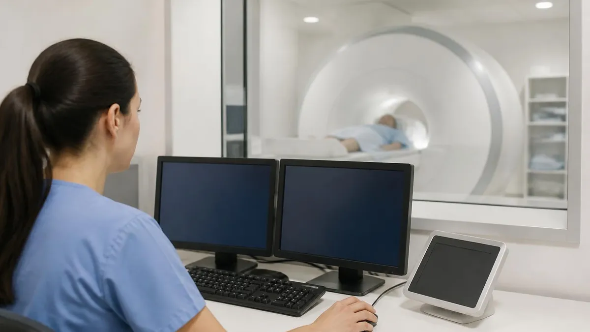
How MRI and fMRI Work: Core Principles
Uses static T1, T2, and proton-density sequences to map tissue anatomy. Hydrogen protons align with the main magnetic field, are tipped by an RF pulse, and release energy as they relax — producing signal differences that reflect tissue composition and water content.
Exploits the magnetic susceptibility difference between oxyhemoglobin and deoxyhemoglobin. Active neurons trigger local blood flow increase, raising oxyhemoglobin concentration and producing a small but measurable T2* signal enhancement captured by gradient-echo EPI sequences.
fMRI relies on EPI to acquire a full brain volume in 1–3 seconds — far faster than conventional spin-echo or gradient-echo sequences. This speed enables time-series sampling of hemodynamic responses but introduces susceptibility and geometric distortion artifacts near air-tissue interfaces.
The BOLD signal peaks 5–8 seconds after neuronal firing due to neurovascular coupling delays. Statistical models (GLM-based) correlate this expected HRF shape with the experimental task timeline, identifying voxels whose signal time-course matches predicted activation patterns.
Task-based fMRI maps activation during a specific stimulus or behavior. Resting-state fMRI measures spontaneous low-frequency BOLD fluctuations in the absence of a task, revealing intrinsic functional connectivity networks such as the default mode, salience, and frontoparietal control networks.
The clinical applications of conventional MRI are extraordinarily broad and well-established. Orthopedic surgeons rely on MRI to evaluate meniscal tears, rotator cuff injuries, anterior cruciate ligament ruptures, and stress fractures invisible on plain radiographs. Neurologists order brain MRI with and without gadolinium contrast to detect multiple sclerosis plaques, stroke infarcts, cerebral neoplasms, and encephalitis. Cardiologists use cardiac MRI to quantify ejection fraction, identify myocardial fibrosis via late gadolinium enhancement, and assess complex congenital heart disease with exceptional soft-tissue contrast that echocardiography cannot match.
Oncologists depend on MRI for staging primary tumors and detecting local invasion of adjacent structures, particularly for prostate, rectal, cervical, and hepatic malignancies where staging accuracy directly determines surgical resectability and treatment planning. The multiplanar capability of MRI — axial, coronal, sagittal, and oblique planes obtainable without repositioning the patient — provides comprehensive volumetric assessment that CT with ionizing radiation cannot replicate with the same soft-tissue differentiation. Spectroscopy sequences can even probe tissue biochemistry, measuring metabolite ratios that help distinguish recurrent tumor from post-treatment necrosis.
Functional MRI has carved out a distinct clinical niche centered on brain mapping. The most established clinical use is pre-surgical language and motor mapping for patients undergoing resection of cortical tumors, epileptic foci, or arteriovenous malformations. Before fMRI, the Wada test — intracarotid sodium amobarbital injection — was the only way to lateralize language dominance. Today, task-based fMRI using verb-generation, picture-naming, or semantic-decision paradigms provides equivalent or superior lateralization in the majority of patients, avoiding an invasive arterial procedure entirely and giving surgeons a precise cortical map overlaid on anatomical images.
Epilepsy surgery centers use fMRI to map eloquent cortex adjacent to seizure foci, informing resection boundaries that balance seizure freedom against functional deficits. A patient whose seizure focus abuts the primary motor hand area requires exquisite pre-operative mapping to determine whether curative resection is feasible without causing permanent hemiparesis. fMRI motor paradigms — typically finger-tapping or hand-clenching tasks — activate the primary motor cortex with high reliability, and the resulting activation maps are integrated into intraoperative neuronavigation systems for real-time surgical guidance.
Psychiatric research has been transformed by resting-state fMRI, which has revealed reproducible disruptions in functional connectivity in conditions including major depressive disorder, schizophrenia, autism spectrum disorder, and post-traumatic stress disorder. The default mode network — a set of midline and lateral parietal regions that activates during self-referential thought and deactivates during externally directed tasks — shows aberrant connectivity in depression, with insufficient suppression during cognitive tasks contributing to ruminative symptomatology. These findings are reshaping how psychiatrists conceptualize mental illness as a disorder of brain circuit dynamics rather than purely a neurochemical imbalance.
Pediatric neuroimaging represents another domain where the differences between structural MRI and fMRI matter enormously. Neonates and infants rarely tolerate task-based paradigms, but resting-state fMRI can be acquired during natural sleep and has revealed remarkably mature functional networks even in premature infants, opening windows into early brain development and the neural substrates of neurodevelopmental disorders such as cerebral palsy and language delay. The absence of ionizing radiation makes repeated MRI and fMRI studies ethically feasible in developing brains, unlike CT-based surveillance.
In research settings, fMRI continues to drive discoveries about the neural basis of perception, memory, decision-making, and social cognition. Multiband EPI sequences now allow simultaneous acquisition of multiple slices, reducing TR to under one second and enabling detection of faster neural dynamics. Ultra-high-field 7T scanners offer BOLD sensitivity high enough to resolve cortical layers and columns, blurring the boundary between human neuroimaging and invasive electrophysiology.
MRI Practice Test Questions
Prepare for the MRI - Magnetic Resonance Imaging exam with our free practice test modules. Each quiz covers key topics to help you pass on your first try.
MRI Knowledge
MRI Exam Questions covering Knowledge. Master MRI Test concepts for certification prep.
MRI Physics
Free MRI Practice Test featuring Physics. Improve your MRI Exam score with mock test prep.
MRI Anatomy and Pathology
MRI Test Prep for MRI Anatomy and Pathology. Practice MRI Quiz questions and boost your score.
MRI Anatomy and Positioning
MRI Questions and Answers on MRI Anatomy and Positioning. Free MRI practice for exam readiness.
MRI Contrast Agents
Free MRI Quiz on MRI Contrast Agents. MRI Exam prep questions with detailed explanations.
MRI Patient Care and Positioning
MRI Practice Questions for MRI Patient Care and Positioning. Build confidence for your MRI certification exam.
MRI vs fMRI: Sequences, Artifacts, and Protocol Design
Conventional MRI uses spin-echo sequences for T1 and T2 contrast, relying on 180-degree refocusing pulses to eliminate field inhomogeneity effects and produce clean anatomical images. Gradient-echo sequences trade some artifact immunity for faster acquisition, enabling dynamic contrast imaging and breath-hold abdominal protocols. The choice of TE and TR determines tissue contrast weighting, with short TR and TE producing T1-weighted images and long TR and TE producing T2-weighted tissue differentiation.
fMRI almost exclusively uses gradient-echo EPI, which reads an entire k-space plane after a single RF excitation by rapidly toggling readout gradients. This makes whole-brain acquisition in 1–3 seconds possible but creates strong susceptibility artifacts near the orbitofrontal cortex, temporal poles, and brainstem — regions where bone-air interfaces distort the local magnetic field. Parallel imaging techniques such as GRAPPA and simultaneous multi-slice (SMS) acceleration help reduce distortion while maintaining temporal resolution adequate for hemodynamic sampling.
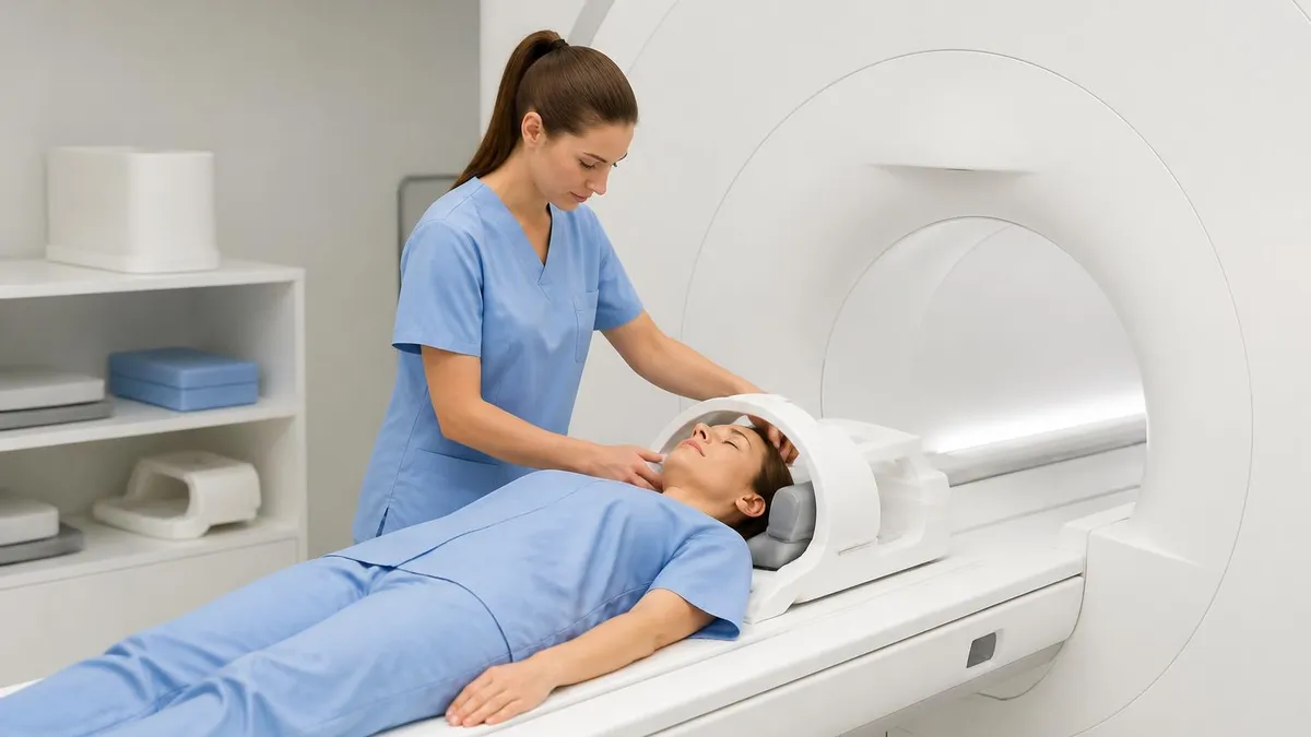
Conventional MRI vs fMRI: Strengths and Limitations
- +Conventional MRI delivers submillimeter anatomical resolution ideal for diagnosing structural pathology
- +Well-established clinical protocols exist for virtually every organ system and body region
- +No ionizing radiation, making repeated imaging safe for pediatric and pregnant patients
- +fMRI provides non-invasive brain mapping that replaces invasive Wada testing for language lateralization
- +fMRI resting-state networks detectable without patient cooperation, enabling infant and sedated-patient studies
- +Both modalities use the same scanner hardware, allowing structural and functional data in one session
- −Conventional MRI cannot assess neuronal function — a structurally normal brain can be severely dysfunctional
- −Long scan times increase motion artifact risk and limit use in claustrophobic or agitated patients
- −fMRI BOLD signal is an indirect measure of neural activity, subject to neurovascular uncoupling in disease
- −EPI sequences produce significant susceptibility artifacts in orbitofrontal and inferior temporal regions
- −fMRI requires complex statistical analysis and specialized post-processing software not available at all centers
- −Clinical fMRI is not yet reimbursed by all payers and remains unavailable at most community hospitals
Pros and Cons at a Glance
| Pros | Cons |
|---|---|
| Conventional MRI delivers submillimeter anatomical resolution ideal for diagnosing structural pathology | Conventional MRI cannot assess neuronal function — a structurally normal brain can be severely dysfunctional |
| Well-established clinical protocols exist for virtually every organ system and body region | Long scan times increase motion artifact risk and limit use in claustrophobic or agitated patients |
| No ionizing radiation, making repeated imaging safe for pediatric and pregnant patients | fMRI BOLD signal is an indirect measure of neural activity, subject to neurovascular uncoupling in disease |
| fMRI provides non-invasive brain mapping that replaces invasive Wada testing for language lateralization | EPI sequences produce significant susceptibility artifacts in orbitofrontal and inferior temporal regions |
| fMRI resting-state networks detectable without patient cooperation, enabling infant and sedated-patient studies | fMRI requires complex statistical analysis and specialized post-processing software not available at all centers |
| Both modalities use the same scanner hardware, allowing structural and functional data in one session | Clinical fMRI is not yet reimbursed by all payers and remains unavailable at most community hospitals |
MRI and fMRI Technologist Preparation Checklist
- ✓Screen all patients for metallic implants, pacemakers, and cochlear devices before entry into Zone III or IV.
- ✓Verify field strength compatibility of any implant using the MRI implant safety database before scanning.
- ✓For fMRI, confirm the patient understands and can perform the cognitive or motor task paradigm reliably.
- ✓Set TR to 2,000 ms or less and use multiband acceleration to achieve adequate temporal sampling for BOLD.
- ✓Apply EPI distortion correction using a field map or reversed phase-encode (B0 shimming) acquisition.
- ✓Monitor head motion in real time and instruct patients to remain completely still throughout fMRI runs.
- ✓Acquire a high-resolution T1 MPRAGE for structural co-registration with functional activation maps.
- ✓Confirm that task-presentation software is synchronized with scanner triggers using a TTL pulse interface.
- ✓After fMRI acquisition, review motion parameters and flag runs exceeding 1.5 mm translation or 1.5-degree rotation.
- ✓Archive raw DICOM data and processed activation maps in PACS with appropriate clinical fMRI series labels.
The BOLD Signal Lags Neural Activity by 5–8 Seconds
One of the most important concepts for both registry exam preparation and clinical practice is that the fMRI BOLD signal does not directly measure neuronal electrical activity. Instead, it reflects neurovascular coupling: the local vasodilation and blood flow increase that neurons trigger through astrocyte signaling. This hemodynamic response peaks approximately 5–8 seconds after the onset of neural firing, meaning fMRI has poor temporal resolution compared to EEG or MEG — but unmatched spatial coverage and resolution for whole-brain mapping of function.
Pre-surgical fMRI planning has become one of the most clinically impactful applications of functional neuroimaging. When a glioma or cavernous malformation lies near eloquent cortex — the regions responsible for language production, language comprehension, primary motor function, or primary sensory processing — the surgical team must understand the precise spatial relationship between the lesion and functional tissue before operating. Resecting into eloquent cortex carries the risk of permanent neurological deficit, yet leaving tumor behind compromises patient survival and quality of life.
Language mapping fMRI typically employs a battery of paradigms targeting Broca's area (inferior frontal gyrus, pars triangularis and opercularis) and Wernicke's area (posterior superior temporal gyrus) independently, because these regions have distinct roles in language production and comprehension and may be differentially at risk depending on tumor location. Common paradigms include silent verb generation, picture naming, semantic decision tasks, and sentence completion — each designed to maximally activate specific components of the language network while minimizing activation of non-linguistic regions that could confound localization.
Motor mapping fMRI using finger-tapping, foot-tapping, or tongue-movement paradigms reliably activates the primary motor cortex somatotopic representations — the famous homunculus — allowing surgeons to identify the central sulcus even when overlying gyral anatomy is distorted by mass effect. Combined with diffusion tensor imaging tractography of the corticospinal tract, pre-surgical fMRI provides a multi-modal functional roadmap that intraoperative neuronavigation systems can display in real time as the surgeon works millimeters from critical tissue.
The American Society of Functional Neuroradiology (ASFNR) and the American College of Radiology (ACR) have both published guidance documents on clinical fMRI standards, emphasizing the importance of paradigm validation, statistical threshold selection, and clinical interpretation frameworks that appropriately communicate uncertainty to surgical teams. An fMRI activation map is a statistical probability map, not a sharp anatomical boundary — this nuance must be conveyed in radiology reports so surgeons do not rely on activation coordinates with false precision.
Epilepsy surgery programs represent the other major clinical consumer of pre-surgical fMRI. Patients with drug-resistant focal epilepsy are candidates for surgical resection, but success depends on complete removal of the seizure-onset zone without damaging eloquent cortex. Beyond language and motor mapping, fMRI can assess memory lateralization — a critical determinant of post-operative memory risk — by using paradigms that engage the hippocampus and parahippocampal gyrus, such as scene encoding, object recognition, or verbal encoding tasks that have been validated against intracarotid amobarbital testing outcomes in large patient series.
Connectivity-based fMRI approaches, including resting-state functional connectivity analysis and graph-theoretic network metrics, are increasingly used in pre-surgical planning to assess how the seizure focus is embedded within broader network topology. Centers have demonstrated that the degree to which the ictal zone is connected to the language and memory networks predicts post-operative cognitive outcomes, suggesting that network-level analysis provides information beyond simple activation mapping. This represents a frontier where basic neuroscience methodology is being translated into clinical decision support.
Insurance coverage for clinical fMRI varies by payer and indication. Medicare currently covers pre-surgical language mapping under specific criteria, and many private insurers follow CMS guidance. Technologists and radiologists entering clinical fMRI programs must be familiar with documentation requirements, including the clinical indication, paradigm used, statistical threshold applied, and explicit statement of lateralization index or activation locus relative to the surgical target — documentation standards that directly mirror topics tested on advanced MRI registry examinations.
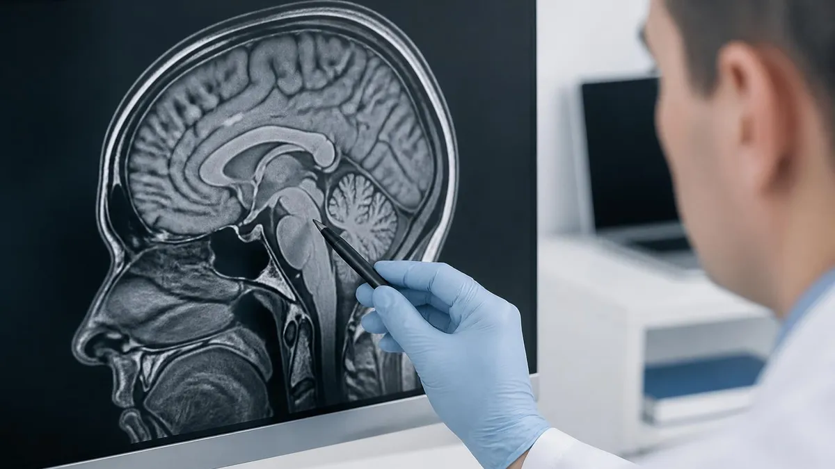
Neurovascular uncoupling — the breakdown of the normal relationship between neuronal firing and hemodynamic response — can occur in brain tumors, vascular malformations, severe cerebrovascular disease, and some medications. In these cases, absent fMRI activation does NOT confirm that eloquent cortex is absent. Surgical teams must integrate fMRI findings with direct cortical stimulation (DCS), transcranial magnetic stimulation (TMS), and clinical neurological assessment rather than relying on fMRI activation alone as the sole determinant of resection boundaries.
For MRI registry candidates, the physics and clinical science of fMRI represent a growing portion of examination content as the technology moves from research into routine clinical practice. The ARRT MRI examination blueprint includes sequences, artifacts, image quality, and patient care domains where fMRI concepts intersect with broader knowledge of gradient-echo physics, echo-planar imaging characteristics, and neurovascular physiology. Understanding why EPI produces susceptibility artifacts — and how shimming and distortion correction mitigate them — connects directly to the physics of gradient-echo sequences tested in the MRI board examination.
Gradient-echo sequences are sensitive to T2* relaxation, which reflects both true T2 spin-spin relaxation and additional dephasing caused by local magnetic field inhomogeneities. Tissues near air-tissue interfaces (sinuses, ear canals, skull base) contain field gradients that accelerate dephasing and reduce signal, producing signal voids on gradient-echo images. The same susceptibility sensitivity that makes gradient-echo sequences prone to artifacts is precisely what makes them sensitive to the BOLD effect — the changing magnetic susceptibility of hemoglobin as it transitions between oxygenated and deoxygenated states is detectable because gradient-echo sequences are not insulated from field inhomogeneity effects the way spin-echo sequences are.
T2* weighting in EPI is controlled by the echo time (TE). For optimal BOLD sensitivity at 3T, TE is typically set near the T2* of gray matter, approximately 28–35 ms. Shorter TE reduces BOLD sensitivity because the signal hasn't had time to reflect susceptibility differences; longer TE allows excessive T2* decay and reduces overall SNR. At 7T, where T2* is shorter due to increased B0 field strength, optimal TE shortens to approximately 20–25 ms — a relationship that illustrates the field-strength dependence of BOLD sensitivity that registry candidates may encounter in advanced physics questions.
Parallel imaging acceleration — GRAPPA, SENSE, or their vendor-specific implementations — is nearly universal in clinical fMRI protocols. By acquiring only a subset of k-space lines and using coil sensitivity profiles to reconstruct the full image, parallel imaging reduces echo train length and therefore geometric distortion in EPI. Understanding k-space, Fourier encoding, and parallel imaging reconstruction principles is central to both conventional MRI physics and fMRI optimization, and these topics appear prominently in the ARRT examination across multiple content categories.
Signal-to-noise ratio (SNR) considerations differ between structural MRI and fMRI. In structural MRI, SNR is primarily a function of voxel volume, field strength, receive coil, and acquisition bandwidth. In fMRI, the relevant metric is temporal SNR (tSNR) — the mean signal divided by the standard deviation across time-points — which reflects both thermal noise and physiological noise from cardiac pulsation, respiratory motion, and scanner instability.
At high spatial resolution, thermal noise dominates and increasing voxel size improves tSNR; at lower resolution, physiological noise becomes dominant and spatial resolution gains plateau. This noise regime transition at approximately 2–3 mm isotropic resolution at 3T is a key consideration in protocol optimization.
Contrast agents are rarely used in fMRI studies because gadolinium-based contrast agents shorten T2* and reduce baseline BOLD signal, interfering with the susceptibility contrast mechanism. This is an important practical point for technologists: if a patient is scheduled for a combined structural contrast-enhanced MRI and fMRI session, the fMRI acquisition must be completed before gadolinium injection, not after. Failure to sequence the protocol correctly results in attenuated BOLD sensitivity and potentially non-diagnostic functional data — a patient safety and quality issue with direct implications for pre-surgical decision-making.
Radiofrequency coil selection also matters for fMRI quality. Dense phased-array head coils with 32 or 64 channels provide higher SNR and greater parallel imaging acceleration factors than older 8-channel coils, improving tSNR and enabling higher spatial resolution or faster acquisition. For registry candidates studying coil technology, the principles of phased-array combination — noise-weighted sum-of-squares — apply equally to structural and functional acquisitions, reinforcing the conceptual unity underlying both modalities despite their very different clinical purposes.
Building practical competence with both MRI and fMRI requires a study strategy that integrates physics principles, clinical application knowledge, and hands-on protocol experience. For registry candidates, the most efficient approach is to anchor each physics concept — T2* contrast, echo-planar imaging, parallel imaging — to a specific clinical or technical scenario where that concept determines diagnostic quality. Abstract physics memorization fades quickly; applied understanding embedded in real clinical examples persists and generalizes to novel examination questions.
When studying BOLD physiology, practice explaining the hemodynamic response function from first principles: neurons fire, glutamate release triggers astrocyte calcium signaling, astrocytes release vasoactive mediators, arterioles dilate, blood flow increases, the oxyhemoglobin-to-deoxyhemoglobin ratio rises, T2* lengthens locally, gradient-echo EPI signal increases. Tracing this causal chain in both directions — from stimulus to signal, and from signal back to limitations — reveals why neurovascular uncoupling matters and why fMRI cannot directly measure action potentials.
Comparing conventional MRI artifacts with fMRI-specific artifacts reinforces understanding of both. Conventional spin-echo MRI minimizes susceptibility effects; gradient-echo EPI maximizes susceptibility sensitivity for BOLD but introduces distortion and dropout. Motion artifacts differ in character: in structural MRI, motion during a single acquisition blurs or ghosts one image; in fMRI, motion across a 6-minute time-series introduces volume-to-volume misalignment that propagates through statistical modeling. Understanding these distinctions prepares candidates for artifact recognition questions on the registry examination.
Protocol design questions test whether candidates understand acquisition trade-offs. A question might present an fMRI protocol with TR of 5,000 ms and ask whether BOLD sensitivity or temporal sampling is compromised — the answer is temporal sampling, since sampling the hemodynamic response every 5 seconds misses the peak of the 5–8 second HRF and aliases cardiac pulsation into the BOLD frequency band.
Alternatively, a protocol with TE of 80 ms at 3T would sacrifice BOLD SNR due to excessive T2* decay past the gray matter T2* of 30–35 ms. Recognizing these trade-offs requires understanding the physics deeply enough to reason from principles, not just recall memorized facts.
Practice quizzes that present fMRI scenarios alongside conventional MRI physics questions are the most effective preparation tool because they train the contextual switching that the actual registry examination requires. You may encounter a question about spin-echo refocusing followed immediately by a question about BOLD contrast mechanisms — the exam does not segregate topics in the logical sequence of a textbook. Regular timed practice across mixed topic banks builds the cognitive flexibility needed to perform well under examination conditions.
Study groups and peer teaching are underutilized preparation strategies. Explaining the difference between T2 and T2* relaxation to a study partner — and then explaining how that difference makes gradient-echo sequences BOLD-sensitive while spin-echo sequences are not — consolidates understanding more deeply than passive reading. Teaching others forces articulation of logical connections that passive study can leave implicit, and peer questions surface gaps in understanding that solo study misses entirely.
Finally, integrate your examination preparation with clinical observation whenever possible. If your training program includes exposure to a neuroimaging research center or a hospital with a pre-surgical fMRI program, request permission to observe an fMRI acquisition and post-processing session. Watching an activation map emerge from raw EPI time-series data and understanding the statistical steps that produced it connects textbook physics to real-world practice in a way that makes both more memorable and more meaningful when you encounter related questions in an examination environment.
Sample MRI - Magnetic Resonance Imaging Practice Questions
Try these questions from our free MRI - Magnetic Resonance Imaging practice tests. The correct answer and an explanation follow each question.
In MRI, what does the term 'bandwidth' typically refer to in clinical practice?
- A. The range of flip angles used in a sequence
- B. The range of frequencies sampled during readout, affecting SNR and chemical shift
- C. The number of phase-encode steps acquired
- D. The RF pulse duration in milliseconds
Answer: B. The range of frequencies sampled during readout, affecting SNR and chemical shift
Receiver bandwidth is the range of frequencies digitized during readout; wider bandwidth reduces chemical shift and distortion but decreases SNR.
What is the minimum number of clinical MRI hours required by ARRT before a candidate can sit for the MRI registry examination?
- A. 200 hours
- B. 500 hours
- C. 1,000 hours
- D. 1,500 hours
Answer: B. 500 hours
ARRT requires candidates to document at least 500 hours of clinical MRI experience as part of eligibility requirements.
Which gradient is primarily responsible for frequency encoding in MRI?
- A. Readout gradient
- B. Phase-encoding gradient
- C. Slice-select gradient
- D. Spoiler gradient
Answer: A. Readout gradient
The readout (frequency-encoding) gradient is applied during signal acquisition, causing spins at different positions to precess at different frequencies.
Wraparound (aliasing) artifact in MRI is caused by:
- A. Structures outside the FOV contributing signal that is folded back into the image
- B. Gradient non-linearity at the edges of the bore
- C. T2* dephasing from metallic objects
- D. Phase errors from pulsatile flow
Answer: A. Structures outside the FOV contributing signal that is folded back into the image
When anatomy extends beyond the selected FOV in the phase-encode direction, it is aliased and appears superimposed on the opposite side of the image.
Take the full MRI - Magnetic Resonance Imaging practice test
MRI Questions and Answers

MRI of Cervical Spine: Complete Guide to Imaging, Anatomy, Pathology, and Patient Preparation

Knee MRI Images: A Complete Guide to Reading, Understanding, and Interpreting Knee Scans
Ace Your Exam with MRI Practice Test 2026 Prep

MRI Tech School 2026 — Programs, Requirements, and What to Expect
MRI Procedure: What to Expect Before, During and After Your Scan
How this page was made: written with AI tools and published by Nathan Beja, founder of Practice Test Geeks, who is responsible for its content. Exam facts are checked against the official sources listed below. Not reviewed by a subject-matter expert. Editorial policy
Sources & references
Exam facts on this page (fees, format, dates, passing scores) are checked against these official sources; the date is our last check. Editorial policy
- arrt.org — Passing score, score release, fees, exam information · verified Oct 2026
- assets-us-01.kc-usercontent.com — Renewal, score validity, test delivery, eligibility, exam format, test dates, time limit · verified Oct 2026