
DTI MRI: A Complete Guide to Diffusion Tensor Imaging, Tractography, and Clinical Applications
❓ dti mri explained: how diffusion tensor imaging maps white matter tracts, FA and ADC values, tractography, clinical uses, and exam-ready prep tips.
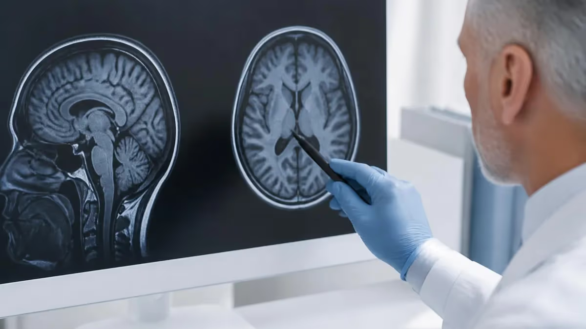
A dti mri, short for diffusion tensor imaging magnetic resonance imaging, is a specialized technique that maps the movement of water molecules along white matter pathways in the brain and spinal cord. Unlike conventional sequences that show anatomy by signal intensity alone, dti mri measures the direction and magnitude of water diffusion, revealing the organized fiber bundles that connect different regions of the central nervous system. For technologists and radiologists, understanding this method is essential to modern neuroimaging.
The physics behind dti mri rests on a concept called diffusion anisotropy. In free fluid such as cerebrospinal fluid, water diffuses equally in all directions, a condition known as isotropic diffusion. Inside tightly packed nerve fibers, however, water moves more freely parallel to the axon than across the myelin sheath. This directional preference is called anisotropic diffusion, and the diffusion tensor model captures it mathematically using a three-by-three matrix that describes diffusion along multiple axes.
To build a diffusion tensor, the scanner applies motion-probing diffusion gradients in at least six non-collinear directions, and frequently in 30, 60, or more directions for higher angular resolution. Each direction produces a diffusion-weighted image, and software combines them to calculate eigenvalues and eigenvectors at every voxel. From these values, radiologists derive scalar maps such as fractional anisotropy and mean diffusivity that quantify tissue microstructure with remarkable sensitivity to subtle changes.
One of the most striking outputs of dti mri is tractography, the three-dimensional reconstruction of white matter tracts. By following the principal eigenvector from voxel to voxel, algorithms trace continuous fiber pathways such as the corticospinal tract, the arcuate fasciculus, and the corpus callosum. These colorful renderings are not just visually impressive; they guide neurosurgeons who must preserve critical connections when removing tumors located near eloquent brain regions.
Clinically, dti mri has become a workhorse for evaluating conditions that disrupt white matter integrity. Stroke, traumatic brain injury, multiple sclerosis, and neurodegenerative diseases all alter the way water diffuses through neural tissue. Because these microstructural changes often precede visible abnormalities on standard sequences, diffusion tensor imaging frequently detects damage earlier, offering clinicians a window into disease processes that conventional imaging may miss entirely.
This guide walks through every practical aspect of dti mri, from acquisition parameters and the meaning of fractional anisotropy to artifacts, clinical indications, and study strategies for registry and physics exams. Whether you are a student preparing for boards, a technologist refining protocols, or a clinician interpreting tractography reports, the sections below break down the technique into clear, exam-ready knowledge you can apply immediately at the scanner.
Throughout, we emphasize the connection between physics principles and real patient care. Mastering diffusion tensor imaging means understanding b-values, gradient directions, echo-planar acquisition, and the trade-offs between scan time and image quality, all while recognizing how each setting affects the final tractography that a surgeon may rely upon during an operation.
DTI MRI by the Numbers
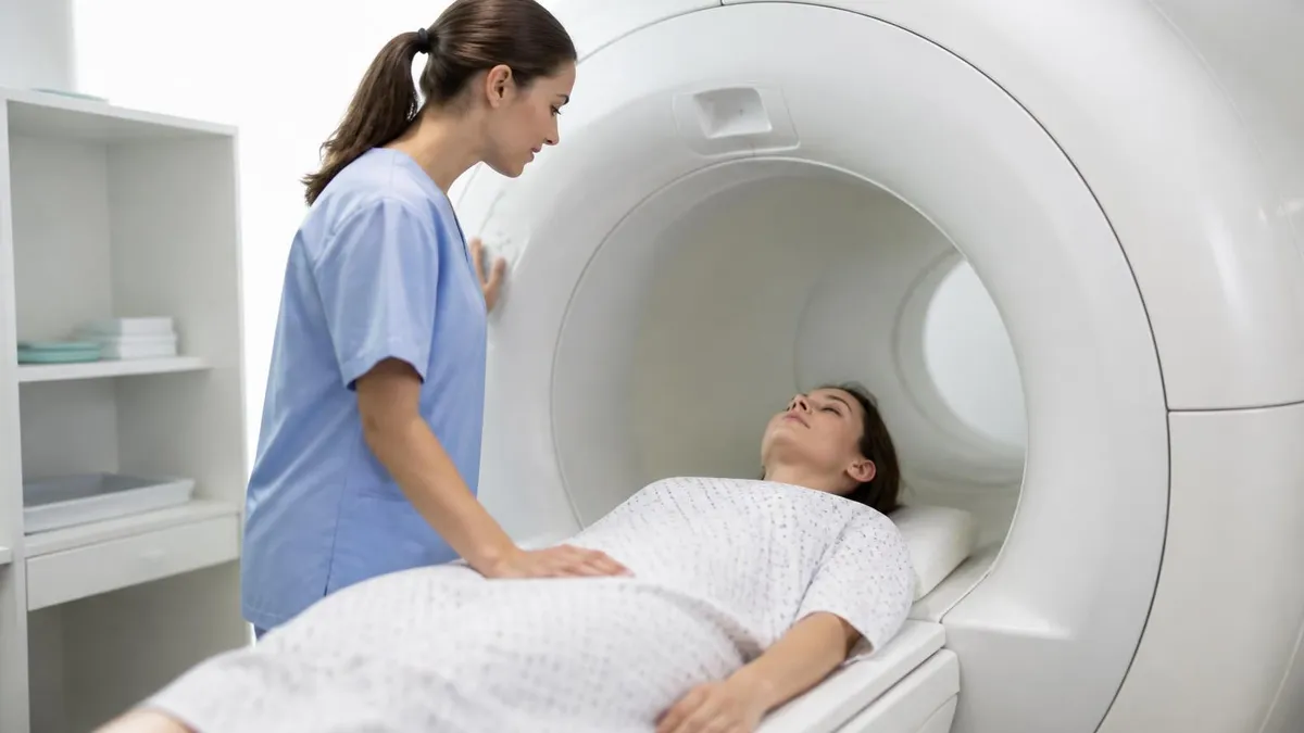
How DTI Acquisition Works Step by Step
Strong motion-probing gradients are applied in at least six non-collinear directions. Each sensitizes the signal to water movement along a specific axis, providing the raw directional data the tensor model requires.
DTI almost always uses single-shot echo-planar imaging to freeze motion and capture each diffusion-weighted volume in a fraction of a second, minimizing blur from patient movement and pulsation.
A b-value near 1000 s/mm² balances diffusion sensitivity against signal-to-noise. A baseline b0 image with no diffusion weighting is acquired for comparison and tensor calculation.
Software fits the directional measurements to a tensor, deriving eigenvalues and eigenvectors at each voxel. These feed the scalar maps and fiber-tracking algorithms used in interpretation.
Once the diffusion tensor is calculated for every voxel, radiologists rely on a family of scalar metrics to interpret the data. The most widely cited is fractional anisotropy, abbreviated FA, which ranges from zero to one. A value near zero indicates isotropic diffusion, as seen in cerebrospinal fluid, while values approaching one reflect highly organized, directional diffusion typical of dense white matter tracts such as the corpus callosum and the corticospinal tract.
Fractional anisotropy is sensitive but not specific. A drop in FA can signal demyelination, axonal loss, edema, or simply the natural crossing of fibers within a voxel. Because of this, experienced readers interpret FA alongside other measures rather than in isolation. Color-coded FA maps add directional information, conventionally rendering left-right fibers in red, anterior-posterior in green, and superior-inferior in blue, a scheme that helps identify specific tracts at a glance.
Mean diffusivity, sometimes reported as the apparent diffusion coefficient or ADC, describes the overall magnitude of water movement regardless of direction. Improved mean diffusivity often accompanies tissue breakdown, vasogenic edema, or chronic infarction, whereas restricted diffusion with a low ADC is the hallmark of acute ischemic stroke. Reading FA and ADC together gives a fuller picture of whether tissue is structurally disorganized, edematous, or acutely injured.
Two additional metrics, axial diffusivity and radial diffusivity, separate diffusion parallel to the axon from diffusion perpendicular to it. Research suggests that reduced axial diffusivity may correlate with axonal damage, while increased radial diffusivity tends to reflect myelin disruption. Although these distinctions remain partly investigational, they illustrate how diffusion tensor imaging can probe specific aspects of white matter microstructure that no other noninvasive method can reach.
Quantitative analysis takes several forms. Region-of-interest measurements place a sampling area on a tract and report average FA or ADC, which is simple but operator-dependent. Tract-based spatial statistics align data across subjects to compare groups, a method common in neuroscience studies of aging, schizophrenia, and traumatic brain injury. Each approach carries assumptions, and technologists who understand them can better anticipate the questions clinicians and researchers will ask.
Normal FA values vary by location and age. Mature white matter in the splenium of the corpus callosum may show FA above 0.7, while peripheral subcortical fibers read considerably lower. FA rises during childhood as myelination progresses and declines gradually in late adulthood. Knowing these expected ranges prevents misreading normal developmental or aging patterns as pathology, a frequent pitfall for newcomers to diffusion tensor analysis.
Finally, reproducibility matters. Different scanners, field strengths, and gradient schemes produce slightly different FA and ADC numbers, so longitudinal studies ideally use identical protocols. When a report cites quantitative diffusion values, the reader should confirm the acquisition matched prior exams. This attention to consistency separates reliable clinical interpretation from misleading comparisons across mismatched datasets.
MRI Practice Test Questions
Prepare for the MRI - Magnetic Resonance Imaging exam with our free practice test modules. Each quiz covers key topics to help you pass on your first try.
MRI Knowledge
MRI Exam Questions covering Knowledge. Master MRI Test concepts for certification prep.
MRI Physics
Free MRI Practice Test featuring Physics. Improve your MRI Exam score with mock test prep.
MRI Anatomy and Pathology
MRI Test Prep for MRI Anatomy and Pathology. Practice MRI Quiz questions and boost your score.
MRI Anatomy and Positioning
MRI Questions and Answers on MRI Anatomy and Positioning. Free MRI practice for exam readiness.
MRI Contrast Agents
Free MRI Quiz on MRI Contrast Agents. MRI Exam prep questions with detailed explanations.
MRI Patient Care and Positioning
MRI Practice Questions for MRI Patient Care and Positioning. Build confidence for your MRI certification exam.
Tractography and Diffusion Tensor Imaging Fiber Mapping
Tractography is the computational process of reconstructing white matter pathways from diffusion tensor data. Algorithms follow the principal eigenvector, the direction of greatest diffusion, from one voxel to the next, building continuous simplifies that approximate real nerve fiber bundles. The result is a three-dimensional map of tracts such as the arcuate fasciculus, optic radiations, and corticospinal tract.
These reconstructions are estimates, not direct visualizations of axons. Simplifies represent the most probable orientation of fibers averaged within each voxel. Despite this limitation, tractography offers clinicians an unprecedented view of connectivity, transforming abstract diffusion numbers into intuitive anatomical pictures that support diagnosis, surgical planning, and research into brain networks.
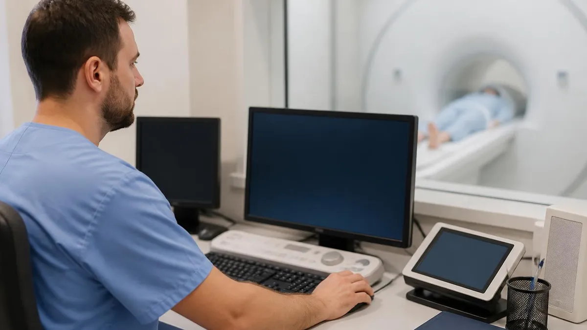
Is DTI MRI Worth the Added Scan Time?
- +Detects white matter damage before it appears on conventional MRI sequences
- +Enables three-dimensional tractography for neurosurgical planning
- +Quantifies microstructure with FA, ADC, and diffusivity metrics
- +Noninvasive with no contrast agent or ionizing radiation required
- +Valuable in stroke, trauma, MS, and neurodegenerative research
- +Adds only a few minutes to a standard brain MRI protocol
- −Sensitive to motion, requiring patient cooperation and fast acquisition
- −Susceptible to susceptibility and eddy-current distortions near bone and air
- −Cannot resolve crossing fibers reliably with simple tensor models
- −FA changes are sensitive but nonspecific to a single pathology
- −Quantitative values vary across scanners and protocols
- −Tractography produces estimates that may not reflect true axons
Pros and Cons at a Glance
| Pros | Cons |
|---|---|
| Detects white matter damage before it appears on conventional MRI sequences | Sensitive to motion, requiring patient cooperation and fast acquisition |
| Enables three-dimensional tractography for neurosurgical planning | Susceptible to susceptibility and eddy-current distortions near bone and air |
| Quantifies microstructure with FA, ADC, and diffusivity metrics | Cannot resolve crossing fibers reliably with simple tensor models |
| Noninvasive with no contrast agent or ionizing radiation required | FA changes are sensitive but nonspecific to a single pathology |
| Valuable in stroke, trauma, MS, and neurodegenerative research | Quantitative values vary across scanners and protocols |
| Adds only a few minutes to a standard brain MRI protocol | Tractography produces estimates that may not reflect true axons |
DTI MRI Acquisition and Quality Checklist
- ✓Confirm the clinical indication and which tracts must be evaluated
- ✓Acquire a baseline b0 image with no diffusion weighting
- ✓Apply diffusion gradients in at least six non-collinear directions
- ✓Select a b-value near 1000 s/mm² for routine brain DTI
- ✓Use single-shot echo-planar imaging to minimize motion blur
- ✓Maximize patient comfort and immobilization to reduce movement
- ✓Check for and correct eddy-current and susceptibility distortions
- ✓Verify adequate signal-to-noise before ending the acquisition
- ✓Generate FA, ADC, and color-coded directional maps
- ✓Review tractography for anatomically plausible tract pathways
- ✓Match parameters to any prior exams for valid comparison
- ✓Document gradient scheme and b-value in the technical notes
Fractional anisotropy is sensitive but never specific
A reduced FA value tells you white matter organization has changed, but not why. Demyelination, axonal loss, edema, and crossing fibers can all lower FA. Always interpret FA together with ADC, axial and radial diffusivity, and conventional sequences before attributing a finding to a single disease process.
No imaging technique is flawless, and diffusion tensor imaging carries a distinctive set of artifacts that every technologist must recognize. The most pervasive arise from the echo-planar readout that DTI depends on for speed. Echo-planar imaging is exquisitely sensitive to magnetic field inhomogeneity, producing geometric distortion and signal pile-up near interfaces between tissue and air, such as the skull base, paranasal sinuses, and temporal lobes. These susceptibility artifacts can stretch or compress anatomy and corrupt local diffusion measurements.
Eddy currents form another common problem. The rapid switching of strong diffusion gradients induces residual currents in the scanner hardware, which subtly shift and shear the diffusion-weighted images relative to the baseline b0 volume. If left uncorrected, this misregistration smears the tensor calculation and produces spuriously high or low FA values along tract edges. Modern reconstruction software applies eddy-current correction automatically, but technologists should verify it ran before signing off on a study.
Patient motion is perhaps the most clinically significant limitation. Because DTI combines many separate volumes acquired sequentially, even small head movements between volumes cause misalignment that degrades both scalar maps and tractography. Restless, pediatric, or cognitively impaired patients are especially challenging. Fast acquisition, careful immobilization, clear coaching, and in some cases sedation help, but a single jerk can render an entire diffusion dataset unusable and require a repeat sequence.
The tensor model itself imposes a fundamental ceiling on accuracy. A single tensor assumes one dominant fiber orientation per voxel, yet roughly a third of white matter voxels contain crossing, kissing, or fanning fibers. In these regions, the simple tensor underestimates anisotropy and tractography algorithms may stop short or wander into incorrect pathways. Advanced models such as high angular resolution diffusion imaging and q-ball imaging address this, but they demand more directions and longer scans.
Signal-to-noise is a constant concern because diffusion weighting inherently reduces signal. Pushing the b-value higher increases sensitivity to diffusion but lowers signal, while adding more gradient directions improves angular resolution at the cost of scan time. Technologists must balance these competing demands against patient tolerance. Averaging repeated acquisitions can recover signal but lengthens the exam and raises the risk of motion contaminating the data.
Partial volume effects further complicate interpretation, particularly at tract boundaries and near cerebrospinal fluid. When a voxel straddles white matter and CSF, the mixed signal lowers measured FA and can mimic disease. Free-water elimination techniques attempt to model and remove this contamination, improving the reliability of quantitative values in periventricular regions where so many clinically important tracts run close to the ventricles.
Recognizing these limitations does not diminish the value of DTI; it sharpens its use. A skilled team anticipates distortion near the skull base, corrects eddy currents and motion, chooses appropriate b-values and directions, and reads quantitative metrics with healthy skepticism. That disciplined approach turns a technically demanding sequence into a dependable clinical and research tool rather than a source of misleading artifacts.
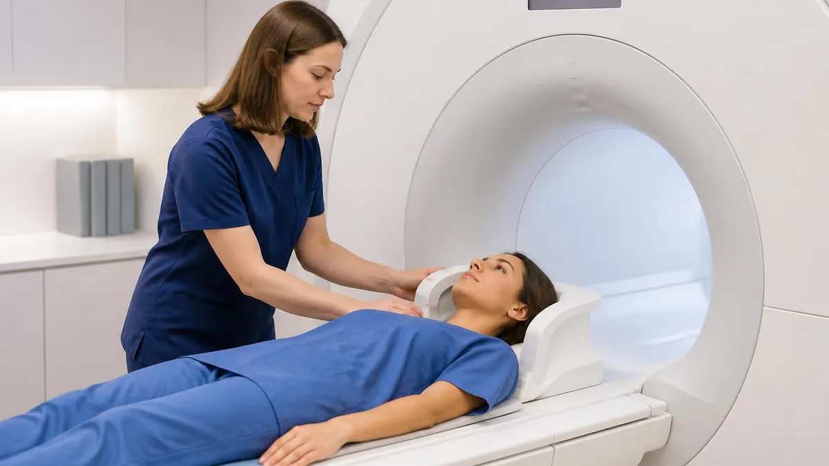
Because DTI stitches together many sequential volumes, even slight head movement between acquisitions misaligns the dataset and corrupts FA maps and tractography. Optimize immobilization, coach the patient, and consider sedation for restless cases. A single uncorrectable motion event may force a complete repeat of the sequence.
The clinical reach of diffusion tensor imaging now extends across neurology, neurosurgery, psychiatry, and research. In acute stroke, diffusion-weighted imaging, the simpler cousin of DTI, identifies ischemic tissue within minutes of onset by detecting restricted water movement, while the tensor analysis adds insight into how an infarct disrupts downstream white matter tracts over time. This makes diffusion imaging central to modern stroke triage and thrombectomy decision-making.
Traumatic brain injury is a major application where conventional MRI often appears normal despite clear cognitive symptoms. Diffuse axonal injury shears delicate fibers throughout the white matter, lowering FA in tracts such as the corpus callosum and superior longitudinal fasciculus. DTI can reveal this microstructural damage, supporting diagnosis, prognosis, and research into concussion, blast injury, and chronic traumatic encephalopathy when standard sequences offer no visible explanation for a patient's deficits.
In multiple sclerosis and other demyelinating diseases, diffusion tensor metrics track the integrity of myelinated pathways. Lesions show altered FA and diffusivity, and so-called normal-appearing white matter often harbors subtle abnormalities invisible on routine sequences. Researchers use these measures to monitor disease progression and treatment response, while clinicians correlate tract damage with specific functional impairments such as visual, motor, or cognitive decline along affected pathways.
Brain tumors represent the application with the clearest immediate impact on patient care. Before resecting a glioma near the motor strip or language cortex, neurosurgeons use tractography to map the corticospinal tract and arcuate fasciculus relative to the lesion. This guides the surgical approach, helping preserve movement and speech. Tumors may displace, infiltrate, or destroy tracts, and recognizing which pattern is present shapes both the operative plan and the prognosis.
Neurodegenerative and psychiatric conditions form a rapidly growing research frontier. Alzheimer disease, Parkinson disease, and amyotrophic lateral sclerosis each show characteristic white matter changes that DTI can quantify, sometimes before atrophy becomes obvious. In psychiatry, studies of schizophrenia, depression, and autism use diffusion metrics to probe disrupted connectivity, advancing the view of these disorders as conditions of altered brain networks rather than isolated regions.
Pediatric and developmental imaging benefits as well. Because FA rises predictably as myelination matures during childhood, diffusion tensor imaging can assess normal brain development and detect delays or injuries in premature infants and children with disorders of white matter. This longitudinal sensitivity makes DTI a valuable tool for understanding how the developing brain wires itself and where that process goes awry.
Across all these uses, the common thread is that DTI sees what conventional imaging cannot: the organized microstructure of white matter and the connections that bind brain regions into functioning networks. As acquisition speeds improve and advanced models replace the simple tensor, the technique continues to expand from a specialized research method into a routine clinical instrument that informs diagnosis, surgery, and the monitoring of disease over a patient's lifetime.
For students and technologists preparing to master diffusion tensor imaging, a structured study approach pays off both on registry exams and at the scanner. Begin with the underlying physics: understand that diffusion weighting comes from paired gradients straddling a refocusing pulse, that the b-value encodes the strength and timing of those gradients, and that anisotropy reflects how axonal architecture restricts water movement. A firm grasp of these fundamentals makes every downstream concept easier to retain.
Memorize the scalar metrics and their typical ranges. Know that fractional anisotropy spans zero to one, that dense tracts like the splenium read high while gray matter and CSF read low, and that ADC rises with tissue breakdown but falls in acute ischemia. Practice associating each metric with the pathology it best detects, because exam questions frequently present a scenario and ask which diffusion measure changes and in which direction.
Learn the color-coding convention cold: red for left-right fibers, green for anterior-posterior, and blue for superior-inferior, following the standard right-anterior-superior scheme. Examiners love directional color FA images, and being able to instantly name the corpus callosum, corticospinal tract, and major association fibers from their colors will earn quick points and build real interpretive confidence in clinical practice.
Connect physics to artifacts. Be ready to explain why echo-planar imaging causes susceptibility distortion at the skull base, how eddy currents misregister images, and why patient motion is so devastating to a multi-volume acquisition. Questions often test whether you can match an artifact to its cause and to the correction strategy, so build a mental table linking each problem to its source and its remedy.
Use active recall and practice questions rather than passive rereading. Quizzes that mix DTI with broader MRI physics and safety reinforce how diffusion fits into the larger field. After each set, review every missed item until you can explain not only the correct answer but why each distractor is wrong. This habit converts shallow familiarity into the durable understanding exams demand.
Reinforce learning with real cases whenever possible. Pull up DTI studies in your department, identify the b0 and diffusion-weighted volumes, examine the FA and ADC maps, and trace a tractography reconstruction. Seeing how acquisition choices translate into final images cements abstract parameters into practical knowledge, and it prepares you to troubleshoot when a scan returns distorted or noisy data on a difficult patient.
Finally, space your study sessions and revisit core concepts repeatedly over weeks rather than cramming. Diffusion tensor imaging blends physics, anatomy, and clinical reasoning, and that breadth rewards steady review. Combine reading, diagrams, hands-on scanner experience, and timed practice tests, and you will walk into both the exam room and the control room equipped to handle DTI with genuine competence and confidence.
Sample MRI - Magnetic Resonance Imaging Practice Questions
Try these questions from our free MRI - Magnetic Resonance Imaging practice tests. The correct answer and an explanation follow each question.
The b-value in diffusion-weighted imaging controls the degree of diffusion weighting. Increasing the b-value:
- A. Increases overall image SNR
- B. Increases diffusion contrast but decreases SNR
- C. Decreases T2 contribution
- D. Has no effect on image quality
Answer: B. Increases diffusion contrast but decreases SNR
Higher b-values provide greater diffusion contrast by increasing the dephasing of mobile water protons, but at the cost of reduced SNR due to greater signal attenuation from all spins.
Diffusion-weighted imaging (DWI) measures the random Brownian motion of water molecules. Restricted diffusion (e.g., in acute stroke) appears as:
- A. Low signal on DWI, high ADC
- B. High signal on DWI, low ADC
- C. High signal on DWI, high ADC
- D. Low signal on DWI, low ADC
Answer: B. High signal on DWI, low ADC
In acute ischemia, cytotoxic edema restricts water movement; restricted diffusion shows high signal on DWI (b=1000) and low signal on ADC map (low diffusivity).
Echo planar imaging (EPI) is used in which MRI techniques due to its extremely fast acquisition speed?
- A. Conventional T1 spin echo and MRCP
- B. DWI, fMRI, and perfusion imaging
- C. MR arthrography and spectroscopy
- D. STIR and inversion recovery sequences
Answer: B. DWI, fMRI, and perfusion imaging
EPI acquires an entire k-space in a single excitation, enabling the speed needed for diffusion-weighted imaging, fMRI BOLD imaging, and dynamic perfusion studies.
For pituitary gland MRI, which plane is considered the primary imaging plane?
- A. Axial
- B. Sagittal
- C. Coronal
- D. Oblique axial
Answer: C. Coronal
Coronal thin-slice images are the primary plane for pituitary imaging because they best demonstrate lateral extension, cavernous sinus invasion, and microadenomas.
Take the full MRI - Magnetic Resonance Imaging practice test
MRI Questions and Answers

MRI of Cervical Spine: Complete Guide to Imaging, Anatomy, Pathology, and Patient Preparation
MRI Medical Abbreviation: What MRI Stands For and Why It Matters

Knee MRI Images: A Complete Guide to Reading, Understanding, and Interpreting Knee Scans
ICD 10 Codes for MRI: Complete Guide to Abnormal MRI Coding, Documentation, and Reimbursement
Is Nickel Titanium MRI Compatible? A Complete Guide to MRI Safety Materials
About the Author

Medical Laboratory Scientist & Clinical Certification Expert
Johns Hopkins UniversityDr. Sandra Kim holds a PhD in Clinical Laboratory Science from Johns Hopkins University and is certified as a Medical Technologist (MT) and Medical Laboratory Scientist (MLS) through ASCP. With 16 years of clinical laboratory experience spanning hematology, microbiology, and molecular diagnostics, she prepares candidates for ASCP board exams, MLT, MLS, and specialist certification tests.
Join the Discussion
Connect with other students preparing for this exam. Share tips, ask questions, and get advice from people who have been there.
View discussion (8 replies)