
MRI Internal Auditory Canal: Complete Guide to IAC Imaging, Anatomy, Indications, and Interpretation
Learn everything about MRI internal auditory canal imaging: anatomy, indications, protocols, sequences, and how to interpret IAC scan findings. 📗
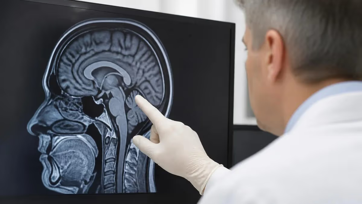
MRI internal auditory canal (IAC) imaging is one of the most specialized and clinically important examinations in neuroradiology. The internal auditory canal is a narrow bony channel in the petrous portion of the temporal bone that carries the vestibulocochlear nerve (cranial nerve VIII) and the facial nerve (cranial nerve VII), along with their accompanying blood vessels. Because these structures are small, closely packed, and enclosed within dense bone, MRI remains the gold-standard modality for evaluating pathology in this region, offering unmatched soft-tissue contrast without radiation exposure.
Patients referred for an internal auditory canal mri typically present with symptoms such as sudden or progressive sensorineural hearing loss, unilateral tinnitus, vestibular dysfunction, facial nerve palsy, or a combination of these findings. The most feared diagnosis prompting IAC imaging is vestibular schwannoma — formerly called acoustic neuroma — a benign tumor arising from Schwann cells of the vestibulocochlear nerve that can cause permanent hearing loss if detected late. Early, accurate imaging is therefore essential for guiding clinical management and surgical or radiosurgical planning.
Modern IAC MRI protocols typically combine high-resolution T2-weighted sequences, such as FIESTA (Fast Imaging Employing Steady-State Acquisition) or CISS (Constructive Interference in Steady State), with gadolinium-enhanced T1-weighted sequences. The fluid-filled labyrinth and cisternal portions of the nerves appear bright on heavily T2-weighted images, while enhancing lesions stand out dramatically against the dark background on post-contrast T1 images. Together, these sequences allow radiologists to identify even sub-millimeter intracanalicular tumors with high sensitivity and specificity.
Understanding the anatomy of the IAC is fundamental to interpreting MRI findings correctly. The canal averages approximately 8 to 10 millimeters in length and 4 to 5 millimeters in diameter. At its lateral end, the falciform crest (transverse crest) divides the canal into superior and inferior compartments, and the vertical crest (Bill's bar) further subdivides the superior compartment. These bony landmarks help localize which nerve is involved when a lesion is identified, since the facial nerve occupies the anterior-superior quadrant while the superior and inferior vestibular nerves occupy the posterior quadrants and the cochlear nerve runs anteriorly and inferiorly.
The clinical relevance of IAC MRI extends far beyond tumor detection. Radiologists also evaluate the scan for signs of labyrinthitis ossificans, cochlear and semicircular canal anomalies, vascular loops in the IAC that may cause neurovascular compression syndromes, inflammatory conditions such as Bell's palsy and Ramsay Hunt syndrome, and postoperative changes in patients who have undergone cochlear implantation or acoustic neuroma resection. Each of these conditions has distinct MRI characteristics that require familiarity with both normal anatomy and a range of pathological appearances.
For radiology students, sonography technologists cross-training into MRI, and allied health professionals preparing for registry examinations, a thorough understanding of IAC MRI is indispensable. Questions about cranial nerve anatomy, posterior fossa pathology, and specialized head MRI protocols appear frequently on the ARRT MRI examination and on advanced registry assessments. Mastering this topic builds a strong foundation not only for test success but also for clinical competence in a setting where precision directly affects patient outcomes.
This guide walks through the complete picture of internal auditory canal MRI: the anatomy you need to know, the clinical indications that drive referrals, the imaging sequences used in practice, how to systematically interpret findings, and practical tips for exam preparation. Whether you are a student studying for boards or a practicing technologist seeking a deeper understanding of the procedures you perform daily, this resource provides the depth and clarity you need to excel.
IAC MRI by the Numbers
Key Anatomical Structures of the Internal Auditory Canal
Occupies the anterior-superior quadrant of the IAC lateral segment. It exits via the facial canal and is critical to identify on MRI because even small compressive lesions can cause facial palsy, requiring urgent surgical evaluation and careful pre-operative planning.
Comprises the superior vestibular, inferior vestibular, and cochlear nerve branches. These fill the remaining quadrants of the IAC. The cochlear nerve is the target of cochlear implant surgery, so its integrity on pre-operative MRI is a critical prognostic factor for hearing rehabilitation success.
The falciform (transverse) crest divides the lateral IAC into superior and inferior compartments. Bill's bar further divides the superior compartment anteriorly and posteriorly, helping localize which nerve a lesion involves. These bony landmarks appear as low-signal dividers on high-resolution T2 sequences.
A terminal branch of the anterior inferior cerebellar artery (AICA) that enters the IAC alongside the nerves. Vascular loops from AICA can cause neurovascular compression syndromes mimicking vestibular schwannoma symptoms, and are identifiable on high-resolution 3D MRI sequences with careful vascular analysis.
While technically part of the inner ear rather than the IAC itself, these fluid-filled structures are routinely evaluated on the same high-resolution T2 sequences used for the canal. Abnormal signal in the cochlea or canals helps diagnose labyrinthitis, labyrinthitis ossificans, and congenital inner ear anomalies.
The range of clinical conditions that prompt a referral for IAC MRI is broad, reflecting the diverse functions of the nerves housed within this small canal. The most common indication is asymmetric sensorineural hearing loss — defined by most guidelines as a difference of 15 dB or more at two or more consecutive frequencies, or 20 dB or more at a single frequency between ears.
In patients with this audiometric pattern, MRI is recommended to exclude a retrocochlear mass, with vestibular schwannoma being the primary concern. The American Academy of Otolaryngology recommends MRI as the preferred screening tool over auditory brainstem response testing due to its significantly higher sensitivity for small tumors.
Unilateral tinnitus, particularly when it is pulsatile or of sudden onset, is another strong indication for IAC MRI. Pulsatile tinnitus may indicate a vascular lesion such as a paraganglioma (glomus tympanicum or glomus jugulare), an arteriovenous malformation, or an aberrant carotid artery, all of which have characteristic MRI appearances. Non-pulsatile unilateral tinnitus is more often associated with sensorineural hearing loss and retrocochlear pathology, prompting evaluation for schwannoma or other mass lesions of the cerebellopontine angle (CPA) cistern, which extends medially to the IAC opening and is the most common site of schwannoma origin.
Vestibular dysfunction presenting as true vertigo, disequilibrium, or episodic falls also warrants IAC MRI, especially when symptoms are persistent or progressive. While the differential diagnosis for dizziness is vast, MRI helps exclude posterior fossa tumors, demyelinating plaques affecting the vestibular nuclei or pathways, and structural inner ear abnormalities. Superior semicircular canal dehiscence — a thinning or absence of bone overlying the superior canal — is better assessed with CT but can sometimes be inferred from MRI signal patterns in the right clinical context.
Facial nerve palsy, whether acute or recurrent, is a critical indication for IAC and temporal bone MRI. In patients with Bell's palsy, enhancement of the facial nerve on post-gadolinium T1-weighted images reflects inflammatory edema and breakdown of the blood-nerve barrier. The pattern and extent of enhancement help differentiate viral neuritis from other causes such as herpes zoster oticus (Ramsay Hunt syndrome, which also involves the geniculate ganglion and may show vesicular changes in the external auditory canal), Lyme neuroborreliosis, sarcoidosis, or perineural tumor spread along the nerve from parotid malignancy.
Pre-operative and post-operative evaluation represents a growing indication for IAC MRI. Before cochlear implantation, MRI assesses cochlear nerve integrity — absence or severe hypoplasia of the cochlear nerve is associated with poor hearing outcomes even after successful implantation. After acoustic neuroma resection via a translabyrinthine, retrosigmoid, or middle fossa approach, surveillance MRI detects residual or recurrent tumor. The timing and frequency of surveillance imaging are guided by tumor size and surgical approach, with gadolinium-enhanced MRI typically performed at 6 months, 12 months, and then annually for several years.
Evaluation of congenital inner ear malformations in pediatric patients is another important indication, particularly in children with congenital sensorineural hearing loss or failed newborn hearing screening. MRI without radiation is preferred in this population. Common congenital anomalies visible on IAC MRI include cochlear hypoplasia, incomplete partition types I through III (Michel deformity, Mondini malformation, and incomplete partition III), aplasia of the semicircular canals, and a narrow or absent IAC associated with cochlear nerve deficiency. These findings directly affect candidacy and outcome for hearing rehabilitation devices.
Finally, patients being evaluated for suspected neurofibromatosis type 2 (NF2) require dedicated bilateral IAC MRI. NF2 is an autosomal dominant tumor suppressor gene disorder causing bilateral vestibular schwannomas, along with meningiomas, ependymomas, and other tumors throughout the nervous system. Screening MRI of the brain and spine with gadolinium enhancement is recommended beginning in childhood for known NF2 gene carriers, and IAC findings — particularly bilateral enhancing intracanalicular lesions — are pathognomonic when present in the right clinical context.
MRI Practice Test Questions
Prepare for the MRI - Magnetic Resonance Imaging exam with our free practice test modules. Each quiz covers key topics to help you pass on your first try.
MRI Knowledge
MRI Exam Questions covering Knowledge. Master MRI Test concepts for certification prep.
MRI Physics
Free MRI Practice Test featuring Physics. Improve your MRI Exam score with mock test prep.
MRI Anatomy and Pathology
MRI Test Prep for MRI Anatomy and Pathology. Practice MRI Quiz questions and boost your score.
MRI Anatomy and Positioning
MRI Questions and Answers on MRI Anatomy and Positioning. Free MRI practice for exam readiness.
MRI Contrast Agents
Free MRI Quiz on MRI Contrast Agents. MRI Exam prep questions with detailed explanations.
MRI Patient Care and Positioning
MRI Practice Questions for MRI Patient Care and Positioning. Build confidence for your MRI certification exam.
MRI Sequences Used for Internal Auditory Canal Imaging
The cornerstone of IAC MRI protocols is the high-resolution 3D T2-weighted sequence — known as CISS on Siemens scanners and FIESTA on GE systems, with DRIVE on Philips. These sequences exploit the very high signal of fluid (endolymph and perilymph) to create bright-fluid images that outline the cranial nerves as dark linear structures against a bright background within the IAC. With slice thicknesses as thin as 0.5 to 0.8 mm, these sequences can detect even very small intracanalicular tumors and reliably display all four nerve bundles within the lateral IAC segment.
Reformatted images in oblique planes aligned along the long axis of the IAC — sometimes called the Pöschl and Stenvers planes — give radiologists the best visualization of individual nerve branches and help confirm whether a lesion involves one or multiple divisions of cranial nerve VIII. At 3 Tesla field strength, the signal-to-noise advantage allows even greater resolution, and some centers reduce slice thickness to 0.4 mm or less for optimal nerve delineation in pre-operative cochlear implant candidates. The main limitation is susceptibility artifact near the temporal bone air cells, which can degrade image quality in some patients.
MRI vs. CT for Internal Auditory Canal Evaluation
- +Superior soft-tissue contrast to visualize cranial nerves directly within the IAC
- +No ionizing radiation — safe for children, pregnant patients, and repeated surveillance
- +Gadolinium enhancement detects even tiny intracanalicular enhancing tumors below 3 mm
- +CISS/FIESTA sequences resolve individual nerve bundles in the lateral IAC segment
- +Simultaneous evaluation of the entire posterior fossa, brainstem, and inner ear fluid spaces
- +Gold standard for vestibular schwannoma detection with sensitivity exceeding 95%
- −Cannot directly visualize fine bony detail of the otic capsule and ossicular chain
- −Contraindicated in patients with non-MRI-compatible implants, certain pacemakers, or cochlear implant magnets
- −Longer acquisition time (30–45 minutes) compared to CT (seconds), with more motion artifact risk
- −Higher cost and more limited availability in rural and low-resource settings
- −Gadolinium contrast carries a small risk of nephrogenic systemic fibrosis in patients with severe renal impairment
- −Claustrophobia affects a significant minority of patients, requiring sedation or open MRI alternatives
Patient Preparation Checklist for IAC MRI
- ✓Screen for all metallic implants, especially cochlear implants, hearing aids, and middle ear prostheses before scheduling.
- ✓Verify eGFR or creatinine level if gadolinium contrast is planned, particularly in patients over 60 or with renal disease.
- ✓Instruct the patient to remove all hearing aids and external processors before entering the MRI suite.
- ✓Confirm the patient can lie still for 30–45 minutes; arrange sedation or anesthesia consultation for young children or severely anxious adults.
- ✓Ask specifically about claustrophobia history and discuss anxiolytic premedication options with the ordering provider if needed.
- ✓Review prior imaging to choose appropriate sequences — post-operative patients may need modified protocols with longer fat-sat T1 runs.
- ✓Ensure the patient has reviewed and signed the MRI safety screening form, including implant model and serial number verification.
- ✓Brief the patient on the expected tapping and banging noises from the gradient coils to reduce anxiety during the scan.
- ✓Position the patient supine with a dedicated head coil centered over the external auditory meatus for optimal temporal bone coverage.
- ✓Confirm IV access is patent before injecting gadolinium, and observe the patient for at least 20 minutes for any adverse reaction.
The 1 cm Rule for Vestibular Schwannoma Management
Vestibular schwannomas smaller than 1 cm confined to the IAC are typically managed with active surveillance MRI rather than immediate surgery or radiosurgery, as growth rates are highly variable and many tumors remain stable for years. Imaging surveillance intervals and treatment thresholds depend on tumor growth rate, patient age, hearing status, and institutional preference — understanding this management paradigm helps MRI technologists appreciate why patients return for serial IAC studies.
Systematic interpretation of an IAC MRI begins with establishing a mental roadmap of the relevant anatomy before searching for pathology. Radiologists and trainees are taught to evaluate the study in a structured sequence: first review the axial and coronal high-resolution T2 images to assess the fluid signal in the cochlea, vestibule, and semicircular canals; then examine the IAC lumen bilaterally for filling defects or asymmetric nerve thickness; next scrutinize the CPA cisterns for mass lesions at the porus acusticus; and finally review the post-contrast T1 images for any abnormal enhancement along the cranial nerves, meninges, or labyrinthine structures.
Vestibular schwannoma is the most common finding on IAC MRI, accounting for approximately 80% of CPA-IAC tumors. On T2 CISS/FIESTA sequences, smaller intracanalicular schwannomas appear as a focal region of decreased signal filling part of the IAC, displacing the surrounding bright fluid. Larger tumors extending into the CPA cistern appear as lobulated masses with heterogeneous T2 signal, sometimes containing cystic areas.
On gadolinium-enhanced T1 images, enhancement is invariably present — a small, uniformly enhancing nodule within the IAC fundus is highly specific for intracanalicular schwannoma. The ice cream cone appearance — a rounded CPA component continuous with a cylindrical intracanalicular extension — is a classic descriptor seen on coronal images.
Meningioma is the second most common CPA-IAC tumor, representing about 10 to 15% of cases. Unlike schwannomas, meningiomas have a broad dural base, tend to be more uniformly T2 hypointense relative to gray matter, and often demonstrate a dural tail sign on post-contrast images — linear enhancement extending along the adjacent dura beyond the tumor margin. Meningiomas rarely enter the IAC fundus, which helps differentiate them from schwannomas when the tumor straddles the porus acusticus. Calcification, which occurs in up to 20% of meningiomas, may be visible as foci of signal void on T2 sequences.
Epidermoid cysts are the third most common CPA lesion. They follow CSF signal on T1 and T2 sequences, making them easy to miss on routine brain MRI, but restricted diffusion on DWI distinguishes them definitively from arachnoid cysts. Epidermoids tend to insinuate around neurovascular structures rather than displace them, giving them an irregular, cauliflower-like morphology that distinguishes them from the rounded profile of schwannomas. They do not enhance, and their growth is slow but inexorable — clinical symptoms often become apparent only after years of asymptomatic expansion.
Labyrinthitis ossificans deserves special attention because it has a direct impact on cochlear implant candidacy. This condition involves progressive fibrosis and bony obliteration of the membranous labyrinth following an insult such as bacterial meningitis, viral labyrinthitis, trauma, or autoimmune disease. The radiologic hallmark on T2 sequences is loss of the normal bright fluid signal within the cochlear turns — initially the basal turn is affected, and in severe cases the entire cochlea becomes signal-void. Pre-operatively, radiologists stage the degree of cochlear obliteration to guide surgical approach (standard vs. drill-out) and to counsel patients and families on expected implant performance.
Vascular lesions of the CPA and IAC constitute an important differential category. A high-riding jugular bulb can project into the floor of the IAC and mimic a mass on clinical examination (visible as a blue-tinged mass behind an intact tympanic membrane), but MRI readily demonstrates the flowing blood signal and connection to the jugular vein.
Paragangliomas (glomus jugulare and glomus tympanicum tumors) are highly vascular and show the classic salt-and-pepper pattern on T1 images — multiple punctate flow voids (pepper) interspersed with areas of hemorrhagic bright signal (salt) — with intense enhancement on post-contrast sequences. These findings are virtually diagnostic and guide preoperative embolization planning.
Inflammatory and infectious conditions affecting the IAC and CPA include viral neuritis, bacterial labyrinthitis, leptomeningeal carcinomatosis, and neurosarcoidosis. Bilateral IAC enhancement in a patient with a known primary malignancy strongly suggests leptomeningeal metastatic disease and triggers further workup with lumbar puncture for CSF cytology. Sarcoidosis affecting the cranial nerves can produce enhancement of multiple nerve branches simultaneously and may be associated with pachymeningeal enhancement, hypothalamic involvement, and posterior fossa lesions, all of which are visible on the same gadolinium-enhanced study that reveals the IAC findings.
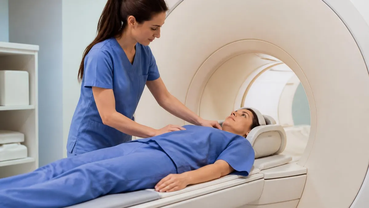
Patients with cochlear implants present a unique challenge for IAC MRI. Most modern implants are conditionally MRI-compatible at 1.5T with the internal magnet in place (after bandaging), but the magnet can rotate or demagnetize during scanning, and significant artifact degrades image quality near the implanted side. In such cases, the contralateral IAC can still be evaluated, but CT may be needed to assess the implanted cochlea. Always verify the specific implant model against the manufacturer's MRI compatibility specifications before proceeding — never rely on patient report alone.
For MRI technologists and radiologic technologists preparing for the ARRT MRI registry examination, knowledge of the internal auditory canal occupies a disproportionately important niche within the broader anatomy and pathology content domain. The ARRT MRI exam blueprint allocates a significant portion of questions to head and neck MRI, including temporal bone anatomy, cranial nerve identification, and posterior fossa pathology. Understanding why specific sequences are chosen for IAC imaging — not just their names but their physical principles — bridges the gap between rote memorization and genuine clinical understanding that the exam rewards.
A practical approach to studying for registry questions about IAC MRI begins with mastering the anatomy: be able to identify the four nerve bundles in the IAC fundus, name the bony landmarks that separate them, trace the course of the facial nerve from the pons through the parotid gland, and describe the vascular supply to the inner ear. Many exam questions are anatomy-based and require not only knowing the names of structures but understanding their clinical relevance — for example, why damage to the cochlear nerve division specifically causes sensorineural rather than conductive hearing loss.
Sequence physics relevant to IAC MRI is another high-yield area for registry preparation. CISS and FIESTA are steady-state free precession (SSFP) sequences that use very short TR and TE values with a refocusing flip angle to maintain transverse magnetization across repetitions. Their high T2/T1 contrast ratio and relative insensitivity to field inhomogeneity make them ideal for small structure imaging in the posterior fossa.
Understanding how changes in flip angle, TR, and bandwidth affect the signal-to-noise ratio and tissue contrast on SSFP sequences is the kind of conceptual physics question that appears on advanced registry assessments and distinguishes top scorers from average test-takers.
Contrast agent pharmacology and safety are also tested on the registry. The six gadolinium-based contrast agents currently approved in the United States differ in their thermodynamic and kinetic stability, with macrocyclic agents (gadoterate meglumine, gadobutrol) considered more stable and less likely to deposit gadolinium in tissues including the brain.
While the clinical significance of gadolinium brain deposition remains under active investigation, technologists should understand current FDA guidance, which recommends using the lowest effective dose of contrast and considering non-contrast protocols when diagnostic quality can be maintained. IAC MRI is one area where non-contrast CISS/FIESTA sequences alone are often sufficient for tumor screening in low-risk patients.
Registry questions about patient safety in the context of IAC MRI frequently focus on metallic implants in the temporal bone and skull base region. Cochlear implants, stapedectomy prostheses, tympanostomy tubes, and brainstem auditory implants all require verification before entering the MRI environment.
The ACR guidance document on MRI safe practices provides a tiered framework for evaluating implant safety: MR Safe (no known hazard), MR Conditional (safe under specific conditions), and MR Unsafe (contraindicated). Test questions may ask you to identify which category applies to a described implant or to determine the appropriate action when a patient presents with an unknown implant.
Understanding the relationship between MRI findings and clinical management is another differentiating factor for high-scoring registry candidates. The exam increasingly tests applied knowledge rather than isolated facts. For example, a question might describe an enhancing IAC lesion in a patient with NF2 and ask which management approach is most appropriate — active surveillance with serial MRI, stereotactic radiosurgery (Gamma Knife), or microsurgical resection. Knowing that small NF2-associated bilateral schwannomas are often managed conservatively to preserve residual hearing as long as possible requires integrating anatomy, pathology, and clinical decision-making in a way that purely memorization-based study does not support.
One of the most effective ways to consolidate IAC MRI knowledge for registry preparation is through systematic case review — studying actual scan images alongside the corresponding radiology reports and clinical outcomes. Online case repositories, textbook atlases, and structured question banks that include image-based items are invaluable for building the visual pattern recognition skills that translate directly to exam performance. Pairing image review with active recall practice — explaining aloud what you see and why it matters — reinforces memory consolidation far more effectively than passive re-reading of notes or textbook chapters on the topic.
Optimizing image quality during an IAC MRI scan requires attention to several technical factors that fall squarely within the technologist's scope of practice. Field of view (FOV) selection is critical: a smaller FOV increases spatial resolution by distributing the available matrix over a smaller area, but if the FOV is too small, signal aliasing (wrap-around artifact) can obscure the IAC and adjacent structures. Most protocols use a FOV of 16 to 20 cm centered on the temporal bones, with phase-encoding direction set to minimize wrap artifact from the overlying brain tissue outside the FOV.
Gradient coil selection and orientation also matter for IAC MRI. Dedicated eight-channel or 32-channel head coils provide substantially better signal-to-noise ratios than body or surface coils, and the increased SNR can be traded for higher resolution or shorter acquisition time. For 3T scanners, parallel imaging acceleration factors of 2 to 3 are commonly used on CISS/FIESTA sequences, reducing scan time and associated motion artifact without appreciable loss of diagnostic quality. The technologist should confirm that the correct coil and acceleration parameters are loaded from the institutional protocol before beginning the scan.
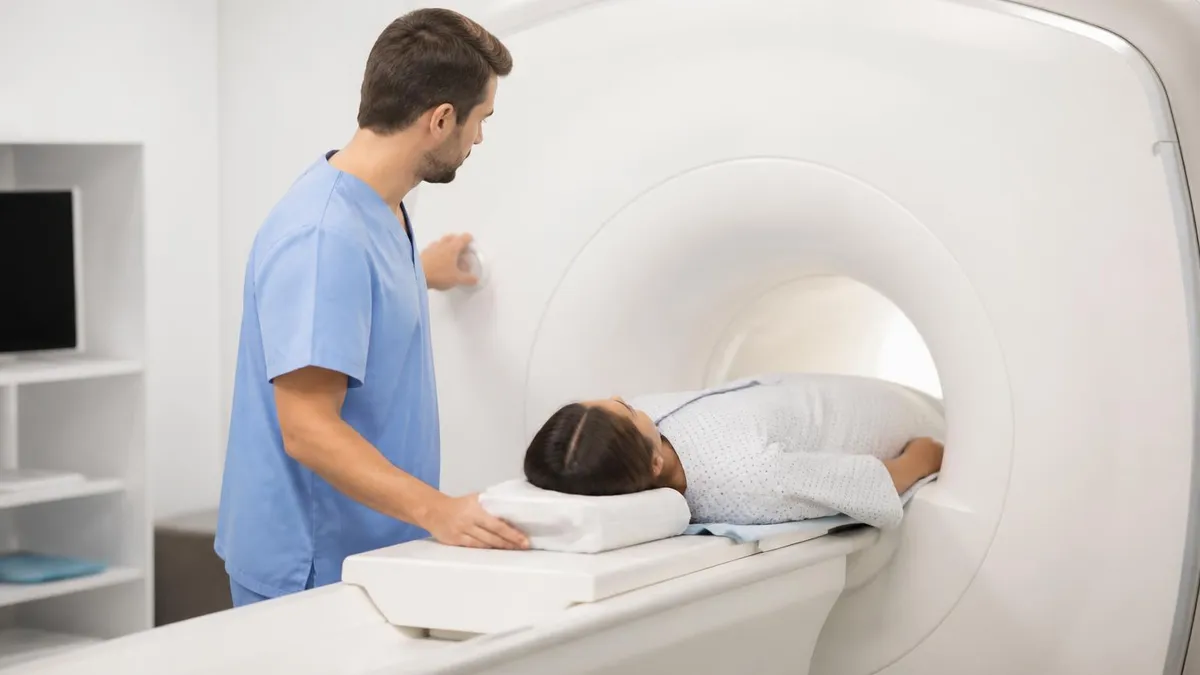
Patient positioning requires precision because even slight head tilt can make oblique reformats less accurate and hamper side-to-side comparisons. The external auditory meatus should be aligned with the isocenter of the magnet, and a small foam pad under the occiput can help maintain a neutral head position throughout the scan. Gentle restraint with tape or foam padding around the head coil reduces motion without causing discomfort, which is especially important for older patients or children who may be restless during the longer acquisition windows of high-resolution 3D sequences.
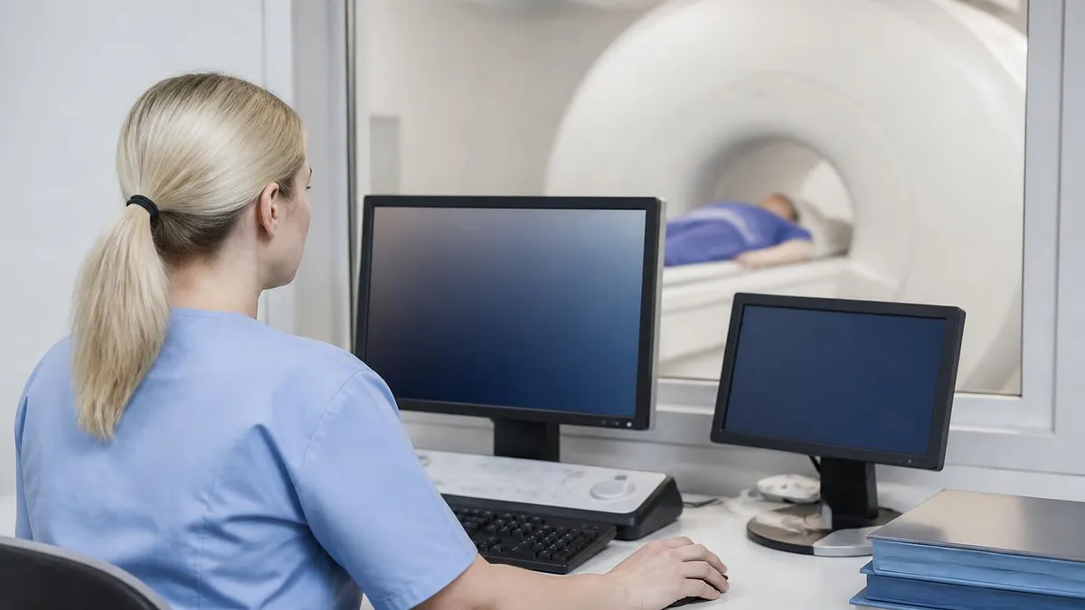
Communication between the technologist and the radiologist before and after the scan is a best practice that directly affects diagnostic yield. A brief pre-scan review of the clinical indication allows the technologist to anticipate which sequences are most critical — for example, if the indication is specifically to evaluate an NF2 patient for bilateral schwannoma growth, both IACs must be imaged with equal quality rather than focusing on the symptomatic side. After the scan, flagging technically suboptimal sequences before the patient leaves the scanner allows for immediate repeat acquisitions, avoiding the need for a return visit and delayed diagnosis.
Documentation and quality assurance in IAC MRI extend to contrast injection records, technical parameters, and patient tolerance notes. Technologists should document the specific contrast agent used, the dose in millimoles per kilogram, the injection site, the rate, and any patient reactions. Recording acquisition parameters — slice thickness, TR, TE, flip angle, matrix, bandwidth, and number of averages — in the scan worksheet allows retrospective quality review and enables consistent protocol compliance across different technologists and scanner maintenance cycles. These records also have medicolegal importance if questions arise later about contrast exposure.
Advanced MRI applications are expanding the capabilities of IAC imaging beyond conventional anatomical sequences. Diffusion tensor imaging (DTI) with fiber tractography can depict the three-dimensional course of the cochlear and vestibular nerve bundles with remarkable clarity, providing surgeons with a spatial map of nerve anatomy before acoustic neuroma resection or cochlear implant surgery.
Functional MRI of the auditory cortex, combined with audiometric testing, can assess central hearing pathway integrity in patients with severe deafness prior to auditory brainstem implant placement. While these techniques are not yet routine in most centers, awareness of their existence and applications is relevant for technologists working in academic or tertiary referral settings.
The future of IAC MRI is likely to involve artificial intelligence-assisted image analysis, including automated nerve segmentation, lesion detection, and volumetric tumor measurement. Early deep learning systems trained on large IAC MRI datasets have already demonstrated performance comparable to expert radiologists for vestibular schwannoma detection on screening studies.
As these tools move into clinical practice, technologists will play an important role in ensuring that input images meet the technical quality standards required for reliable AI performance — reinforcing the central importance of protocol optimization and image quality assurance skills that remain fundamentally within the technologist's domain regardless of how much image interpretation shifts toward automated tools.
MRI Questions and Answers

MRI of Cervical Spine: Complete Guide to Imaging, Anatomy, Pathology, and Patient Preparation

Knee MRI Images: A Complete Guide to Reading, Understanding, and Interpreting Knee Scans
Ace Your Exam with MRI Practice Test 2026 Prep

MRI Tech School 2026 — Programs, Requirements, and What to Expect
MRI Procedure: What to Expect Before, During and After Your Scan
About the Author

Medical Laboratory Scientist & Clinical Certification Expert
Johns Hopkins UniversityDr. Sandra Kim holds a PhD in Clinical Laboratory Science from Johns Hopkins University and is certified as a Medical Technologist (MT) and Medical Laboratory Scientist (MLS) through ASCP. With 16 years of clinical laboratory experience spanning hematology, microbiology, and molecular diagnostics, she prepares candidates for ASCP board exams, MLT, MLS, and specialist certification tests.
Join the Discussion
Connect with other students preparing for this exam. Share tips, ask questions, and get advice from people who have been there.
View discussion (8 replies)