
Brachial Plexus MRI: Complete Guide to Imaging, Anatomy, Pathology, and Clinical Applications
🟢 Master brachial plexus MRI with this complete guide covering anatomy, protocols, pathology, and clinical applications for radiologists and MRI technologists.
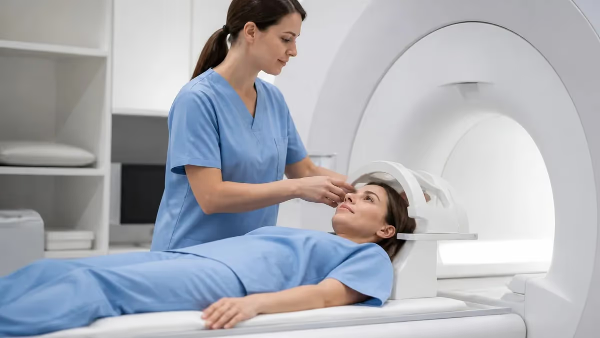
Brachial plexus MRI has become the gold standard imaging modality for evaluating complex nerve network injuries, tumors, and inflammatory conditions affecting the shoulder and upper extremity. The brachial plexus is a remarkably complex web of nerve roots arising from C5 through T1, weaving together to form the major nerves that control arm, hand, and shoulder function. When something goes wrong with this network — whether from trauma, tumor infiltration, or radiation damage — MRI provides the anatomical detail necessary to guide surgical planning and predict patient outcomes. Practice with the free MRI - Magnetic Resonance Imaging practice test on PracticeTestGeeks (200+ questions with answer explanations).
Understanding the technical demands of brachial plexus mri is essential for MRI technologists and radiologists alike. Unlike routine spine or extremity imaging, brachial plexus studies require specialized coil configurations, precise patient positioning, and a thorough knowledge of regional anatomy. The nerves course through multiple fascial compartments and fat planes, making high-resolution, high-contrast imaging absolutely critical. A suboptimal scan can obscure a surgically correctable lesion or delay a cancer diagnosis by months.
In clinical practice, brachial plexus MRI is ordered for a diverse range of presentations. Trauma patients who present after motorcycle accidents, birth injuries, or sports collisions may have traction injuries ranging from mild neuropraxia to devastating avulsion from the spinal cord. Oncology patients with breast, lung, or lymphoma diagnoses may develop plexopathy from direct tumor invasion or as a delayed consequence of radiation therapy. Differentiating malignant from radiation-induced plexopathy is one of the most clinically impactful roles that MRI plays in this anatomical region.
From a technical standpoint, brachial plexus imaging typically employs a 3 Tesla magnet to maximize signal-to-noise ratio, though high-quality studies can be performed at 1.5T with appropriate sequence optimization. Phased-array surface coils centered over the neck and shoulder are preferred, and many institutions combine a neurovascular coil with a spine coil to cover the full course of the plexus from the neural foramina to the infraclavicular fossa. Fat suppression techniques, including STIR and spectral fat saturation, are indispensable for highlighting edematous or infiltrated nerve fascicles.
The sequences that form the backbone of a brachial plexus MRI protocol are carefully chosen to answer specific clinical questions. Coronal STIR images provide an excellent overview of nerve signal and symmetry. Axial T1-weighted images map the anatomical relationships of the plexus to adjacent muscles, vessels, and lymph nodes. Three-dimensional isotropic sequences, such as SPACE or CUBE, allow multiplanar reformations and can be processed into neurography images that beautifully depict individual nerve fascicles. Diffusion-weighted neurography is an emerging technique that quantifies nerve microstructure through fractional anisotropy and apparent diffusion coefficient measurements.
For MRI technologists preparing for registry examinations or clinical practice, brachial plexus imaging represents a high-yield area that bridges anatomy, physics, and pathology. Knowing why each sequence is chosen — not just how to run it — is what distinguishes a competent technologist from an expert one. The interplay between repetition time, echo time, inversion recovery, and field strength directly determines whether a subtle nerve injury will be visible or missed, making technical mastery clinically consequential.
This comprehensive guide covers everything from brachial plexus anatomy and normal MRI appearances to pathology recognition, protocol design, patient preparation, and practical examination tips. Whether you are a student preparing for boards, a technologist expanding your clinical skills, or a clinician seeking to better understand what MRI can and cannot tell you about brachial plexus disease, this resource provides the depth and clarity you need.
Free MRI - Magnetic Resonance Imaging Study Tools
Interactive MRI - Magnetic Resonance Imaging resources on PracticeTestGeeks, free to start:
- MRI - Magnetic Resonance Imaging practice test — 200+ questions with instant scoring and answer explanations
- MRI - Magnetic Resonance Imaging flashcards — review key terms and concepts card by card
- MRI - Magnetic Resonance Imaging study guide — topic-by-topic overview of what the exam covers
- MRI - Magnetic Resonance Imaging cheat sheet — one-page printable summary for last-minute review
Brachial Plexus MRI by the Numbers
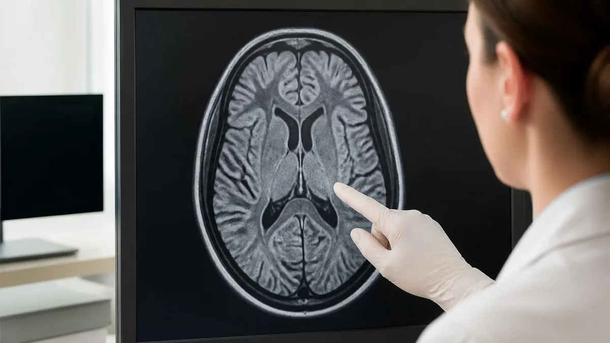
Brachial Plexus Anatomy: Key Divisions and Their MRI Landmarks
The five nerve roots exit the intervertebral foramina and are best seen on axial and coronal MRI. Root avulsions — traumatic separation from the spinal cord — appear as pseudomeningoceles or absent rootlets on high-resolution T2 and myelographic sequences.
Formed by root combinations, the three trunks traverse the posterior cervical triangle. On coronal STIR images they appear as oval nerve structures flanked by scalene muscle fat planes. Abnormal signal or enlargement here suggests proximal plexopathy.
Each trunk splits into anterior and posterior divisions behind the clavicle, then reunites as three cords — lateral, medial, and posterior — named by their relationship to the axillary artery. Cords are best evaluated on axial images at the axillary level.
The five major terminal nerves (musculocutaneous, median, ulnar, radial, axillary) arise from the cords. MRI of these distal nerves evaluates denervation changes in target muscles — fatty infiltration and STIR hyperintensity reveal which nerve segment is compromised.
The subclavian and axillary arteries run alongside the plexus and serve as reliable anatomical guides on MRI. Vascular anomalies or perineural fibrosis from prior hematoma can mimic or compress the plexus, making familiarity with normal vascular anatomy essential.
Designing an effective brachial plexus MRI protocol requires balancing anatomical coverage, spatial resolution, and scan time — three variables that are perpetually in tension. The full plexus spans from the cervical neural foramina to the axillary fossa, a distance that may exceed 20 centimeters in a large patient. Covering this field of view while maintaining the submillimeter resolution needed to visualize individual fascicles demands thoughtful sequence selection and often requires two overlapping acquisitions with careful coil repositioning between stations.
Coronal plane imaging is the single most valuable orientation for brachial plexus assessment. A coronal STIR sequence with a large field of view captures the entire nerve network in just a few image slices, making it ideal for screening and for comparing the symptomatic and asymptomatic sides side by side. Nerve signal on STIR should be mildly hyperintense relative to muscle — markedly increased signal indicates edema, inflammation, or active injury. Asymmetric signal between sides is one of the most reliable indicators of pathology, even when individual nerve caliber appears normal.
T1-weighted sequences without fat suppression are indispensable for evaluating nerve caliber, fatty infiltration of muscles, and the relationship of masses to the plexus. On T1, normal nerves appear isointense to muscle with a faintly striated internal architecture corresponding to fascicular bundles. When a nerve is infiltrated by tumor, its internal architecture is replaced by homogeneous or irregular soft tissue signal. Perineural fibrosis from radiation or prior surgery appears as T1 isointense thickening that encases the nerve without the mass effect or T2 signal elevation seen with active tumor.
Three-dimensional isotropic sequences have transformed brachial plexus neurography over the past decade. Sequences marketed under vendor-specific names — SPACE (Siemens), CUBE (GE), VISTA (Philips) — acquire a single high-resolution volume that can be reconstructed in any plane post-acquisition. When combined with a background suppression technique such as DWIBS (diffusion-weighted whole-body imaging with background body signal suppression), these sequences produce striking maximum intensity projection images that highlight nerves against a dark background, dramatically improving the detection of focal lesions and architectural distortion.
Contrast-enhanced MRI with gadolinium plays a selective but important role in brachial plexus imaging. Normal peripheral nerves do not enhance, so avid or nodular enhancement strongly suggests an inflammatory process (such as Parsonage-Turner syndrome in the acute phase) or a neoplastic lesion. Perineural tumor spread from head and neck cancers can be elegantly mapped with fat-saturated T1 post-contrast sequences, showing enhancement tracking along nerve sheaths toward the neural foramina. However, gadolinium is not routinely administered for trauma cases where the clinical question is limited to mechanical injury.
Diffusion tensor imaging (DTI) and tractography represent the frontier of brachial plexus MRI. By measuring the directional diffusivity of water molecules along nerve fascicles, DTI can generate color-encoded fractional anisotropy maps and three-dimensional tractograms that visualize individual nerve fiber bundles. Studies comparing DTI metrics to surgical and electrophysiological findings show that fractional anisotropy drops significantly at sites of nerve injury, sometimes before conventional sequences reveal abnormality. While DTI is not yet standard of care, it is increasingly available at academic centers and will likely become routine within the next decade.
Patient positioning and coil selection are as important as sequence choice. Patients lie supine with the arm at the side in the neutral or slightly abducted position. Some protocols ask the patient to place the arm overhead in the swimmer's position to open the costoclavicular space and reduce vascular pulsation artifact near the subclavian vessels. A neurovascular phased-array coil centered over the ipsilateral neck and shoulder provides the best signal coverage for unilateral studies. For bilateral assessment, a large-field spine array or two overlapping coils positioned symmetrically across the upper chest and neck is preferred.
MRI Practice Test Questions
Prepare for the MRI - Magnetic Resonance Imaging exam with our free practice test modules. Each quiz covers key topics to help you pass on your first try.
MRI Knowledge
MRI Exam Questions covering Knowledge. Master MRI Test concepts for certification prep.
MRI Physics
Free MRI Practice Test featuring Physics. Improve your MRI Exam score with mock test prep.
MRI Anatomy and Pathology
MRI Test Prep for MRI Anatomy and Pathology. Practice MRI Quiz questions and boost your score.
MRI Anatomy and Positioning
MRI Questions and Answers on MRI Anatomy and Positioning. Free MRI practice for exam readiness.
MRI Contrast Agents
Free MRI Quiz on MRI Contrast Agents. MRI Exam prep questions with detailed explanations.
MRI Patient Care and Positioning
MRI Practice Questions for MRI Patient Care and Positioning. Build confidence for your MRI certification exam.
Brachial Plexus MRI Pathology: Trauma, Tumors, and Inflammation
Traumatic brachial plexus injuries are classified as preganglionic (proximal to the dorsal root ganglion, involving nerve roots) or postganglionic (distal to the ganglion, involving trunks, divisions, or cords). Root avulsions — the most severe preganglionic injury — appear on MRI as pseudomeningoceles, which are CSF-filled pouches extending laterally from the thecal sac into the neural foramen. High-resolution T2 or myelographic sequences reveal absent nerve rootlets within the dural sleeve, confirming avulsion. These injuries are surgically irreparable at the root level, making accurate MRI characterization critical for realistic counseling and operative planning.
Postganglionic injuries, including neurotmesis, axonotmesis, and neuropraxia, are characterized by nerve discontinuity, fusiform swelling, or STIR signal elevation at the injury site. Neuromas-in-continuity appear as focal fusiform enlargements with heterogeneous T2 signal, sometimes with surrounding scar tissue. Denervation changes in target muscles — seen as diffuse STIR hyperintensity in acute injury and fatty muscle infiltration in chronic cases — help localize the level of injury and estimate the duration of nerve dysfunction. Serial MRI at three to six month intervals can track reinnervation by demonstrating normalization of muscle signal as axons regenerate.
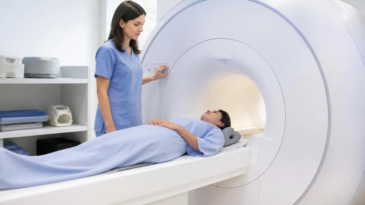
Brachial Plexus MRI vs. Other Imaging Modalities
- +Excellent soft tissue contrast visualizes individual nerve fascicles without ionizing radiation
- +Multiplanar capability covers the entire plexus from cervical roots to axillary branches in one study
- +STIR and neurography sequences detect nerve edema and injury before muscle atrophy develops
- +Post-contrast sequences differentiate active inflammation from fibrosis and tumor infiltration
- +3D isotropic acquisitions allow unlimited retrospective reformatting and neurography post-processing
- +Functional sequences like DTI provide quantitative microstructural data unavailable with any other modality
- −Long scan times (45–60+ minutes) increase motion artifact risk, especially in acute trauma patients
- −Vascular pulsation artifact from the subclavian and carotid vessels can obscure adjacent nerve structures
- −Claustrophobia and body habitus limitations affect image quality in a meaningful subset of patients
- −Gadolinium contraindicated in severe renal impairment, limiting post-contrast assessment when needed
- −Electrodiagnostic studies (EMG/NCS) provide complementary functional data MRI alone cannot offer
- −High cost and limited availability of 3T scanners with dedicated neurography capability in rural settings
Pros and Cons at a Glance
| Pros | Cons |
|---|---|
| Excellent soft tissue contrast visualizes individual nerve fascicles without ionizing radiation | Vascular pulsation artifact from the subclavian and carotid vessels can obscure adjacent nerve structures |
| Multiplanar capability covers the entire plexus from cervical roots to axillary branches in one study | Claustrophobia and body habitus limitations affect image quality in a meaningful subset of patients |
| STIR and neurography sequences detect nerve edema and injury before muscle atrophy develops | Gadolinium contraindicated in severe renal impairment, limiting post-contrast assessment when needed |
| Post-contrast sequences differentiate active inflammation from fibrosis and tumor infiltration | Electrodiagnostic studies (EMG/NCS) provide complementary functional data MRI alone cannot offer |
Brachial Plexus MRI Patient Preparation Checklist
- ✓Screen the patient for all metallic implants, pacemakers, neurostimulators, and cochlear implants before room entry.
- ✓Review prior imaging reports and surgical history to identify expected post-operative changes that may mimic pathology.
- ✓Obtain eGFR within 30 days if gadolinium contrast is ordered, and confirm no history of nephrogenic systemic fibrosis.
- ✓Instruct the patient to remove all jewelry, hair clips, and underwire bras before entering the magnet room.
- ✓Position the patient supine with the head neutral and the symptomatic arm gently tucked at the side.
- ✓Place the phased-array neurovascular coil centered over the neck and ipsilateral shoulder, securing with provided straps.
- ✓Provide earplugs and a communication squeeze bulb, and coach the patient on the importance of remaining still during acquisition.
- ✓Acquire localizer images first to confirm coil placement covers the C4 level superiorly through the axillary fossa inferiorly.
- ✓Check early scout images for vascular pulsation artifact and reposition the saturation band over the subclavian vessels if needed.
- ✓Document the clinical indication, symptom side, and any prior nerve conduction studies to guide sequence prioritization.
Benign Nerve Sheath Tumors Show a Pathognomonic Target Sign
On T2-weighted MRI, schwannomas and neurofibromas characteristically display a target sign — a central area of low signal intensity (fibrous collagen core) surrounded by a rim of high signal intensity (myxoid peripheral stroma). Recognizing this sign on brachial plexus MRI can confidently distinguish a benign peripheral nerve sheath tumor from a malignant soft tissue sarcoma or a lymph node metastasis, potentially sparing the patient an unnecessary biopsy.
Interpreting a brachial plexus MRI requires a systematic approach that begins with clinical context and proceeds through each anatomical segment from root to terminal branch. Before reviewing images, the interpreting radiologist should know the patient's dominant hand, the mechanism and timing of injury or symptom onset, any prior surgeries or radiation fields, and the results of electrodiagnostic studies. This clinical correlation transforms image interpretation from pattern recognition into true diagnostic reasoning — the same finding may have very different implications in a trauma patient versus a breast cancer survivor.
Begin review with the coronal STIR sequence, which provides the clearest overview of plexus signal and symmetry. Systematically compare the affected side to the contralateral normal side at each level — roots, trunks, and cords. Note any focal or diffuse signal elevation, asymmetric nerve caliber, or complete nerve discontinuity. Pay particular attention to the supraclavicular plexus (roots and trunks), where traumatic traction injuries and most nerve sheath tumors occur, and to the infraclavicular plexus (cords and branches), where thoracic outlet compression and lymph node involvement are most commonly encountered.
Axial T1-weighted images are next, reviewed from the C4-5 disk level through the mid-axillary fossa. At each level, identify the expected nerve structures and assess whether they maintain their normal oval morphology, signal intensity, and relationship to adjacent anatomical landmarks. Asymmetric fatty replacement of muscles served by specific plexus segments provides powerful clues about the location and chronicity of injury. For example, isolated denervation of the serratus anterior (supplied by the long thoracic nerve, C5-7) suggests a specific nerve injury distinct from a complete trunk-level lesion.
Post-contrast T1 fat-saturated images should be reviewed with enhancement pattern as the primary diagnostic criterion. Normal nerve fascicles are not expected to enhance. Any enhancement within the expected nerve course demands explanation — possibilities include active inflammation (Parsonage-Turner syndrome), perineural tumor spread, or nerve sheath tumor. The distribution, avidity, and homogeneity of enhancement help narrow the differential. Thin, smooth perineural enhancement extending along multiple nerve segments toward the foramina is highly suggestive of perineural tumor spread from a head and neck primary.
Three-dimensional neurography images, when available, should be reviewed as maximum intensity projections and multiplanar reformations rather than exclusively as source images. The MIP images depict the nerve network as a branching structure similar to an angiogram, making it immediately apparent when a segment is absent, focally thickened, or displaced by an adjacent mass. Source images, however, are essential for characterizing internal nerve architecture, assessing fascicular detail, and measuring lesion dimensions for surgical planning or follow-up comparison.
DTI-derived metrics add a quantitative dimension to brachial plexus assessment that is particularly valuable for longitudinal monitoring. In healthy peripheral nerves, fractional anisotropy values typically range from 0.4 to 0.7, reflecting the highly organized parallel arrangement of axons. At sites of compression, crush injury, or inflammation, FA drops significantly — sometimes to values below 0.2. Mean diffusivity, conversely, tends to rise at injury sites as the ordered microstructure breaks down and water diffusion becomes less restricted and more isotropic. Tracking these metrics on serial studies correlates better with clinical recovery than conventional sequence findings in some published series.
Reporting a brachial plexus MRI requires structured communication that directly addresses the clinical question. A useful report format identifies the affected plexus segment (using standard anatomical nomenclature), characterizes the MRI findings at that level (signal change, caliber, morphology, enhancement), identifies target muscle denervation changes and their chronicity, and provides a differential diagnosis ranked by probability given the clinical context. For surgical cases, explicitly state whether the injury is preganglionic or postganglionic, as this distinction fundamentally determines operative strategy — avulsion cannot be repaired at the root level and requires nerve transfer procedures from intact donor nerves.
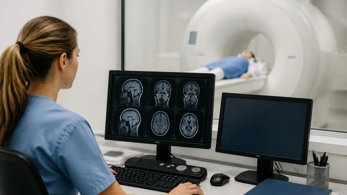
Preganglionic root avulsions and postganglionic traction injuries require entirely different surgical strategies. Avulsed roots cannot be reconnected to the spinal cord — treatment relies on nerve transfers from intact donors such as the intercostal, phrenic, or contralateral C7 nerve. Failure to distinguish these injury types on MRI can result in futile surgical exploration. Always assess for pseudomeningoceles and absent rootlets on thin-slice axial and coronal T2 sequences before characterizing injury level in any traumatic brachial plexus case.
For MRI technologists and students preparing for the ARRT registry examination, brachial plexus imaging represents a convergence of anatomy, physics, and pathology that is heavily tested across multiple content domains. The ability to name and locate each component of the plexus — roots, trunks, divisions, cords, and terminal branches — is foundational. Equally important is understanding the dermatomal and myotomal distributions of each root level, since denervation changes in specific muscle groups on MRI directly map to root-level localization.
On the registry examination, MRI physics questions related to brachial plexus imaging most commonly address fat suppression techniques and their trade-offs. STIR suppresses fat by exploiting its short T1 relaxation time using an inversion recovery pulse, making it insensitive to B0 field inhomogeneity — a major advantage in the neck where field distortion is significant near air-tissue interfaces.
Spectral fat saturation (CHESS or SPIR) uses a frequency-selective pulse to null fat signal and is faster than STIR but fails at regions of field inhomogeneity, which is precisely why STIR is preferred for brachial plexus imaging despite its lower signal-to-noise ratio and longer acquisition time.
Understanding the clinical rationale for gadolinium in brachial plexus imaging is another registry-relevant concept. The blood-nerve barrier, analogous to the blood-brain barrier, normally prevents gadolinium from entering the endoneurial compartment of healthy peripheral nerves. Breakdown of this barrier in inflammation or tumor invasion allows contrast to accumulate within the nerve, producing visible enhancement. This physiological principle explains why enhancement is always pathological in a peripheral nerve context and why non-contrast sequences are sufficient for most traumatic plexus injuries where inflammatory breakdown of the blood-nerve barrier is not the primary diagnostic concern.
Motion artifact management is a key technical skill that registry candidates should master conceptually. Cardiac and respiratory motion propagates as ghosting artifact in the phase-encoding direction and can obscure plexus anatomy in the axillary and supraclavicular regions. Technologists use multiple strategies to minimize this artifact: placing the phase-encoding direction in the foot-head orientation (so breathing motion ghosts away from the plexus), applying spatial saturation bands over the chest and heart, using respiratory gating or triggering for sensitive sequences, and prescribing cardiac gating for axial sequences at the level of the subclavian vessels.
Gradient coil performance and its relationship to resolution is another physics concept relevant to neurography. Achieving sub-millimeter in-plane resolution requires strong, fast-switching gradients that generate maximum amplitude slew rates. Modern 3T systems designed for neurography can achieve gradient amplitudes of 80 mT/m or higher, enabling the short echo spacing necessary for high-resolution 3D SPACE or CUBE acquisitions without excessive T2 blurring. Understanding why resolution has improved so dramatically over the past decade requires appreciating these hardware advances — not just the sequence design.
Contraindication screening for brachial plexus patients deserves special mention because this population often includes trauma victims who may be unconscious, intoxicated, or unable to provide an accurate implant history. Institutional protocols for emergent MRI in unresponsive patients typically require rapid x-ray screening of the chest and potentially the orbital region to exclude ferromagnetic foreign bodies. Peripheral nerve stimulators and spinal cord stimulators — increasingly common in chronic pain patients — require individual implant verification before 3T scanning and may necessitate downgrade to 1.5T or exclusion from MRI entirely depending on device labeling.
For exam candidates, the most effective way to solidify brachial plexus knowledge is to practice with case-based questions that integrate anatomy, physics, and pathology into realistic clinical scenarios. Knowing that a pseudomeningocele indicates root avulsion is useful, but knowing why — the dural sleeve tears, CSF extravasates laterally through the foramina, and the displaced nerve root loses its connection to the cord — is what cements the concept and allows application to novel cases. Active recall through quiz-based practice, rather than passive re-reading, is the evidence-based strategy that most efficiently converts knowledge into durable exam-ready understanding.
Practical success in brachial plexus MRI — whether measured by diagnostic yield, patient throughput, or examination performance — comes down to preparation, protocol discipline, and the habit of systematic review. The following practical tips represent the distilled wisdom of experienced MRI technologists and radiologists who have built high-volume brachial plexus imaging programs. Incorporating these practices into your workflow will improve image quality, reduce repeat examinations, and sharpen the diagnostic precision of your interpretations.
Always perform a brief pre-scan patient interview even when the order provides clinical history. Ask specifically which arm is symptomatic, whether symptoms are purely sensory or include weakness, how long symptoms have been present, and whether there has been any prior surgery or radiation to the neck or axilla. This two-minute conversation frequently reveals information not captured in the electronic order — a previous mastectomy, a forgotten prior MRI at another institution, or a history of childhood plexus birth injury — that directly changes which sequences you prioritize or which side you center the coil on.
For trauma cases, where patient cooperation may be limited, front-load the most diagnostically critical sequences. Acquire the coronal STIR and axial T2 or STIR sequences first, before the patient fatigues or becomes unable to cooperate with breath-hold instructions. Save the longer 3D neurography acquisitions for later in the protocol when they are less urgent. Having the two highest-yield sequences completed within the first fifteen minutes of scanning ensures that even a truncated examination yields actionable diagnostic information.
Optimizing fat suppression in the neck requires careful attention to shimming. After coil placement and patient positioning, always run the local shim over a volume that includes the entire plexus coverage area — from C4 to the axillary fold. A poorly shimmed volume will produce heterogeneous fat suppression that creates bright fat patches masquerading as nerve signal on STIR or creates dark signal dropout regions that mimic nerve injury on spectral fat-saturated sequences. Spending two extra minutes optimizing shim before acquiring fat-suppressed sequences consistently pays off in diagnostic quality.
When reviewing coronal neurography images, use the windowing controls actively rather than accepting the default display settings. Narrowing the window width to enhance nerve-to-background contrast often reveals subtle signal asymmetries that are invisible on default settings. Conversely, widening the window is useful for assessing the background tissue signal and ensuring that fat suppression is homogeneous across the field of view. Most PACS workstations allow saving custom window presets — creating a neurography-specific window preset for your institution saves time and ensures consistent display across readers.
Keep a personal log of interesting brachial plexus cases encountered in clinical practice or during study. Each case should record the clinical presentation, key MRI findings, final diagnosis, and one teaching point. Over time, this case library becomes an invaluable resource for registry preparation and for developing the pattern recognition skills that textbooks cannot fully convey. Reviewing twenty real cases of Parsonage-Turner syndrome — noting the variation in nerve segments affected, the presence or absence of enhancement, and the evolution of findings on follow-up — builds the experiential foundation that allows confident diagnosis in the clinical setting.
Collaboration between the MRI technologist and the radiologist before scanning complex cases improves outcomes measurably. A brief three-minute conversation about the clinical question — is this a trauma case where root avulsion needs exclusion, or an oncology case where perineural spread needs assessment — allows the technologist to intelligently adjust the protocol rather than running a generic default. At institutions where this practice is formalized through pre-scan radiologist sign-off on complex neurological studies, repeat examination rates for brachial plexus MRI drop significantly and referring physicians consistently report higher satisfaction with report actionability.
Finally, stay current with the rapidly evolving literature on brachial plexus neurography. New sequences, new post-processing techniques, and new clinical applications are published regularly. Following radiology journals focused on neuroradiology and musculoskeletal imaging, attending hands-on neurography workshops at major radiology meetings, and participating in online case-sharing communities are all effective strategies for maintaining expertise in a field where the state of the art advances faster than most textbooks can track. The combination of strong foundational knowledge — built through systematic study and registry-level preparation — with curiosity-driven continuous learning is what sustains excellence in brachial plexus MRI over a career.
Sample MRI - Magnetic Resonance Imaging Practice Questions
Try these questions from our free MRI - Magnetic Resonance Imaging practice tests. The correct answer and an explanation follow each question.
Which property of hydrogen makes it the most commonly used nucleus in clinical MRI?
- A. High natural abundance and large magnetic moment
- B. Low atomic mass and zero spin
- C. Absence of electron cloud interference
- D. High atomic number and radioactivity
Answer: A. High natural abundance and large magnetic moment
Hydrogen (¹H) is abundant in the body and has a large gyromagnetic ratio, producing strong MRI signals.
Which MRI sequence is most sensitive to hemorrhage detection because of its sensitivity to blood products?
- A. Gradient echo (GRE)
- B. Inversion recovery (IR)
- C. Fast spin echo (FSE)
- D. Diffusion-weighted imaging (DWI)
Answer: A. Gradient echo (GRE)
Gradient echo sequences are sensitive to magnetic susceptibility differences caused by hemosiderin and deoxyhemoglobin, making them ideal for detecting hemorrhage.
The corpus callosum is best evaluated on which MRI plane for assessing callosal agenesis?
- A. Axial
- B. Coronal
- C. Sagittal
- D. Oblique
Answer: C. Sagittal
The sagittal midline plane provides the best single view of the entire corpus callosum from genu to splenium for assessing agenesis or dysgenesis.
Which gradient is responsible for determining slice position in a 2D MRI acquisition?
- A. Frequency-encoding gradient
- B. Phase-encoding gradient
- C. Slice-selection gradient
- D. Spoiler gradient
Answer: C. Slice-selection gradient
The slice-selection gradient is applied simultaneously with the RF pulse to excite only protons in the desired anatomical plane.
Take the full MRI - Magnetic Resonance Imaging practice test
MRI Questions and Answers

MRI of Cervical Spine: Complete Guide to Imaging, Anatomy, Pathology, and Patient Preparation

Knee MRI Images: A Complete Guide to Reading, Understanding, and Interpreting Knee Scans
Ace Your Exam with MRI Practice Test 2026 Prep

MRI Tech School 2026 — Programs, Requirements, and What to Expect
MRI Procedure: What to Expect Before, During and After Your Scan
About the Author

Medical Laboratory Scientist & Clinical Certification Expert
Johns Hopkins UniversityDr. Sandra Kim holds a PhD in Clinical Laboratory Science from Johns Hopkins University and is certified as a Medical Technologist (MT) and Medical Laboratory Scientist (MLS) through ASCP. With 16 years of clinical laboratory experience spanning hematology, microbiology, and molecular diagnostics, she prepares candidates for ASCP board exams, MLT, MLS, and specialist certification tests.
Join the Discussion
Connect with other students preparing for this exam. Share tips, ask questions, and get advice from people who have been there.
View discussion (8 replies)