MRI of the Cervical Spine: Procedure, Findings, Cost 2026 July
📚 MRI of the cervical spine explained: what it shows, how long it takes, costs, claustrophobia tips, and how to read your radiology report.
A magnetic resonance imaging scan of the cervical spine looks inside your neck without a single drop of radiation. It uses a strong magnet, radio waves, and a computer to build sharp, layered pictures of the seven vertebrae stacked between your skull and your shoulders — plus the discs, ligaments, nerves, and spinal cord weaving through them. If your doctor has ordered one, you probably have neck pain that won’t quit, numbness running into an arm, a recent injury, or a strange weakness that needs explaining.
This guide walks you through everything: why doctors choose this test over X-rays or CT, what happens during the appointment, how to read the report your radiologist writes, and what the typical findings actually mean. You’ll also see real cost ranges, claustrophobia tips, and which red flags push doctors to recommend the scan urgently. By the end, you’ll know how to walk into the appointment ready — and how to make sense of whatever shows up on the images.
A quick orientation first. Magnetic resonance imaging beats other scans for soft tissue. X-rays show bones. CT shows bones and bleeding well, but soft structures blur together. MRI lights up nerves, discs, and the spinal cord like a high-definition map — which is exactly what your doctor needs when symptoms point to a pinched nerve, disc herniation, or cord compression.
Why Doctors Order an MRI of the Cervical Spine
Most referrals come from one of five reasons. Persistent neck pain that hasn’t responded to four to six weeks of conservative treatment tops the list. Radiculopathy — pain, tingling, or numbness shooting down an arm — is another big one, because the nerve root squeezed at C5, C6, or C7 produces a predictable pattern your doctor can map.
Trauma cases come in fast. A car accident, a fall from a ladder, a sports collision — anything that loads the neck suddenly can damage discs, ligaments, or the cord itself. CT catches fractures quickly in the ER, but MRI is the follow-up when soft tissue injury is suspected. Then there’s myelopathy, a slower-burning problem where the cord itself gets compressed. Symptoms include clumsy hands, balance problems, or trouble with fine motor tasks like buttoning a shirt. That one needs imaging fast.
Tumors, infections, and demyelinating disease (like multiple sclerosis) also bring patients in. Multiple sclerosis lesions in the cervical cord show up clearly on MRI — which is why neurologists use the scan to diagnose and monitor the condition over years.
Red Flag Symptoms That Speed Things Up
Some symptoms aren’t wait-and-see. Severe weakness in both arms, loss of bladder or bowel control, fever with neck pain, or progressive difficulty walking — these push the scan from routine to urgent. If you have any of those alongside neck symptoms, expect imaging within days, not weeks.
What Happens During the Scan
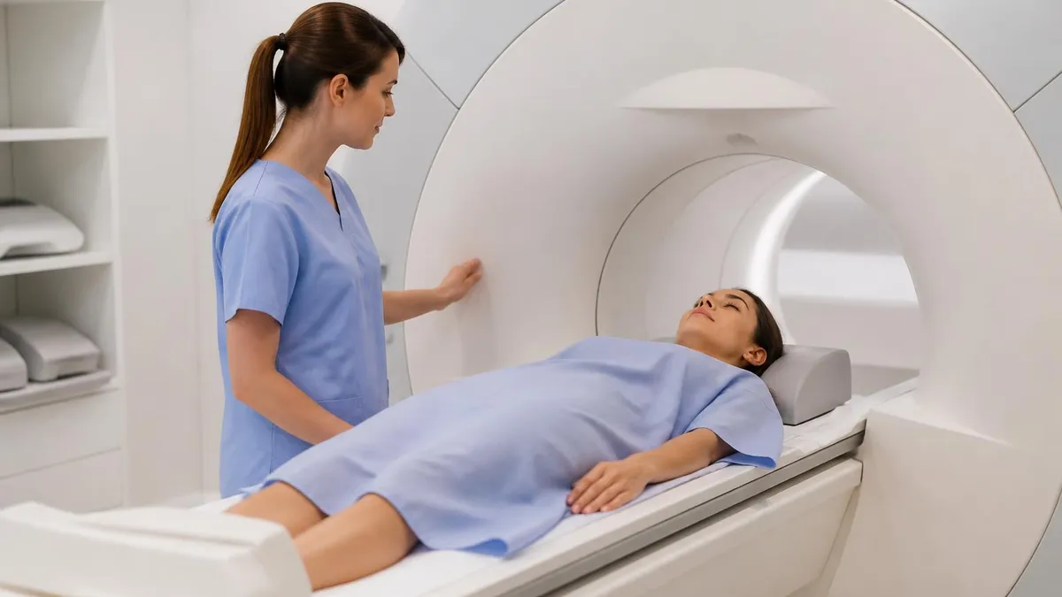
You’ll arrive 15 to 30 minutes early to change into a gown, fill out a metal screening form, and remove anything magnetic — jewelry, hairpins, hearing aids, watches. Even underwire bras come off. The tech asks about implants, pacemakers, surgical clips, shrapnel, and prior surgeries. This isn’t bureaucratic — the magnet is roughly 30,000 times stronger than Earth’s magnetic field, and metal becomes a real hazard inside the room.
For the scan itself, you lie on your back on a padded table. A coil — basically a curved antenna — sits around your neck to pick up the radio signals. The tech slides you headfirst into the bore of the magnet. The opening is wider than people expect (about 60 to 70 cm across), but it’s still a tube, and you’re inside it for 20 to 45 minutes. Earplugs or headphones help with the noise — and the machine is loud, like rapid knocking or buzzing during each sequence.
You need to stay completely still. Any movement blurs the images. The tech can see and hear you the entire time, and you get a squeeze ball if you need to stop. If contrast is needed, an IV in your arm delivers gadolinium dye partway through — it lights up areas of inflammation, infection, or tumor activity. Most cervical scans don’t need contrast, but tumor follow-ups and infection workups usually do.
Worried about tight spaces? You’re not alone. About 5 to 10% of patients struggle with claustrophobia inside the bore. Tell the tech in advance. Options include a mild oral sedative (ask your doctor a few days ahead), an open mri machine if image quality allows, headphones with music or a podcast, and an eye mask so you don’t see the tube above you. Wide-bore 3T systems also help — they’re standard now in most newer imaging centers.
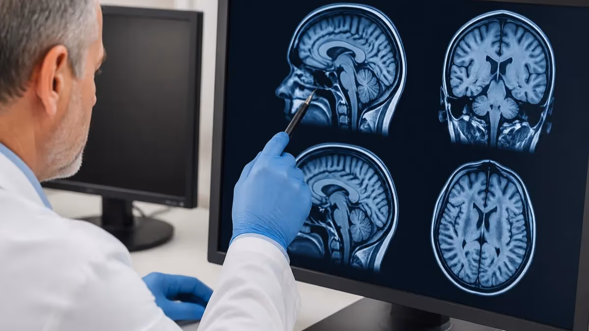
Reading Your Cervical Spine MRI Report
The report has a predictable structure: clinical history, technique, findings (level by level), and impression. Skip to the impression first — that’s where the radiologist sums up what matters. Then work backward into the findings if you want detail.
Radiologists describe each level from C2-C3 down through C7-T1. At every level, they comment on the disc (normal, bulging, protruded, extruded, or sequestered), the central canal (the tube the cord runs through), the neural foramen (the side openings where nerve roots exit), and any signal changes in the cord or vertebrae. A normal disc is fully hydrated; a desiccated disc has lost water and looks darker on T2 sequences.
Common Terms You’ll See
Disc bulge means the disc has spread out evenly but stayed within its normal boundary. Almost everyone over 40 has at least one. Disc protrusion means a focal piece pokes out farther — this can press on a nerve. Extrusion means a fragment has broken through the outer ring. Sequestration means that fragment has detached completely. Each step is more serious, but only matters if it correlates with symptoms.
Foraminal stenosis means the side opening for a nerve root has narrowed. Central canal stenosis means the main tube the cord runs through is tighter than normal. Cord signal change — especially myelomalacia — can indicate chronic compression damage. The impression section ties findings to your symptoms: a C6 root entrapment matches arm pain running into the thumb and index finger; a C7 root issue matches the middle finger.
What Findings Are Common (and Often Meaningless)
Studies of pain-free adults consistently show that 30 to 50% have disc bulges, mild facet arthritis, or minor protrusions on MRI — with zero symptoms. That’s why the report alone doesn’t diagnose anything. Your symptoms have to match the imaging. A C5-C6 disc bulge with weakness in your biceps and tingling in your thumb? Real. The same finding with no matching symptoms? Likely incidental and not the source of your pain.
Open MRI vs. Closed MRI for the Cervical Spine
Closed-bore (the traditional tube) gives the sharpest images because the magnet field is stronger and more uniform. For the cervical spine, where small details matter, closed 1.5T or 3T machines are preferred. Open machines — with open sides or a wider C-shape — are friendlier for claustrophobic or larger patients but use weaker magnets (0.3T to 1.0T). Resolution drops noticeably.
If you’re seriously claustrophobic, the trade-off may still be worth it. For a follow-up scan after surgery, or a tumor workup where every millimeter matters, your doctor will push for closed. Some newer wide-bore 3T machines split the difference — 70cm bores with full strength — and they handle most claustrophobia cases without losing image quality.
Cost, Insurance, and Getting the Scan Done Faster
Cost varies wildly. In the US, the same scan costs $400 at a standalone imaging center in Texas and $3,500 at a Manhattan hospital. Call ahead, ask for the cash price, and compare. With insurance, the deductible and copay structure determines your real out-of-pocket. Many insurers require prior authorization — meaning your doctor submits paperwork and waits for approval before scheduling. Plan a 5 to 10 day delay for that step unless your case is flagged urgent.
To get scanned faster, ask your doctor to write urgent on the referral when symptoms warrant it, call imaging centers directly to find cancellation slots, and consider standalone imaging centers over hospitals — they often have same-week availability. Mobile MRI units (yes, they exist — trailer-mounted scanners) visit rural areas weekly.
Preparing for Your Appointment
Day before: take any prescribed sedative as directed. Eat normally unless contrast is planned — some centers ask for a 4-hour fast for contrast scans, but rules vary, so ask. Bring previous imaging on a disc if available; comparison helps the radiologist spot changes.
Day of: arrive 30 minutes early. Wear clothes without metal — no zippers, no underwire. Leave jewelry, hearing aids, and metal hairpins at home. Drink water normally. If you take medication, take it as usual. Empty your bladder right before going in — 45 minutes lying still can otherwise feel long.
After: drive home as usual unless you took a sedative. Resume normal activity immediately. If contrast was used, drink extra water for 24 hours to help your kidneys clear the gadolinium. Watch for any rare delayed reactions like itching or rash — report them to your doctor.
How This Scan Fits the Bigger MRI Picture
The cervical spine is one of many body parts that get imaged this way. If you’re curious about the technology behind it, what an MRI is covers the basics in detail. Different body parts need different scan times — how long an MRI takes by region varies from 15 minutes for a quick knee scan to over an hour for a full cardiac study. If you need related neck imaging, the cervical spine MRI reference covers more depth on positioning and protocol. For broader spine imaging or brain conditions affecting the neck, MRI brain studies are sometimes ordered together.
An MRI scan guide covers what to expect across the most common scan types. Following the right prep and screening keeps the procedure smooth and the images useful — which is, in the end, the whole point. For a primer on safety screening before any scan, mri safety walks through implants, foreign bodies, and pregnancy considerations. If money is the bigger question, mri cost breaks down pricing by region and insurance.
About the Author

Medical Laboratory Scientist & Clinical Certification Expert
Johns Hopkins UniversityDr. Sandra Kim holds a PhD in Clinical Laboratory Science from Johns Hopkins University and is certified as a Medical Technologist (MT) and Medical Laboratory Scientist (MLS) through ASCP. With 16 years of clinical laboratory experience spanning hematology, microbiology, and molecular diagnostics, she prepares candidates for ASCP board exams, MLT, MLS, and specialist certification tests.
Join the Discussion
Connect with other students preparing for this exam. Share tips, ask questions, and get advice from people who have been there.
View discussion (6 replies)