Multiple Sclerosis MRI: McDonald Criteria, Lesion Patterns and Protocols (2026 July Guide)
🎯 Multiple sclerosis MRI explained: McDonald 2017 criteria, typical lesion patterns, brain vs spinal cord scans, gadolinium contrast use and monitoring
If you or someone you love is being worked up for multiple sclerosis, the words your neurologist keeps using are probably the same three: brain MRI, spinal cord MRI, and McDonald criteria. There is a reason for that. MRI is the single most important imaging test for diagnosing MS, and it is also the test that follows you for the rest of your life if the diagnosis is confirmed.
This guide walks you through what a multiple sclerosis MRI actually shows, how radiologists describe lesions, why gadolinium contrast matters, and how doctors apply the 2017 McDonald criteria to turn pictures into a diagnosis. We have kept it practical so you can read your own report with a bit more confidence on the next neurology visit.
You will see plenty of jargon along the way — Dawson's fingers, FLAIR hyperintensities, dissemination in space, NEDA-3 — and we promise to translate every single one. By the end you should know what to expect on scan day, what your report is actually telling you, and what the difference between an old plaque and an active flare looks like on screen. Bookmark this page. You will reference it again.
MRI is the most sensitive test for detecting MS plaques (areas where myelin has been damaged). Around 95% of people with clinically definite MS have visible lesions on brain MRI. No other test, including blood work, can show the demyelinating lesions directly. That is why an MRI brain scan is almost always the first imaging study ordered when MS is suspected, often paired with cervical spine MRI.
Multiple sclerosis is an autoimmune disease in which the immune system attacks myelin, the insulating coating around nerve fibers in your brain, spinal cord, and optic nerves. The damaged patches are called plaques or lesions, and they can disrupt nerve signaling in unpredictable ways. About one million Americans live with MS today. Most are diagnosed between ages 20 and 40, and women are affected roughly three times more often than men.
The disease behaves like a series of slow-motion electrical short-circuits. One person might lose vision in an eye for a few weeks (optic neuritis). Another might have numb feet, double vision, or a sudden loss of balance. Symptoms come and go, often for months, before someone connects the dots. MRI is what links those scattered episodes to a single, unifying diagnosis.
It can show that lesions exist in different parts of the central nervous system and have appeared at different times — the two ideas at the heart of every modern MS diagnosis. Without imaging, doctors would be left guessing whether a numb hand, a blurry eye, and a wobbly gait all belong to the same disease. MRI removes most of that guesswork, and the McDonald criteria turn the picture into a defensible diagnosis.
MS MRI Sequences By Region
Brain MRI is the workhorse of MS imaging. Standard sequences include T1 (anatomy and black holes), T2 (fluid-sensitive), FLAIR (the gold standard for picking up periventricular plaques because it suppresses cerebrospinal fluid), DWI (rules out stroke), GRE or SWI (microbleeds), and a post-contrast T1 after gadolinium to highlight active lesions. A typical brain protocol runs 30 to 45 minutes on a 3T scanner.
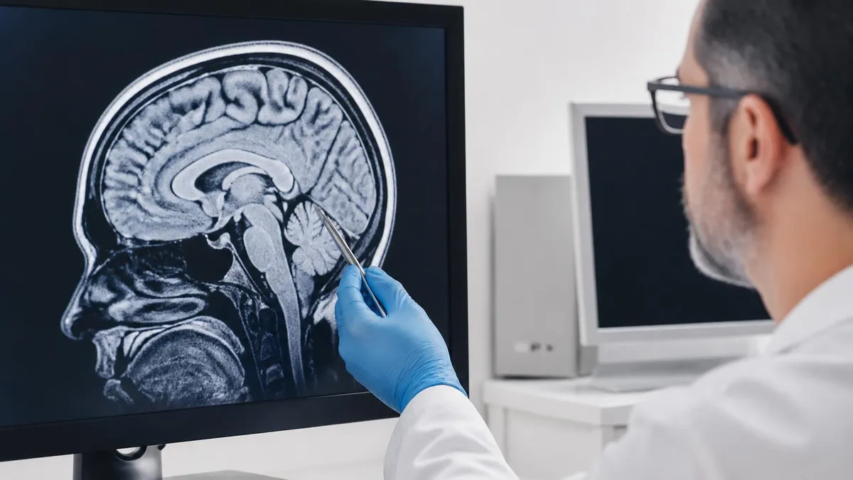
Where the lesions sit matters as much as how many there are. MS plaques have a strong preference for certain neighborhoods of the brain. Periventricular lesions, hugging the lateral ventricles, are classic. The famous Dawson's fingers — ovoid lesions radiating perpendicular from the ventricles along small veins — are highly specific for MS and are best seen on sagittal FLAIR images of the brain.
Compared to a normal brain MRI, the difference is striking once you have seen a few examples. A healthy scan shows clean, dark white matter on FLAIR with only faint normal signal around the ventricles. An MS scan looks dotted with bright spots in classic locations: around the ventricles, touching the cortex, and tucked into the brainstem or cerebellum.
Juxtacortical lesions sit right up against the gray matter, while infratentorial lesions live in the brainstem or cerebellum. Cortical lesions, harder to see without specialized sequences like double inversion recovery, are increasingly recognized as drivers of long-term disability and cognitive change. Radiologists check all four neighborhoods because the McDonald criteria reward you for diversity of locations, not raw volume of plaques.
That is why a single very large lesion in one spot is less diagnostic than four small lesions scattered across periventricular, juxtacortical, infratentorial, and spinal cord regions. The geography of the disease tells doctors more than the headcount, and a careful radiologist will list each anatomical neighborhood the lesions affect in the impression line of the report.
What MS Lesions Look Like On MRI
- Brain involvement: 90-95% of MS patients
- Spine-only disease: 10-15% of patients
- Optic nerve: Common, especially in CIS
- Typical brain zones: Periventricular, juxtacortical, infratentorial
- Shape: Ovoid (egg-shaped), perivenular
- Typical size: 3 mm to 15 mm
- Spinal cord lesions: Usually 3-8 mm, short segment
- Hallmark sign: Dawson's fingers
- T2 / FLAIR: Hyperintense (bright)
- T1 (chronic): Hypointense (black holes)
- Active lesions: Enhance with gadolinium
- Chronic plaques: Do not enhance
- Highly specific: Periventricular ovoid, callosal-septal
- Less specific: Subcortical dots
- Spine specific: Short, eccentric, peripheral
- Optic nerve: Bright on T2, enhances when active
The 2017 McDonald criteria are the rulebook neurologists use to translate MRI findings into a formal MS diagnosis. The criteria are built on two ideas: dissemination in space (lesions in more than one typical MS area) and dissemination in time (lesions of different ages). You can satisfy both on a single scan if your MRI shows a contrast-enhancing lesion right next to a non-enhancing one.
That single observation captures both old and new disease activity, which is why a thorough first MRI matters so much. If you do not meet both criteria on the first scan, do not panic. A follow-up MRI three to twelve months later that shows a new T2 lesion or a new enhancing lesion clinches dissemination in time. Many patients hit the criteria on the second scan rather than the first.
Cerebrospinal fluid oligoclonal bands can also substitute for time in some scenarios. That is why your neurologist may suggest a lumbar puncture if the MRI is suggestive but not yet diagnostic. The 2024 and 2025 revisions to the criteria expand the picture further, formally including optic nerve lesions and giving more diagnostic weight to spinal cord findings — good news for patients with atypical presentations who would have lingered in diagnostic limbo under the older versions of the rules.
Typical MS MRI Protocol Step By Step
Pre-scan checklist
Brain MRI without contrast
Gadolinium injection
Cervical spine MRI
Thoracic spine (if indicated)
Optic nerve sequences (if needed)
Total scan duration
Not every MS scan is the same. Newly-diagnosed patients tend to get the most thorough imaging — brain plus cervical spine, with contrast, on a 3T magnet. Once you are stable on a disease-modifying therapy, your annual surveillance scan can sometimes drop contrast and stick to brain-only protocols. The decision depends on stability, kidney function, and whether you have had any new symptoms in the months before the appointment.
Some centers also use the upright MRI when patients cannot tolerate lying flat. Open and upright scanners are friendlier for claustrophobia and for people with back pain that makes the closed bore unbearable. The trade-off is resolution: small periventricular plaques can be harder to spot on lower-field open systems, so most MS specialists still steer toward closed bore 3T machines when available.
Pediatric protocols deserve a quick note. Children often need sedation for MRI because staying motionless for an hour is asking a lot of a small body. Pediatric MS tends to be more inflammatory than adult-onset disease, with higher lesion burdens and more frequent tumefactive (very large) plaques. Imaging schedules and sedation plans are individualized at children's hospitals, and parents are usually allowed to stay in the control room or even hold a hand during portions of the scan.
MS MRI By The Numbers (2026, US)
People often ask how MS MRI is different from a scan they had for, say, knee pain or pelvic discomfort. The hardware is the same magnet, but the protocols and contrast use are tuned for the central nervous system. A pelvic MRI, for example, focuses on soft tissue contrast in the bowel, bladder, and reproductive organs and does not need the FLAIR or DWI sequences that hunt for brain plaques.
The decision tree from your doctor for MS, by contrast, is built entirely around white matter and cord anatomy. You might also wonder why MRI gets the call over CT scanning. The short version: CT is fast and good for bleeding or bone, but its soft-tissue contrast is too coarse to reliably see MS plaques in the brain or spinal cord.
If you want a deeper comparison, the breakdown of MRI vs CT scan covers the strengths and weaknesses of each modality. For MS, MRI is not just better — it is the only imaging test that consistently shows the disease. CT is reserved for emergencies where a fast look at the skull or brain is needed, like ruling out a hemorrhage before a high-dose steroid course is started in the hospital for a suspected acute MS attack.
MS Mimics Your Radiologist Will Try To Rule Out
- ✓Migraine-related white matter changes (non-specific subcortical dots)
- ✓Small vessel ischemic disease (aging-related, deep white matter)
- ✓Vasculitis (inflammatory blood vessel disease)
- ✓Neuromyelitis optica spectrum disorder (longitudinally extensive cord lesions)
- ✓Acute disseminated encephalomyelitis (usually pediatric, monophasic)
- ✓Cerebral amyloid angiopathy (microbleeds in older adults)
- ✓Lupus CNS involvement
- ✓Vitamin B12 deficiency
- ✓HIV-related leukoencephalopathy
- ✓Sjögren's syndrome
- ✓Lyme neuroborreliosis
- ✓Neurosarcoidosis
Beyond the initial diagnosis, MRI keeps doing useful work for years. Disease-modifying therapies (DMTs) are judged on a target called NEDA-3: no clinical relapses, no MRI activity (no new T2 lesions and no enhancing lesions), and no disability progression. Hitting NEDA-3 is a strong signal that your current medication is doing its job and earning its place on your chart.
Falling short — especially with new lesions on follow-up MRI — is the most common trigger for switching to a higher-efficacy therapy. Many neurologists now use MRI activity, not just clinical relapses, as the primary signal for treatment change. A patient feeling fine but showing two new T2 lesions and an enhancing plaque is heading toward a tougher conversation about escalating treatment.
Brain atrophy, measured on annual scans, is the other long-term metric to watch. Healthy adults lose about 0.1 to 0.4 percent of brain volume per year. People with active MS often lose two to three times that much, and accelerated atrophy correlates with future disability. Some research centers now report standardized atrophy measures alongside the lesion count, and newer artificial intelligence tools are starting to make this measurement more reliable in everyday clinical practice.
What MRI Looks Like Across MS Subtypes
- Definition: First MS-like attack
- Typical MRI: >=1 lesion in classic locations
- Conversion risk: Higher if multiple lesions
- McDonald 2017: Can diagnose MS at first scan
- Frequency: Most common form (~85%)
- Lesion behavior: Periodic new T2 and enhancing lesions
- Monitoring: Annual brain MRI
- Treatment goal: NEDA-3 on DMT
- Onset: Evolves from RRMS after years
- MRI activity: Fewer new lesions, more atrophy
- Key marker: Brain volume loss
- Black holes: Often increase in number
- Pattern: Continuous decline from onset
- Brain lesions: Sometimes fewer than RRMS
- Spinal cord: Often heavily involved
- Diagnosis: 1+ year of progression + MRI/CSF support
- Clinical status: No MS symptoms
- MRI finding: MS-like lesions found incidentally
- 5-year risk: ~10% convert to clinical MS
- Management: Watchful waiting + repeat MRIs
- Lesion load: Often higher T2 burden
- Tumefactive lesions: More common than adults
- Recovery: Generally better short-term
- Brain regions: More brainstem and cerebellum
Cost and access are worth a moment too, because access shapes outcomes in MS more than people realize. Brain MRI without contrast in the US typically runs $400 to $2,500 depending on where you live, your insurance plan, the time of year, and whether you go to a hospital outpatient department or to a freestanding imaging center. Adding contrast pushes that to $500 to $3,000.
Tacking on cervical or thoracic spine can add another $500 to $1,500 per region. Insurance almost always covers MRI when a neurologist orders it for suspected or confirmed MS, but pre-authorization is the rule, not the exception. Build in a few extra days when scheduling, and ask your imaging center for cash-pay pricing if you are uninsured.
If your plan denies coverage, ask your neurologist's office to submit a peer-to-peer appeal — these are usually successful for MS workups because the imaging is considered medical necessity, not screening. University hospitals often run charity care programs that cover MS imaging at low or no cost, and some MS specialty centers offer free-scan days as part of research protocols you can ask about during your next visit.
If you have read a few MS reports, you have probably noticed the radiologist describes lesions almost as a checklist. Count, location, signal characteristics, presence or absence of enhancement, and comparison to prior scans. That structure is intentional — it maps directly onto the McDonald criteria. Your neurologist needs to know whether the new picture pushes you past a diagnostic threshold.
Or whether it simply confirms what was already documented. Be aware that newer MRIs are far more sensitive than older ones. If your last brain scan was on a 1.5T magnet five years ago and your latest is on a 3T, do not be alarmed if the lesion count jumped. Some of that is real disease activity. Some of it is the scanner picking up plaques the older one missed.
A good radiologist will note this in the report, but ask the neurologist to compare images side by side rather than just totals. Numbers without context can mislead. Two new periventricular plaques in a young patient who stopped their DMT mean something very different from two new dots in a stable patient who upgraded from 1.5T to 3T at a new clinic.
Strengths And Limits Of MRI For MS
- +Most sensitive test for detecting MS plaques
- +Shows brain and spinal cord with no radiation
- +Distinguishes active lesions from chronic plaques using gadolinium
- +Tracks disease activity year over year on the same protocol
- +Helps rule out look-alike conditions like stroke or tumors
- +Required by the 2017 McDonald criteria for diagnosis
- +Guides treatment decisions and DMT switching
- +Detects brain atrophy as disease progresses
- −Cannot diagnose MS without clinical correlation
- −Lesion count correlates poorly with disability level
- −Gadolinium contrast carries small risks (allergy, NSF, deposition)
- −60 to 90 minute scans are tough for claustrophobic patients
- −1.5T scanners miss small lesions seen on 3T
- −Reading is somewhat subjective — radiologist experience matters
- −Some MS subtypes show few or atypical lesions
- −Cost ranges widely and pre-authorization is often needed
One question that comes up often is how frequently you should get an MRI if you already have MS. For most people on a stable DMT, the answer is once a year, brain only, sometimes with contrast and sometimes without depending on your neurologist's protocol. After any new attack — sudden weakness, visual loss, numbness, balance issues — your team will likely add an unscheduled MRI to look for new active lesions.
Pregnancy generally pauses contrast use but not the scans themselves. Most MS pregnancies see fewer relapses, especially in the second and third trimesters, but postpartum is a high-risk window. Many neurologists schedule a non-contrast brain MRI a few months after delivery to make sure nothing new has crept in while you were sleep-deprived and not paying attention to your body's signals.
Travel and continuity of care matter too. If you move or switch insurance, request a CD or digital export of every prior MRI and bring them to your new neurologist. Comparisons are only as good as the older images you can put side by side. A radiologist working blind on a fresh scan can call a stable plaque "new" simply because there is nothing to compare it against in the system.
Gadolinium-based contrast agents are generally safe, but they are not zero-risk. People with severely reduced kidney function (eGFR under 30) carry a small risk of nephrogenic systemic fibrosis. Long-term gadolinium retention in the brain has been documented, although the clinical significance is still debated. Always tell your tech if you are pregnant, breastfeeding, have kidney disease, or have had a previous contrast reaction. For routine MS monitoring scans, many neurologists now skip contrast when the prior scan was stable.
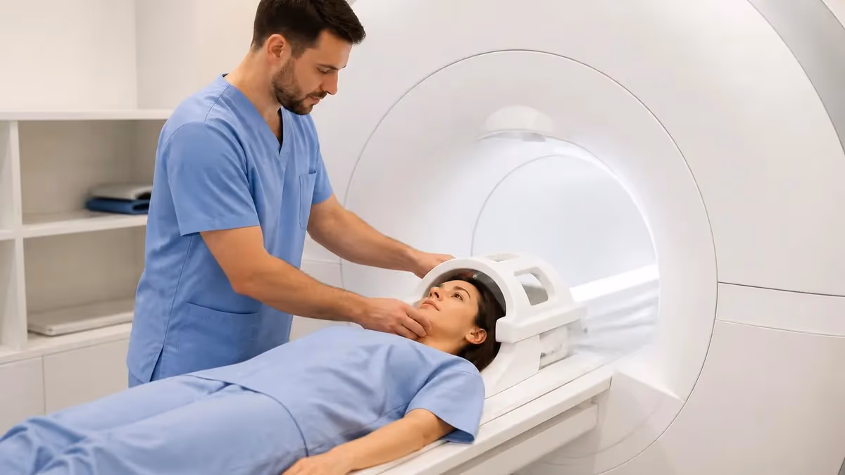
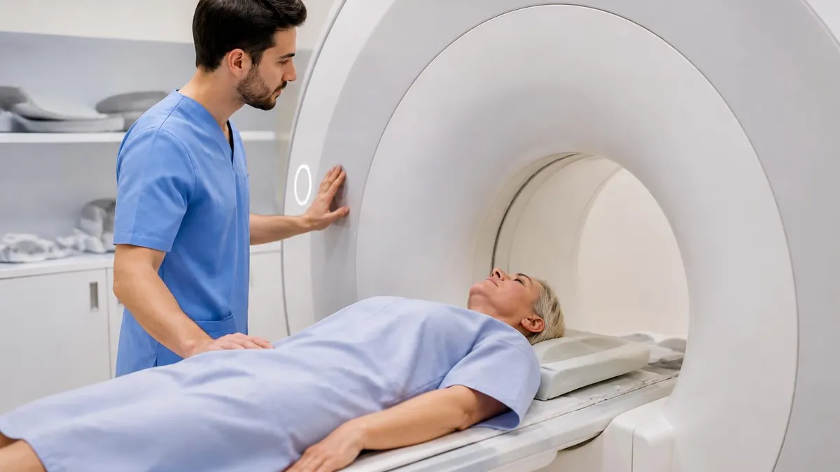
If you are about to walk into your first MS MRI, here is what the day usually looks like. You arrive about 30 minutes early, change into a gown, and answer the metal questionnaire — pacemakers, aneurysm clips, certain implants are all dealbreakers. A nurse places a small IV in your arm if contrast is on the order.
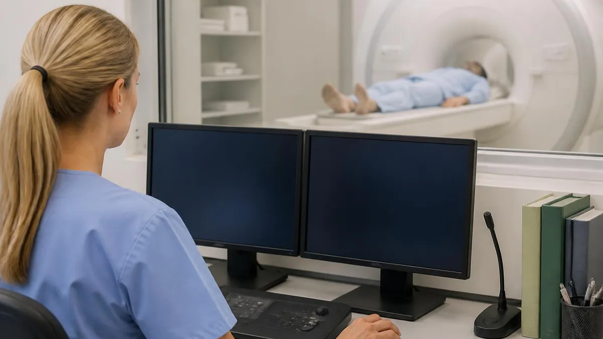
You lie on the table, get a small coil placed around your head, and slide into the bore. The scanner makes loud clanking, knocking, and buzzing sounds, which is why ear protection is mandatory. You will be still for stretches of three to seven minutes per sequence and can communicate with the tech between runs through a microphone.
Most people get through it just fine. If you are claustrophobic, ask ahead about oral anti-anxiety medication, prism glasses that let you see out of the bore, or a referral to an open or wide-bore scanner. Skipping the scan is rarely the right answer — incomplete imaging delays diagnosis, and delayed diagnosis often means delayed treatment with disease-modifying therapy.
Two practical tips: empty your bladder right before the scan, because you will be in the bore for an hour. And wear something with no metal — no underwire bras, no zippers, no metal-threaded yoga pants. The tech will hand you a gown anyway, but skipping the changeroom shuffle saves time and keeps the schedule moving smoothly for everyone else who is waiting their turn in the lobby for that same machine.
Multiple Sclerosis MRI Questions and Answers
The bottom line: a multiple sclerosis MRI is not a single test you pass or fail. It is a long-running conversation between your scans, your symptoms, and your neurologist. The first scan rules in or out MS using the McDonald 2017 criteria. The follow-up scans tell you whether your treatment is winning the long fight against new lesions.
Every report you collect along the way becomes part of a growing picture that helps your team make better decisions for the next decade of your life. Knowing what the words mean — Dawson's fingers, T2 hyperintense, enhancing, NEDA-3 — turns those reports from intimidating documents into useful tools you can actually use in conversations with your doctor.
Bring your MRI CD or imaging portal access to every visit. Ask for a copy of the radiologist's full report, not just the conclusion line. And do not be shy about scheduling a quick MRI review call with your neurologist after a fresh scan. Ten minutes of careful explanation can save weeks of anxious Googling, and your MS team would much rather walk you through the pictures than have you spiral over phrases you misread on the patient portal at midnight.
About the Author

Medical Laboratory Scientist & Clinical Certification Expert
Johns Hopkins UniversityDr. Sandra Kim holds a PhD in Clinical Laboratory Science from Johns Hopkins University and is certified as a Medical Technologist (MT) and Medical Laboratory Scientist (MLS) through ASCP. With 16 years of clinical laboratory experience spanning hematology, microbiology, and molecular diagnostics, she prepares candidates for ASCP board exams, MLT, MLS, and specialist certification tests.
Join the Discussion
Connect with other students preparing for this exam. Share tips, ask questions, and get advice from people who have been there.
View discussion (6 replies)