Brain MRI Scan: What It Shows, Sequences and Preparation Guide 2026 July
Brain MRI scan — 🗨️ what it shows, common indications, key sequences (T1, T2, FLAIR, DWI), gadolinium contrast use and what to expect during the exam.
What a Brain MRI Scan Actually Shows
A brain MRI scan is the imaging study clinicians order when they need a detailed look at the soft tissues inside the skull. Unlike CT, which uses ionizing radiation and is fast but limited in soft-tissue contrast, MRI uses powerful magnetic fields and radiofrequency pulses to map the hydrogen atoms inside the brain at sub-millimetre resolution. The result is a series of cross-sectional images that show gray matter, white matter, the ventricular system, the cerebrospinal fluid spaces, blood vessels, cranial nerves and the meninges with a clarity that no other non-invasive technique can match.
What makes brain MRI uniquely useful is that the same scanner can show several different physical properties of the same tissue by varying the pulse sequence. A T1-weighted image highlights anatomy and fat. A T2-weighted image highlights water and edema. A FLAIR sequence suppresses cerebrospinal fluid so that lesions adjacent to the ventricles become much easier to see. Diffusion-weighted imaging detects acute stroke within minutes of onset. The neuroradiologist reads the same anatomy across multiple sequences and triangulates the diagnosis from how the tissue behaves on each.
Brain MRI is also unique in its ability to characterise tissue abnormalities at a microstructural level that no other technique can reach. Diffusion tensor imaging can map white matter tracts, MR spectroscopy can measure tissue chemistry, perfusion-weighted imaging can quantify blood flow, and functional MRI can localise areas active during specific tasks. Most clinical brain MRI exams do not include these advanced sequences, but they are available at most academic centres for specific clinical questions where conventional imaging falls short.
Brain MRI at a glance
Typical scan length: 30–60 minutes for a full brain protocol. No ionizing radiation. Spatial resolution: 0.5–1 mm. Common sequences: T1, T2, FLAIR, DWI, GRE/SWI, MRA. Gadolinium contrast added when tumour, infection or active demyelination is suspected. Most brain MRIs are reported within 24–48 hours, faster for acute stroke or trauma protocols.
When Doctors Order a Brain MRI
Brain MRI is the workhorse for any neurological complaint where soft-tissue detail matters. Unexplained chronic headaches, especially with red-flag features like new onset after age 50, sudden severe onset, neurological deficit or systemic symptoms, prompt imaging to exclude tumours, vascular malformations or aneurysms. New-onset seizures in adults are imaged routinely to look for cortical malformations, scars, tumours or vascular causes that may explain the seizure focus. A first episode of focal neurological symptoms — numbness, weakness, slurred speech, vision change — usually triggers urgent imaging to differentiate stroke, demyelination and mass effect.
Multiple sclerosis evaluation is one of the most common indications for brain MRI in younger adults. The diagnosis depends on demonstrating lesions disseminated in space and time, and MRI is currently the only practical way to identify those lesions early. Dementia evaluation uses MRI to exclude reversible causes — chronic subdural haematomas, normal pressure hydrocephalus, intracranial tumours — and to look for the regional atrophy patterns that support specific diagnoses like Alzheimer's disease, frontotemporal dementia and posterior cortical atrophy.
Trauma is the one indication where CT comes first and MRI follows. CT is faster and more sensitive for acute haemorrhage and skull fractures. MRI is added later when CT is unrevealing despite persistent symptoms, when diffuse axonal injury is suspected, or when a brainstem lesion is the working hypothesis. The two modalities are complementary rather than competitive in trauma, with imaging protocols specifying when each is appropriate.
Pituitary disorders are another common indication. Hormone abnormalities discovered through endocrine testing — high prolactin, growth hormone excess, Cushing's disease, central diabetes insipidus — almost always trigger a dedicated pituitary MRI with thin-slice sequences and dynamic contrast imaging. The pituitary protocol differs from a routine brain protocol because the gland is small and sits within a cavity that creates challenging imaging artifacts. Specialist neuroradiologists usually read pituitary MRIs because the differential diagnosis hinges on small differences in enhancement patterns.
Common Reasons for a Brain MRI
DWI sequence detects ischaemic stroke within minutes of onset. Identifies the vascular territory affected, evaluates for haemorrhagic transformation, and guides decisions about thrombectomy and antiplatelet therapy. MRA adds vessel imaging.
Detects gliomas, meningiomas, metastases, schwannomas and pituitary lesions. Gadolinium contrast highlights blood-brain-barrier disruption typical of high-grade tumours and active inflammation. Used for diagnosis and post-treatment surveillance.
Detects demyelinating plaques, particularly periventricular, juxtacortical, infratentorial and spinal cord lesions. Tracks lesion burden over time. Guides decisions about disease-modifying therapy.
Identifies cortical malformations, mesial temporal sclerosis, cavernous malformations and gliotic scars that act as seizure foci. Often follows a first seizure to define the underlying cause.
Investigates new headaches with concerning features. Looks for tumours, aneurysms, dural sinus thrombosis, hydrocephalus and Chiari malformation. Most chronic uncomplicated headaches do not need imaging.
Excludes reversible causes such as chronic subdural haematomas, hydrocephalus and tumours. Identifies regional atrophy patterns that support specific dementia diagnoses. Often paired with FDG-PET when available.
The Sequences That Make Up a Brain MRI Protocol
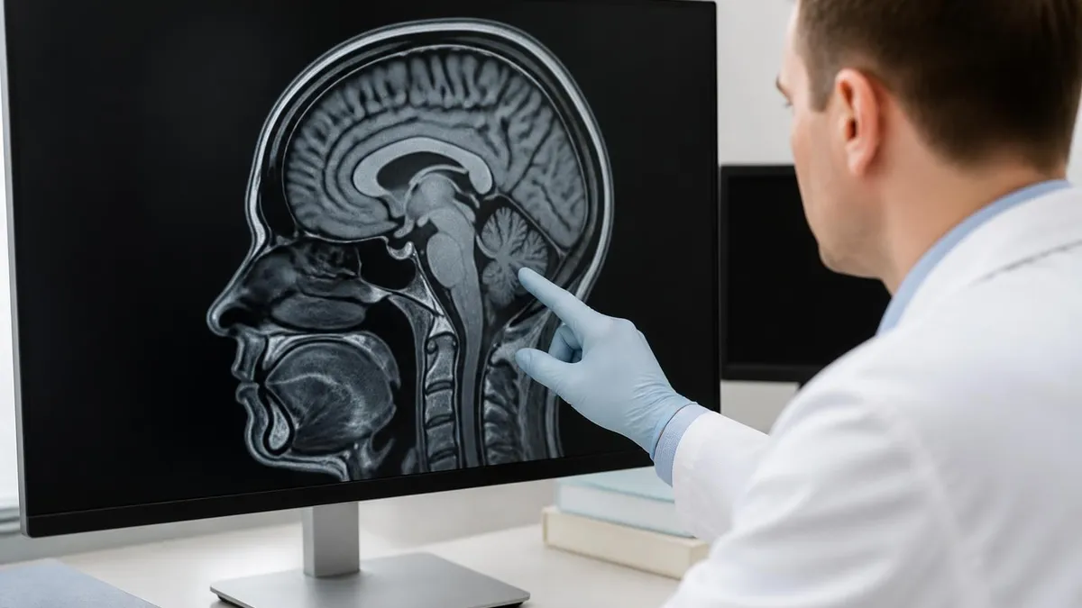
Each sequence in a brain MRI exam answers a different clinical question. T1-weighted images are the anatomical reference, showing gray and white matter contrast in their natural relationship. Fat appears bright, water appears dark. T1 is excellent for assessing volume loss, identifying anatomical landmarks and differentiating gray from white matter on a single image. T2-weighted images flip the contrast — water appears bright, making cerebrospinal fluid, oedema and many pathological lesions stand out from surrounding tissue. T2 is the workhorse for detecting most non-acute brain pathology.
FLAIR (Fluid-Attenuated Inversion Recovery) is essentially T2 with the cerebrospinal fluid signal suppressed. The result is that periventricular lesions, which would otherwise be hidden against the bright CSF on T2, become clearly visible. FLAIR is the single most sensitive sequence for detecting white matter disease, including multiple sclerosis plaques, small vessel ischaemic changes and post-traumatic gliosis. Without FLAIR, many lesions adjacent to the ventricles or sulci would be missed.
Diffusion-weighted imaging measures the random motion of water molecules. In acute stroke, cellular swelling restricts water diffusion within minutes of vessel occlusion, producing a characteristic bright signal on DWI long before any other sequence shows the lesion. DWI is the most time-critical sequence in any acute neurological presentation, and most stroke protocols start with it. Gradient echo (GRE) and susceptibility-weighted imaging (SWI) detect blood products and microbleeds that the other sequences miss — essential for evaluating cerebral amyloid angiopathy, traumatic shear injury and small haemorrhages.
Modern protocols also include three-dimensional volumetric acquisitions that allow the neuroradiologist to reformat the images in any plane after the scan is complete. A 3D T1 MPRAGE sequence acquired at 1 mm isotropic resolution can be sliced sagittally, axially or coronally without losing detail. The same dataset feeds automated volumetric software that quantifies hippocampal and cortical volumes for dementia evaluation, providing percentile comparisons against age-matched normal data.
Key Brain MRI Sequences
Anatomical reference sequence. Fat is bright, water is dark. Shows gray and white matter contrast clearly. Used for volumetric assessment, lesion localization and post-contrast imaging. Often acquired in three planes for surgical planning.
With Contrast or Without?
Many brain MRI exams are performed without contrast and answer the clinical question fully. Routine headache evaluation, stroke workup, MS surveillance in stable patients and basic anatomy assessment do not require contrast. Adding gadolinium-based contrast extends the diagnostic information by showing where the blood-brain barrier is disrupted — which happens in tumours, infections, active demyelinating lesions, post-surgical changes and certain vascular malformations. The decision to add contrast is made by the ordering clinician and the radiologist based on the clinical question.
When contrast is given, the standard pattern is T1-weighted imaging before contrast, then a peripheral IV cannula is placed, gadolinium is administered, and T1-weighted imaging is repeated. Lesions that enhance — meaning they appear brighter on the post-contrast image compared to the pre-contrast image — point toward active disease. The pattern of enhancement carries diagnostic weight: ring enhancement suggests abscess or high-grade glioma, nodular enhancement suggests metastasis, and homogeneous enhancement is more typical of meningioma or pituitary adenoma.
The amount of gadolinium given is calibrated by body weight, with a standard dose around 0.1 mmol per kilogram. Pediatric brain MRIs use the same weight-based calculation. Reduced doses or alternative agents are used in patients with severe kidney impairment, and a few imaging centres now offer contrast-free vascular protocols for patients who cannot tolerate gadolinium at all.
Modern macrocyclic gadolinium agents have an excellent safety profile, but patients with severe kidney impairment (eGFR below 30) face a small risk of nephrogenic systemic fibrosis. Pregnant patients usually avoid gadolinium unless absolutely necessary because it crosses the placenta. Anyone with a prior gadolinium reaction should let the technologist and radiologist know before the scan begins.
What Happens During a Brain MRI
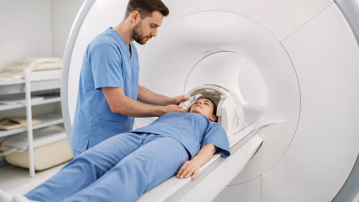
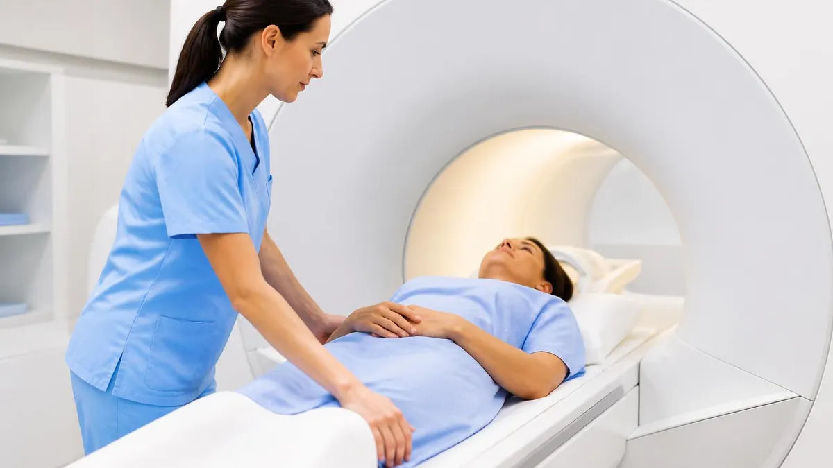
The patient lies supine on the scanner table with the head positioned inside a head coil — a helmet-like radiofrequency receiver that maximises image quality. A bolster and Velcro straps stabilise the head, because even small movements blur the images. A panic ball or call button is placed in the patient's hand. The table moves into the bore of the magnet, and the patient's head ends up roughly at the centre of the magnet's field. Earplugs or headphones are mandatory because the gradient coils generate loud knocking and buzzing during each sequence.
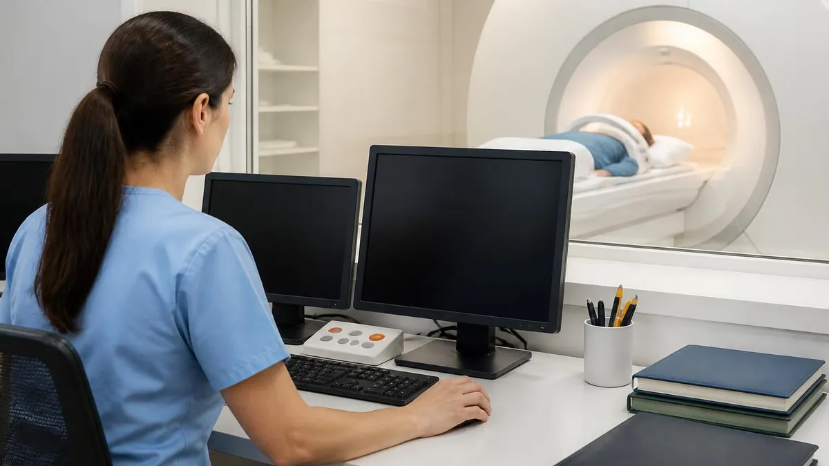
Most brain MRI exams take 30 to 60 minutes from the moment imaging begins. Each sequence runs for two to seven minutes, separated by a brief silence while the scanner reconfigures. The technologist communicates through an intercom system between sequences and reminds the patient to remain still. Many scanners now include a small mirror inside the head coil so the patient can see out of the bore toward the scan room, which reduces the sense of confinement significantly. Some centres also offer in-bore video projection for distraction during longer protocols.
Most centres play music through the headphones during the scan. Patients are usually asked to bring their own playlist on a phone or memory stick. Hearing protection is non-negotiable because peak sound levels exceed 100 decibels during gradient-intensive sequences. Children and anxious adults often benefit from extended pre-scan preparation, including a tour of the scanner room and practice lying still inside the bore.
Sedation is rare for adults but more common in young children, who cannot reliably remain still for the duration of the scan. Most paediatric centres use either oral chloral hydrate, intramuscular ketamine or general anaesthesia depending on the child's age and the scan duration. Adult sedation is reserved for severe claustrophobia and is usually accomplished with short-acting oral benzodiazepines rather than anaesthetic agents.
Brain MRI Preparation Checklist
- ✓Tell the booking team about any metal implants — pacemakers, cochlear implants, aneurysm clips, neurostimulators
- ✓Mention previous metal injuries from welding, gunshots or shrapnel — orbital X-ray screening may be needed
- ✓Disclose pregnancy or possible pregnancy at the time of scheduling
- ✓Tell the radiology team about kidney problems if contrast is ordered
- ✓Wear loose clothing without metal zips or fasteners — gowns provided if needed
- ✓Remove jewellery, hair accessories with metal, hearing aids and dentures
- ✓Empty pockets of phones, keys, coins, wallets and bank cards
- ✓Arrive 30 minutes early to complete the safety questionnaire
- ✓Arrange a driver afterward only if sedation has been planned
- ✓If claustrophobic, request prescribed anxiolytic or wide-bore scanner before the appointment
Claustrophobia and Comfort Options
Brain MRI is the body part where claustrophobia matters most because the head sits at the centre of the magnet bore, surrounded by the coil. Several practical options ease the experience. Wide-bore scanners (70 cm aperture compared to 60 cm conventional) feel substantially more open. Open-bore scanners with C-shaped designs eliminate the tunnel sensation entirely, although image quality is generally lower because field strength is reduced. Some imaging centres offer 3T scanners with shorter bore lengths, so the patient's head emerges from the magnet sooner during head imaging.
Pharmacological options include short-acting oral benzodiazepines like lorazepam, prescribed by the ordering clinician for the morning of the scan. The medication should be taken with a driver arranged for after the appointment. Severe claustrophobia sometimes warrants conscious sedation administered on the day with monitoring by an anaesthesia team, although this is usually reserved for cases where prior attempts have failed. Discussing comfort needs at the time of booking is far more effective than mentioning them at check-in, because rescheduling for sedation often takes additional weeks.
Comfort enhancements are sometimes the difference between a successful and an aborted scan. Eye masks, a thin blanket, foam wedges to support the legs, and a clear understanding of how long each sequence will last all reduce panic. Some centres run a feet-first protocol when clinically possible — entering the scanner feet-first means the head emerges from the magnet first as the scan ends, which feels less confining than head-first entry.
Reading the Results
Brain MRI reports follow a structured format. The header lists the indication and the sequences performed. The findings section describes the brain parenchyma, ventricles, extra-axial spaces, vascular structures and skull base in turn. Each region is described as normal or abnormal with specifics of any pathology. The impression section is the radiologist's conclusion in plain language — typically one or two sentences synthesising the findings and suggesting differential diagnoses or follow-up. Most reports end with a recommendation for further imaging, clinical correlation or simply no follow-up required.
Patients can find normal-variant findings in their reports — small T2 hyperintensities described as nonspecific, small developmental venous anomalies, mucosal thickening of the paranasal sinuses incidentally noted, small pineal cysts. These often appear on the report because the radiologist is meticulous about describing every observation, but they do not always indicate clinically significant disease. The clinician who ordered the scan is the right person to interpret the report's significance to the specific patient. Most centres now offer a patient portal showing the report alongside images, which is helpful for understanding but can also generate anxiety when terminology is unfamiliar.
For complex findings the report often includes recommendations to compare against a prior MRI. Stable findings are far less concerning than ones that have grown or changed. Bringing prior imaging to the appointment, or confirming that the previous scans are accessible to the radiologist, materially improves the report quality. Many institutions can now query each other's electronic systems for prior images, but cross-system access still depends on patient consent and proper paperwork.
Brain MRI Numbers
Conditions Brain MRI Helps Diagnose
DWI lights up affected brain tissue within minutes. Identifies the vascular territory and determines whether the patient is a candidate for thrombolysis or thrombectomy.
Primary gliomas, meningiomas, schwannomas and metastases are characterised by location, signal pattern and contrast enhancement. Surgical and radiation planning depends on MRI.
Demyelinating lesions in characteristic locations meet the dissemination-in-space criterion. Repeat scans demonstrate dissemination in time. Foundation of the McDonald criteria.
MRA shows cerebral arteries without contrast. Detects unruptured aneurysms larger than 3–5 mm and arteriovenous malformations. Sensitivity varies with size and location.
Excludes reversible causes and identifies atrophy patterns. Hippocampal volume loss supports Alzheimer's; frontotemporal atrophy points to FTD; posterior atrophy to PCA variants.
Diffuse axonal injury, cortical contusions, traumatic microbleeds and chronic gliosis after head trauma. Adds detail beyond what acute CT shows in the days and weeks afterward.
Limits of Brain MRI
Brain MRI is not infallible. Some pathologies are missed because the lesion is too small, the sequence is not optimised for the type of pathology, or the patient moved during acquisition. Subtle cortical malformations in epilepsy can hide between sequences, which is why dedicated epilepsy protocols use thinner slices and additional planes. Acute haemorrhage in the very first hours is sometimes better detected on CT than on standard MRI, although GRE and SWI sequences narrow that gap. Image artifacts from dental implants, metallic fragments and even hair products with iron oxide pigments can degrade image quality.
Cost and access are real-world limits. A brain MRI in the United States typically lists at $1,000 to $3,500 depending on the facility and whether contrast is used. Insurance coverage usually requires prior authorization for non-urgent scans, which can delay scheduling by days to weeks. In many parts of the world, MRI access is rationed by waiting lists rather than cost. Knowing which clinical question genuinely requires MRI — versus a question CT could answer faster and cheaper — is part of good ordering practice.
Patient cooperation is the silent variable in many brain MRIs. Even small involuntary movements blur the images, and tremor, restless legs, dystonia, anxiety or pain can all degrade quality enough that sequences must be repeated. Practising stillness for several minutes before arriving, taking pain medication an hour before the scan if pain is an issue, and emptying the bladder immediately before lying down are all simple measures that improve the final image quality without changing the scanner or the protocol.
Brain MRI: Strengths and Limits
- +Highest soft-tissue contrast of any non-invasive brain imaging modality
- +No ionizing radiation — safe for repeated scans over time
- +Multiple sequences answer different clinical questions in one session
- +Detects acute stroke within minutes via DWI
- +Vascular imaging without contrast available through MRA
- −Long scan time (30–60 minutes) requires patient cooperation
- −Claustrophobia can prevent completion in 5–10% of patients
- −Implanted metal devices may be incompatible — careful safety screening required
- −Higher cost than CT; insurance preauthorization often needed
- −Worse than CT for acute haemorrhage in the first hours after onset
MRI Questions and Answers
About the Author
Medical Laboratory Scientist & Clinical Certification Expert
Johns Hopkins UniversityDr. Sandra Kim holds a PhD in Clinical Laboratory Science from Johns Hopkins University and is certified as a Medical Technologist (MT) and Medical Laboratory Scientist (MLS) through ASCP. With 16 years of clinical laboratory experience spanning hematology, microbiology, and molecular diagnostics, she prepares candidates for ASCP board exams, MLT, MLS, and specialist certification tests.
Join the Discussion
Connect with other students preparing for this exam. Share tips, ask questions, and get advice from people who have been there.
View discussion (6 replies)