Difference Between CT Scan and MRI: Key Comparisons Explained 2026 July
Difference between CT scan and MRI: 🏆 technology, uses, cost, time, safety, and when each imaging method is preferred for specific medical conditions.
CT (Computed Tomography) scans and MRI (Magnetic Resonance Imaging) are both major medical imaging technologies but use fundamentally different methods producing different strengths and applications. CT uses X-rays creating cross-sectional images quickly, particularly useful for bone, acute trauma, and emergency situations. MRI uses magnetic fields and radio waves producing detailed soft tissue images without radiation but requiring longer scan times. Whether you're patient comparing imaging options, considering imaging technology career, or simply curious about medical imaging, understanding CT vs MRI differences helps appreciate when each modality is preferred.
For CT vs MRI specifically, several key differences matter. Technology: CT uses X-rays, MRI uses magnetic field plus radio waves. Speed: CT typically 5-15 minutes, MRI typically 20-90 minutes. Best uses: CT for bone/trauma/emergency, MRI for soft tissue/brain/spine/joints. Radiation: CT uses ionizing radiation, MRI uses no radiation. Cost: CT typically $300-$2,000, MRI typically $1,000-$3,000. Each difference affects appropriate use. Quality understanding helps interpret why physicians order specific modality.
For complementary use specifically, CT and MRI sometimes used together for comprehensive evaluation. CT may be initial study with MRI follow-up for soft tissue detail. Both modalities sometimes ordered for complex cases. Specific clinical questions may benefit from both. Each modality contributes specific information. Quality complementary use provides comprehensive imaging diagnostic information beyond either modality alone.
This guide covers CT vs MRI differences comprehensively: technology basics, comparative strengths, when each is used, cost differences, safety considerations, and how physicians choose between them. Whether you're researching for medical decision context or general understanding, you'll find practical context here.
CT scan: X-rays, fast (5-15 min), best for bone/trauma/emergency
MRI: Magnetic field + radio waves, slower (20-90 min), best for soft tissue
Radiation: CT uses ionizing radiation, MRI uses none
Cost: CT $300-$2,000, MRI $1,000-$3,000
Contraindications: CT few, MRI many (implants, claustrophobia)
For specific CT scan technology specifically, CT uses X-rays from rotating source around patient. Multiple X-ray projections from different angles. Computer reconstructs cross-sectional images from projections. Modern CT scanners produce detailed 3D images. Specific contrast agents enhance visualization. Each CT element supports rapid imaging. Quality CT technology produces detailed images quickly making it preferred for emergency situations requiring rapid diagnosis.
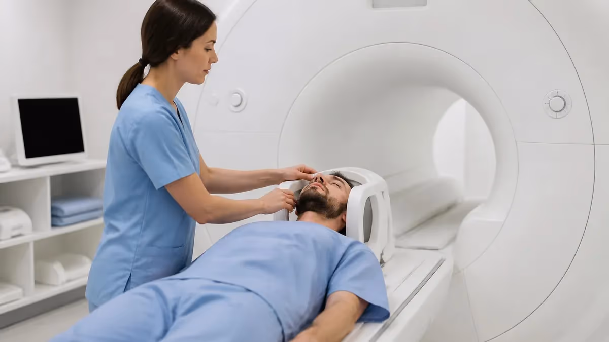
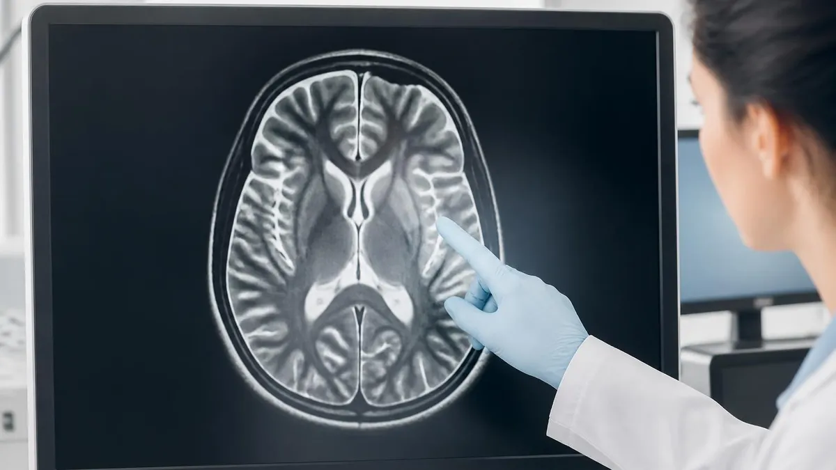
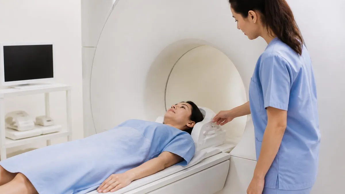
For specific MRI technology specifically, MRI uses powerful magnetic field aligning hydrogen atoms in body. Radio frequency pulses tip atom alignment. Atoms emit signals returning to alignment. Computer processes signals into images. Multiple imaging sequences highlight different tissue types. Each MRI element supports detailed soft tissue imaging. Quality MRI technology provides superior soft tissue detail though requires longer imaging time. The magnetic resonance imaging guide covers MRI in depth.
For specific best CT uses specifically, CT preferred for several clinical situations. Acute trauma evaluation (head injuries, abdominal trauma). Suspected bleeding (acute stroke, internal bleeding). Bone evaluation (fractures, bone disease). Lung imaging. Specific other situations requiring fast detailed imaging. Each preferred CT use leverages CT speed and bone/blood visualization. Quality CT use for these situations substantially better than MRI which may take too long for emergencies.
For specific best MRI uses specifically, MRI preferred for several clinical situations. Brain and spine imaging (excellent soft tissue detail). Joint imaging (ligaments, cartilage). Pelvic imaging. Cardiac imaging. Specific situations requiring detailed soft tissue evaluation. Each preferred MRI use leverages MRI superior soft tissue contrast. Quality MRI use for these situations substantially better than CT which provides less detailed soft tissue visualization. The MRI vs CT scan guide covers comparison details.
For specific overlap situations specifically, both CT and MRI can image many regions. Choice depends on specific clinical question. CT may be initial study followed by MRI for additional detail. Both provide cross-sectional imaging. Specific clinical context affects modality choice. Each overlap situation requires physician judgment. Quality modality choice considers clinical urgency, specific tissue type, patient factors, cost.
CT vs MRI Key Differences
CT uses X-rays from rotating source. MRI uses powerful magnetic field plus radio waves. Different physics produces different strengths and limitations for different tissue types and clinical situations.
CT typically 5-15 minutes total. MRI typically 20-90 minutes. CT speed advantage substantial for emergencies. MRI longer time enables more detailed multi-sequence imaging.
CT uses ionizing radiation (X-rays) creating cumulative exposure concerns for repeated scans. MRI uses no ionizing radiation (just magnetic field and radio waves) safer for repeated imaging.
CT typically $300-$2,000 in U.S. MRI typically $1,000-$3,000. MRI substantially more expensive. Cost factor sometimes affects modality selection in non-urgent situations.
For specific safety comparison specifically, both modalities have specific safety considerations. CT radiation cumulative concern for repeated scans particularly in younger patients. MRI safety concerns include strong magnetic field interactions with implants and metal objects. CT contrast (iodine-based) has allergic reaction risk. MRI contrast (gadolinium-based) small risk in patients with severe kidney disease. Each safety consideration affects appropriate patient selection. Quality safety screening before either modality essential. The MRI safety guide covers MRI safety details.
For specific contraindications specifically, MRI has more contraindications than CT. MRI contraindicated for some metallic implants, certain pacemakers, some other devices. CT generally has few contraindications beyond contrast considerations. Specific patient factors affect modality eligibility. Each contraindication affects patient selection. Quality understanding of contraindications helps physicians choose appropriate modality for specific patients without preventable safety issues.
For specific contrast comparison specifically, both modalities use contrast agents in some scans. CT contrast: iodine-based, intravenous, enhances vessel and organ visualization, allergic reaction possible. MRI contrast: gadolinium-based, intravenous, enhances soft tissue and vessel visualization, kidney function consideration. Specific contrast use depends on clinical question. Each contrast type has specific applications and risks. Quality contrast use enhances diagnostic capability when warranted by specific clinical questions.
For specific radiation comparison specifically, CT radiation exposure varies by scan type. Single CT scan typically equivalent to several months to years of background radiation. Cumulative exposure over multiple CT scans increases concern. Some patients require multiple repeated CT scans. MRI no radiation exposure. Specific radiation comparison favors MRI for repeat imaging when possible. Each radiation consideration affects long-term patient health.
For specific patient experience specifically, patient experiences differ between modalities. CT scan: brief lying still, faster procedure. MRI scan: longer lying still, loud noise, confined space. Both painless procedures. CT typically less anxiety-provoking. MRI may cause claustrophobia. Specific patient comfort varies by modality. Each experience element affects patient satisfaction. Quality understanding helps patients prepare appropriately for chosen modality.
Best Uses Comparison
CT scan ideal applications:
- Acute trauma: Head injuries, abdominal injuries, complex fractures
- Emergency situations: Suspected bleeding, acute stroke evaluation
- Bone evaluation: Complex fractures, bone disease
- Lung imaging: Pulmonary embolism, lung cancer
- Quick screening: Various conditions requiring rapid evaluation
For specific scan time comparison specifically, time differences substantial between modalities. CT scan: 5-15 minutes typical (sometimes faster for emergencies). MRI scan: 20-90 minutes depending on body part and sequences. Time difference critical for emergencies where minutes matter. Specific multi-sequence MRI takes longer than single-sequence CT. Each time difference affects clinical workflow. Quality time understanding helps appreciate why CT preferred for emergencies despite MRI's other advantages.
For specific cost analysis specifically, costs vary substantially between modalities. CT scan: $300-$2,000 typical in U.S. MRI scan: $1,000-$3,000 typical. Insurance coverage varies for both. Specific facility differences (hospital vs imaging center). Each cost factor affects patient out-of-pocket expense. Quality cost awareness helps patients understand financial implications and discuss alternatives with physicians when appropriate. The MRI cost guide covers detailed MRI pricing.
For specific availability specifically, CT scanners more widely available than MRI. Most hospitals have CT capability. MRI scanners less common particularly in smaller hospitals or rural areas. Specific availability affects access. Each availability factor affects scheduling timeframes. Quality availability consideration sometimes affects modality choice particularly when MRI scheduling delays would compromise care timing.
For specific image detail specifically, image detail differs between modalities. CT excellent for bone, acute blood, lung, calcifications. MRI excellent for soft tissue, brain, spinal cord, joints, blood vessels. Specific tissue types better visualized by specific modality. Each modality provides different visualization capability. Quality understanding helps appreciate why specific modality recommended for specific conditions.
For specific physician selection criteria specifically, physicians choose modality based on multiple factors. Specific clinical question being asked. Patient factors (age, kidney function, implants, claustrophobia). Urgency of needed information. Cost considerations. Specific availability. Each criterion affects choice. Quality physician modality selection through systematic consideration of these factors produces appropriate imaging for specific situations rather than reflexive single-modality use.
Physicians order specific imaging modalities based on detailed knowledge of clinical question, patient factors, and modality capabilities. While general knowledge of CT vs MRI helpful for understanding, physicians make informed decisions about which modality serves specific situations. If wondering why CT chosen instead of MRI (or vice versa), ask physician explaining clinical reasoning rather than questioning choice without specific information about your situation. Sometimes both modalities ordered for comprehensive evaluation when each provides complementary information. Quality patient-physician communication about imaging choices produces better patient understanding without second-guessing specialist clinical judgment based on limited general knowledge.
For specific specific conditions specifically, certain conditions strongly favor one modality. Acute stroke: initially CT (rule out bleeding) then MRI for ischemic detail. Suspected fracture: CT excellent for complex fractures, X-ray for simple fractures. Brain tumor: MRI substantially better than CT. Lung cancer: CT primary modality. Specific conditions have established imaging protocols. Quality protocol-based imaging reflects accumulated clinical evidence about best modality for specific conditions.
For specific pediatric considerations specifically, pediatric imaging considerations favor minimizing radiation. MRI preferred when possible to avoid CT radiation in children. CT used when MRI not feasible (need for speed, MRI contraindication). Specific pediatric protocols minimize radiation exposure when CT necessary. Each pediatric consideration emphasizes radiation minimization. Quality pediatric imaging strategy balances clinical needs with radiation minimization through MRI preference when feasible.
For specific emergency department use specifically, emergency departments rely heavily on CT given speed advantage. Trauma evaluation. Suspected stroke. Acute abdominal pain. Specific emergency situations requiring rapid diagnosis. Each emergency context favors CT for speed. Quality emergency CT use produces rapid diagnostic information enabling time-critical treatment decisions impossible to wait for slower MRI scanning.
For specific outpatient imaging specifically, outpatient imaging more often allows MRI choice. Less time pressure than emergencies. Specific outpatient conditions often warrant MRI superior soft tissue detail. CT used in outpatient when specific advantages favor it. Each outpatient context affects modality choice. Quality outpatient modality choice can prioritize image quality over speed compared to emergency situations.
For specific advances specifically, both technologies continue advancing. CT: lower radiation doses, faster scanning, better image quality. MRI: higher field strengths, faster sequences, AI enhancements. Specific technological advances expand capabilities. Each advance affects relative comparison. Quality understanding evolves as technologies advance — comparisons valid today may shift as both technologies continue improving in coming years.
Imaging Modality Considerations
- ✓Trust physician's modality choice based on clinical knowledge
- ✓Discuss any specific concerns (radiation, claustrophobia, cost) with physician
- ✓Disclose all implants and medical history before either CT or MRI
- ✓Understand both procedures painless and routine medical imaging
- ✓Ask physician to explain modality choice if curious about reasoning
For specific CT alternatives specifically, several alternatives to CT exist for various uses. Ultrasound for abdominal and obstetric imaging. X-ray for simple fractures and chest imaging. MRI for soft tissue when CT contraindicated. Specific alternatives serve different uses. Each alternative has specific applications. Quality understanding of alternatives helps patients comprehend why specific modality chosen over alternatives in their situation.
For specific MRI alternatives specifically, alternatives to MRI exist for various uses. CT for many situations when MRI contraindicated. Ultrasound for some soft tissue imaging. PET scan for metabolic imaging. Specific alternatives serve different uses. Each alternative has specific applications. Quality understanding of alternatives helps patients understand imaging options available for their specific clinical situations.
For specific imaging interpretation specifically, both CT and MRI images interpreted by radiologists. Specific subspecialty radiologists for complex cases (neuroradiology, musculoskeletal, etc.). Specific reports communicated to ordering physician. Patient receives results from physician typically. Each interpretation step requires expertise. Quality radiologist interpretation essential for both modalities — specific expertise particularly important for complex cases.
For specific second opinions specifically, complex imaging cases sometimes warrant second opinion. Original images sent to additional radiologist for review. Sometimes different interpretation possible. Specific situations where second opinion valuable. Each second opinion provides independent assessment. Quality second opinion option available particularly for cancer staging or other consequential diagnoses where interpretation accuracy critical for treatment decisions.
For specific patient preparation specifically, both modalities have specific preparation requirements. CT contrast: typically NPO (nothing by mouth) several hours before. MRI: no eating restriction usually unless contrast used. Specific preparation per facility. Each preparation element affects scan quality. Quality preparation following facility instructions produces optimal imaging without procedure delays from inadequate preparation.
For specific PET-CT scans specifically, hybrid PET-CT scanners combine PET (Positron Emission Tomography) with CT. PET measures metabolic activity using radioactive tracers. CT provides anatomical context. Combined produces functional plus anatomical imaging. Specific applications including cancer staging. Each PET-CT element extends imaging capability beyond standalone CT or MRI. Quality PET-CT particularly valuable for cancer staging where both metabolic and anatomical information critical for treatment decisions.
For specific PET-MRI scans specifically, newer hybrid PET-MRI scanners combine PET with MRI. Reduces radiation compared to PET-CT. Provides PET functional imaging with MRI superior soft tissue detail. Specific applications evolving. Each PET-MRI element advances imaging capability. Quality PET-MRI represents emerging technology particularly valuable in oncology and neuroscience research with growing clinical applications.
For specific X-ray comparison specifically, X-ray different from both CT and MRI. X-ray: 2D images from single X-ray exposure, fast, low cost, good for simple bone evaluation. CT: 3D X-ray imaging, more detailed than X-ray. MRI: completely different technology with different applications. Specific X-ray serves different clinical needs than CT/MRI. Each modality has appropriate uses. Quality understanding helps appreciate why X-ray sometimes used instead of CT or MRI for specific simpler evaluation needs.
For specific ultrasound comparison specifically, ultrasound another distinct imaging modality. Uses sound waves rather than X-rays or magnets. Real-time imaging. Common for obstetric, abdominal, vascular imaging. No radiation. Specific applications differ from CT/MRI. Each modality has appropriate uses. Quality understanding shows imaging includes more than just CT vs MRI choice — multiple modalities each with specific strengths.
For specific imaging guideline specifically, professional medical organizations publish imaging guidelines. American College of Radiology (ACR) Appropriateness Criteria provide guidance on best modality for specific clinical situations. Specific guidelines based on accumulated evidence. Physicians follow evidence-based guidelines for modality selection. Each guideline reflects current best practice. Quality evidence-based modality selection through guidelines produces appropriate imaging consistent with current clinical evidence rather than personal preferences alone.
For specific advanced imaging specifically, several advanced imaging techniques extend basic CT and MRI. CT angiography for blood vessels. MR spectroscopy for chemical analysis. Functional MRI for brain function. Specific specialized applications beyond standard imaging. Each advanced technique serves specific needs. Quality advanced imaging substantially extends diagnostic capability for specific complex clinical questions warranting specialized approach beyond standard imaging.
For specific imaging facility selection specifically, choice of imaging facility affects quality. Hospital facilities may handle complex cases better. Imaging centers often lower cost. Specific equipment generation matters (newer scanners typically better). Radiologist subspecialty expertise sometimes available. Each facility selection element affects results. Quality facility selection with appropriate equipment and radiologist expertise produces best diagnostic outcomes for specific imaging needs particularly for complex or unusual clinical situations requiring specialized clinical expertise and modern equipment.
CT vs MRI Quick Facts
Modality Selection Factors
Specific clinical question being asked drives modality choice. Brain tumor: MRI. Acute trauma: CT. Bone evaluation: CT. Soft tissue detail: MRI. Match modality to question.
Patient age (pediatric favors MRI for radiation reasons), implants (may contraindicate MRI), claustrophobia (may favor CT), kidney function (affects contrast options for both).
Emergency situations favor CT speed. Non-urgent situations allow MRI longer scan time. Time-critical decisions about treatment may favor faster CT even when MRI preferred otherwise.
CT more widely available than MRI. CT lower cost typically. Specific facility availability and patient insurance considerations sometimes affect choice when clinical equivalence.
CT vs MRI Comparison
- +CT: faster scanning ideal for emergencies
- +CT: lower cost than MRI typically
- +CT: fewer contraindications than MRI
- +MRI: superior soft tissue detail
- +MRI: no ionizing radiation
- −CT: ionizing radiation cumulative concern for repeated scans
- −CT: limited soft tissue detail compared to MRI
- −MRI: longer scan times problematic for emergencies
- −MRI: more contraindications (implants, claustrophobia)
- −MRI: higher cost than CT
MRI Questions and Answers
About the Author
Medical Laboratory Scientist & Clinical Certification Expert
Johns Hopkins UniversityDr. Sandra Kim holds a PhD in Clinical Laboratory Science from Johns Hopkins University and is certified as a Medical Technologist (MT) and Medical Laboratory Scientist (MLS) through ASCP. With 16 years of clinical laboratory experience spanning hematology, microbiology, and molecular diagnostics, she prepares candidates for ASCP board exams, MLT, MLS, and specialist certification tests.
Join the Discussion
Connect with other students preparing for this exam. Share tips, ask questions, and get advice from people who have been there.
View discussion (6 replies)