MRI Contrast: Gadolinium Agents and Patient Considerations 2026 July
MRI contrast (gadolinium) explained: 💡 how it works, when needed, safety considerations, kidney disease implications, and patient experience.
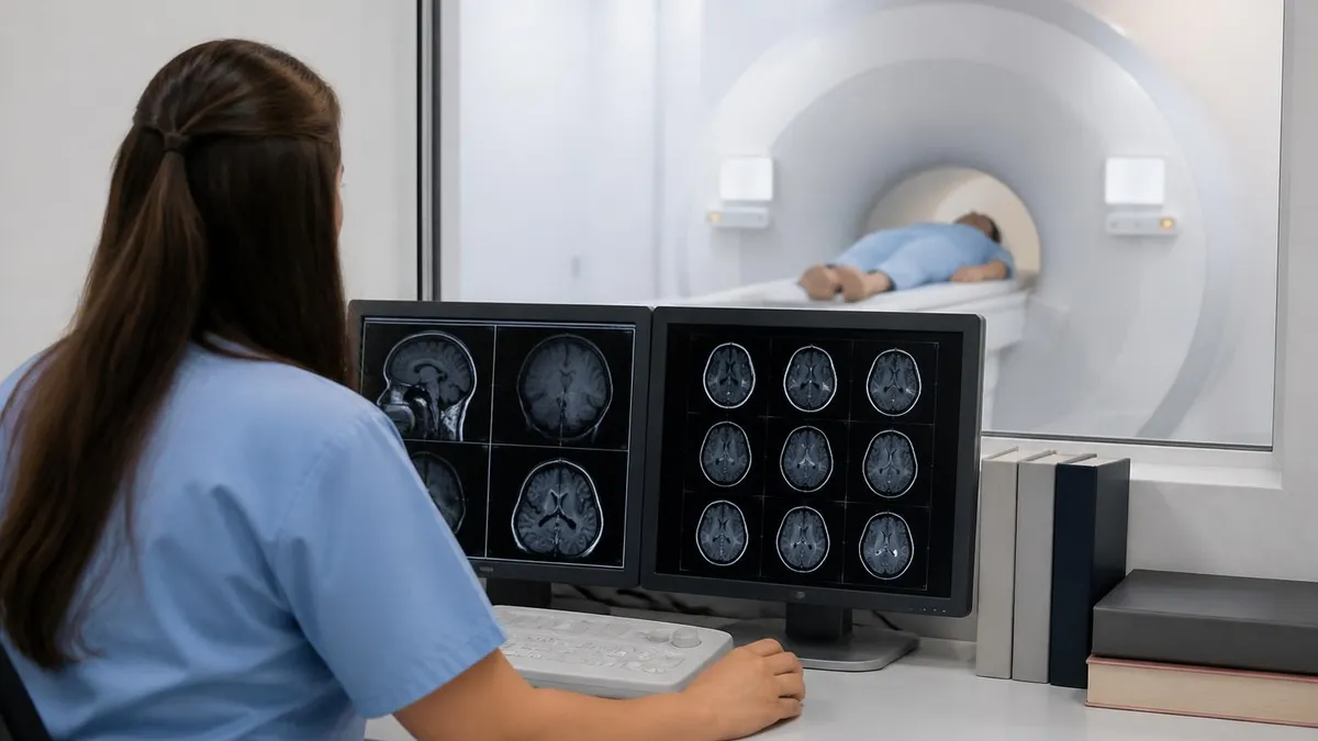
MRI contrast agents enhance the visibility of specific tissues and abnormalities during MRI examinations. The most common contrast agents used in MRI are gadolinium-based contrast agents (GBCAs), which contain the rare earth element gadolinium bound to chelating agents that allow safe administration. When injected through IV during MRI examination, gadolinium contrast accumulates in tissues with disrupted blood-brain barrier (tumors, infections, active multiple sclerosis lesions), areas of active inflammation, and various other pathological tissues, making them appear bright on T1-weighted MRI images. The enhancement provides important diagnostic information beyond what non-contrast MRI alone reveals.
Approximately 30-50% of MRI examinations include contrast injection depending on clinical indication. Some examinations require contrast for adequate diagnosis (most tumor evaluations, MS active disease assessment, vascular evaluations through MR angiography in many cases). Other examinations don't need contrast (most musculoskeletal MRI, basic spine evaluation, certain abdominal applications). The specific examination protocol determines whether contrast is needed based on clinical question being addressed. Patient factors including kidney function, prior contrast reactions, and pregnancy status affect contrast use decisions independently of basic clinical indication.
MRI Contrast Quick Facts
Type: Gadolinium-based contrast agents (GBCAs). Administration: IV injection during MRI examination. Common uses: Tumor evaluation, MS active disease assessment, infection identification, vascular evaluation. Safety: Generally well-tolerated; rare allergic reactions possible. Kidney concern: Severe kidney disease creates rare risk of nephrogenic systemic fibrosis (NSF) with older agents; newer macrocyclic agents safer. Pregnancy: Generally avoided; cross placenta. Frequency: ~30-50% of MRI examinations include contrast.
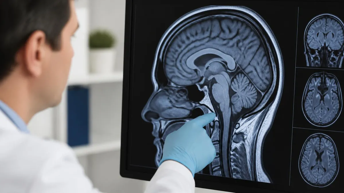
How gadolinium contrast works involves its paramagnetic properties affecting nearby water molecules. Free gadolinium ion would be toxic to tissues but chelating agents (organic molecules binding gadolinium) prevent gadolinium from interacting with body tissues directly. The chelated complex circulates through bloodstream, accumulates briefly in tissues with normal vasculature, and reaches higher concentration in tissues with disrupted blood-brain barrier or increased vascularity. The paramagnetic effect shortens T1 relaxation times causing affected tissues to appear bright on T1-weighted images. Most gadolinium clears through kidneys within 24 hours typically reaching essentially complete elimination within days for normal kidney function patients.
When MRI Contrast Is Used
Most cancer imaging includes contrast highlighting tumor vascularity. Critical for tumor characterization and treatment planning.
Gadolinium enhancement identifies active MS lesions versus inactive scars. Affects treatment decisions.
Brain abscess, spinal infection, soft tissue infection often show contrast enhancement supporting diagnosis.
Contrast-enhanced MR angiography (CE-MRA) for evaluating arteries and veins in many vascular territories.
Distinguishing post-surgical changes from residual or recurrent disease often requires contrast.
Late gadolinium enhancement identifies myocardial scar from prior heart attacks supporting cardiac evaluation.
Multiple gadolinium-based contrast agents (GBCAs) are FDA-approved for clinical use. Modern macrocyclic agents (Gadovist/Gadavist, Dotarem, ProHance) are considered most stable with lowest risk of gadolinium release in tissues. Older linear agents (Magnevist, Omniscan, OptiMARK) have been associated with greater risk of gadolinium retention and have largely been replaced or restricted in use. The macrocyclic versus linear distinction matters for safety considerations particularly for patients receiving multiple contrast administrations over time. Different agents have somewhat different imaging characteristics making them preferred for specific applications by individual radiologists and institutions.
Nephrogenic systemic fibrosis (NSF) was identified in 2006 as rare serious complication occurring in patients with severe kidney disease who received certain gadolinium agents. NSF causes thickening and hardening of skin and connective tissue with potential to cause severe disability.
The condition essentially eliminated through current safety practices: avoiding gadolinium in severe kidney disease (eGFR less than 30), preferring macrocyclic agents over linear agents particularly for any kidney impairment, careful screening for kidney function before contrast administration. Modern incidence of NSF essentially zero through these practices. Patients with mild to moderate kidney disease can usually receive contrast safely particularly when macrocyclic agents used.
Gadolinium retention concerns emerged in recent years through research showing some gadolinium remains in tissues (particularly brain) long after contrast administration. The clinical significance of this retention remains under investigation — no clinical disease has been clearly established as caused by retention. However, FDA recommends consideration of retention risk particularly for patients receiving multiple contrast administrations over lifetimes. Pediatric patients, pregnant women, and patients with chronic conditions requiring repeated MRIs face theoretical concerns about cumulative retention. Macrocyclic agents show lower retention than linear agents supporting modern preference for macrocyclic agents particularly in patients facing potential lifetime exposures across multiple examinations.
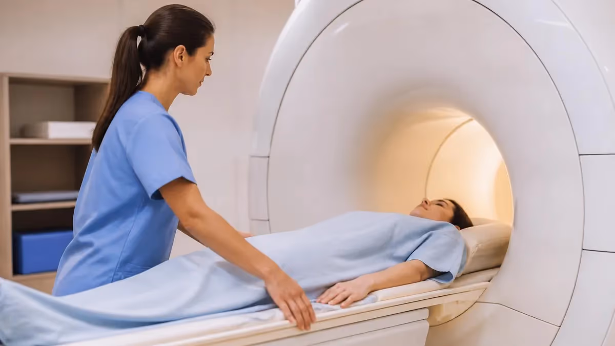
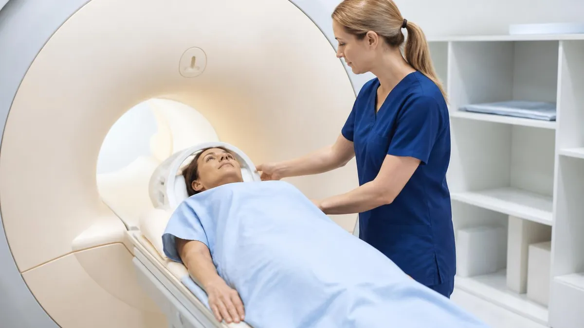
Contrast administration during MRI: IV access established before MRI examination begins (small needle inserted in arm vein). Patient enters scanner and initial non-contrast sequences performed (typically 15-20 minutes of imaging). At appropriate point during examination, contrast injected through IV (small bolus typically 10-20 mL depending on patient weight and agent). Post-contrast sequences performed (typically 5-15 minutes) capturing enhancement pattern. IV removed after examination completion. Patient generally feels brief warm sensation during injection but no other immediate effects.
Patient screening before contrast administration reduces complications. Kidney function assessment through recent eGFR (estimated glomerular filtration rate) testing within 30 days of contrast for at-risk patients. Allergy history including any prior contrast reactions, asthma, or general allergies affecting risk assessment. Current medications including any that might interact. Pregnancy status for women of childbearing age. Detailed medical history particularly focused on kidney disease, autoimmune conditions, and various other relevant factors. Each screening element supports appropriate contrast use decisions matching individual patient circumstances. Quality imaging facilities have systematic screening processes ensuring contrast safety considerations addressed before each administration.
The IV access for contrast administration involves brief needle stick before MRI examination begins. Most patients tolerate IV placement without significant difficulty. Patients with difficult venous access may need multiple attempts or use of veins in less typical locations. Anxiety about needles affects some patients — discussion with technologist about anxiety management supports successful access. The IV remains in place during entire examination (typically 30-45 minutes for standard MRI plus contrast time) for contrast injection at appropriate point in protocol. After examination, IV removed and pressure applied briefly. Most patients have minimal discomfort beyond initial needle stick.
Common patient questions about contrast include whether allergic reactions are possible (rare but possible — alert technologist immediately if symptoms develop), how long contrast stays in body (most cleared through kidneys within 24 hours), whether contrast affects breast milk (small amounts pass into breast milk but breast feeding not contraindicated per current guidelines), whether contrast can affect future MRIs (no significant effect on future imaging), and whether contrast is safe for children (yes with appropriate screening; pediatric protocols typically minimize contrast use when not essential). Patient education before examinations reduces anxiety and supports cooperative participation throughout the imaging process.
Always disclose kidney disease history — affects contrast use decisions substantially. Inform about prior contrast reactions — increases risk of subsequent reactions. Disclose pregnancy or possible pregnancy — contrast generally avoided during pregnancy. Don't skip eGFR testing if requested before contrast — kidney function screening protects safety. Inform about all medications and allergies — supports comprehensive safety assessment before contrast administration.
Adverse reactions to gadolinium contrast occur infrequently but require recognition and prompt response. Mild reactions (nausea, vomiting, hives, mild itching, headache) occur in 1-3% of administrations. Moderate reactions (more extensive hives, mild bronchospasm, vasovagal reactions) occur in less than 1%. Severe reactions (severe bronchospasm, anaphylaxis, cardiovascular collapse) very rare (less than 0.01%) but require immediate emergency response. Imaging facilities maintain emergency protocols and equipment for managing reactions. Patients with prior reactions have higher risk of subsequent reactions and may require pre-medication with steroids and antihistamines, alternative contrast agents, or non-contrast imaging alternatives.
For patients with severe contrast reactions, alternative imaging approaches may suit better. Non-contrast MRI sequences provide substantial diagnostic information without contrast for many applications. Non-contrast MRA techniques (time-of-flight, phase contrast) provide vascular imaging without gadolinium. CT scans use different contrast (iodinated) with different reaction profile — patients allergic to gadolinium may tolerate iodinated contrast and vice versa. Ultrasound provides imaging for some applications without contrast. Each alternative has different advantages and limitations. Discussion with radiologist about appropriate alternatives supports diagnostic information without contrast risk for specific patients.
For specific indications, contrast adds critical diagnostic information beyond non-contrast imaging alone. Brain tumor evaluation depends substantially on contrast enhancement patterns distinguishing tumor types, differentiating tumor from edema, and supporting treatment monitoring. MS active disease requires contrast distinguishing active inflammation from old scars. Brain abscess and infection benefit from contrast highlighting infection extent. Many vascular abnormalities benefit from contrast-enhanced MRA. Each indication uses contrast as essential diagnostic tool with non-contrast alternatives providing inferior information for specific clinical questions. Skipping contrast in these scenarios may produce incomplete diagnosis affecting clinical decisions.
Preparing for Contrast MRI
- ✓Recent eGFR test if requested (kidney function screening)
- ✓Disclose all medical history particularly kidney disease
- ✓Inform about prior contrast reactions
- ✓Disclose pregnancy or possible pregnancy
- ✓List all medications and allergies
- ✓Plan for IV access during examination
- ✓Drink water before and after examination supporting kidney clearance
- ✓Discuss any concerns with imaging facility staff before examination
For breast feeding mothers needing contrast MRI, current guidelines support continued breast feeding without interruption. Small amounts of gadolinium pass into breast milk (less than 1% of administered dose typically) with very small amount actually absorbed by infant from milk. Calculated infant exposure represents tiny fraction of pediatric clinical doses considered safe for direct administration.
American College of Radiology current guidelines state contrast administration is not contraindication to continuing breast feeding. Mothers concerned about exposure may elect to pump and discard milk for 24 hours after administration, though scientific evidence doesn't require this practice. Discussion with radiologist or pediatrician supports individual decisions based on personal preferences.
For pediatric patients, contrast use considerations differ somewhat from adults. Children's kidney function generally excellent supporting safe contrast use. Lifetime exposure considerations matter — children have decades of remaining life for any retained gadolinium. Pediatric protocols typically minimize contrast use when not essential. When contrast needed, macrocyclic agents preferred for lower retention. Sedation may be needed for younger children unable to remain still during MRI — pediatric anesthesia adds complexity but supports successful examination. The combined considerations make pediatric contrast MRI specialty area benefiting from facilities with substantial pediatric experience.
For specific clinical scenarios, contrast use decisions involve careful risk-benefit analysis. Known severe kidney disease (eGFR less than 30) requires substantial benefit to justify contrast use. Pregnancy requires substantial benefit to justify contrast given fetal exposure concerns. Prior severe contrast reactions require careful consideration with possible pre-medication or alternative approaches. Multiple prior contrast administrations raise long-term retention concerns supporting avoiding additional contrast when not essential. Each scenario requires individualized assessment rather than standard protocols. Discussion with referring physician and radiologist supports appropriate decisions matching specific patient circumstances and clinical needs.
Cost considerations for contrast MRI typically modestly higher than non-contrast MRI given contrast cost and additional injection time. Without insurance, contrast MRI costs $500-$3,500 typical depending on facility and region with contrast adding $100-$300 to non-contrast price. Most insurance plans cover contrast MRI for appropriate clinical indications. Some plans require pre-authorization particularly for advanced imaging. Cash-pay rates at outpatient centers often substantially lower than insurance-billed rates. Verify costs and coverage before scheduling. The investment is generally worthwhile when contrast provides important diagnostic information not available through non-contrast imaging alone.
For people considering whether to follow physician's recommendation for contrast MRI when offered, the answer is generally yes for appropriate clinical indications. Modern gadolinium contrast is well-tolerated by most patients. Safety screening reduces complications substantially. Diagnostic information from contrast often substantially affects clinical decisions about diagnosis and treatment. Quality facilities maintain emergency protocols supporting safe contrast administration. The combination of clinical value, generally safe profile with appropriate screening, and typical insurance coverage makes contrast MRI worthwhile component of modern diagnostic imaging when clinically indicated by physician.
For radiologists and imaging facility staff, ongoing contrast administration safety practices require attention. Quality screening processes including eGFR testing before contrast for at-risk patients. Pre-medication protocols for patients with prior reactions. Emergency response protocols and equipment maintenance. Documentation of contrast administration including agent type, dose, and any reactions. Appropriate use of macrocyclic agents preferred for safety. Continuous education about contrast safety as guidelines evolve. The combination of practices supports safe contrast use across substantial volumes of administrations annually at busy imaging facilities.
MRI Contrast Quick Stats
Common Contrast MRI Indications
Most brain tumor MRI uses contrast highlighting tumor vascularity supporting diagnosis and treatment planning.
Gadolinium identifies active MS lesions versus inactive scars affecting treatment decisions.
Brain abscess and other infections often show contrast enhancement supporting diagnosis.
Spine tumor evaluation including primary tumors and metastases benefits from contrast.
Liver tumor characterization through dynamic contrast-enhanced MRI supports diagnosis and treatment planning.
Late gadolinium enhancement identifies myocardial scar from prior heart attacks supporting cardiac evaluation.
For people with prior contrast reactions facing recommendation for additional contrast MRI, several considerations apply. Severity of prior reaction affects subsequent reaction risk — mild prior reactions don't substantially increase severe reaction risk; severe prior reactions warrant careful consideration. Pre-medication with steroids and antihistamines before contrast administration substantially reduces recurrent reaction risk. Alternative contrast agents (different chemistry) sometimes tolerated when prior agent caused reaction. Non-contrast imaging alternatives may provide adequate diagnostic information for some scenarios. Discussion with radiologist about specific prior reaction history and appropriate strategy supports informed decision-making rather than universal avoidance or universal acceptance.
Looking forward at MRI contrast evolution, several trends affect future practice. Continued macrocyclic agent dominance over linear agents reflecting safety profile differences. New contrast agents under development with various potential advantages. AI-assisted reading sometimes enables information extraction from non-contrast images that previously required contrast. Ultra-high-field MRI (7T) sometimes provides information without contrast that lower fields require contrast for. Non-contrast vascular imaging techniques continue improving providing alternatives to contrast-enhanced MRA. Each trend continues evolving contrast use practice supporting both diagnostic capability and patient safety across diverse clinical scenarios.
For ordering physicians considering whether to request contrast MRI versus non-contrast, several factors guide decision. Specific clinical question being addressed matters fundamentally — some questions answered only with contrast (most tumor evaluations, MS active disease, infection identification, vascular imaging in many cases). Other questions answered adequately without contrast (most musculoskeletal MRI, basic spine evaluation, certain abdominal applications). Patient factors including kidney function, prior reactions, and pregnancy status affect contrast use independently of clinical indication. Each consideration matters in clinical judgment about whether contrast adds sufficient diagnostic value to justify use given individual patient circumstances and clinical context.
For radiologists protocoling MRI examinations, several considerations affect contrast use decisions. Standard protocols for specific indications establish baseline contrast use patterns. Patient factors may modify standard protocols when contrast contraindicated or unnecessary. Clinical urgency may affect protocol decisions when extended evaluation impractical. Prior imaging available for comparison may reduce contrast need by providing baseline. Each protocol decision balances diagnostic information needs against contrast risks and additional time/cost. Quality protocols match contrast use to clinical situation rather than applying standard contrast use universally without consideration of individual factors.
Patient education about MRI contrast supports better examination experience and outcomes. Understanding what contrast is and what it provides helps patients accept its use when needed. Knowing what to expect during administration (brief warm sensation, no pain) reduces anxiety. Awareness of safety considerations and screening importance encourages honest disclosure of relevant medical history. Recognition of potential reactions supports prompt reporting if symptoms develop. Quality patient education before examinations supports both patient cooperation and safety through informed engagement with the examination process.
Contrast administration involves clinical care across diverse healthcare settings throughout the United States. MRI imaging is widely available for diagnostic medical care today across many medical specialties. MRI serves as foundational diagnostic tool supporting clinical decision-making for diverse patient populations encountered routinely in modern medicine across hospital, outpatient, and various specialty imaging settings throughout healthcare systems requiring sophisticated diagnostic support for patient evaluation across many clinical disciplines and various patient populations seen daily across modern medicine in clinical practice today across many global regions.
MRI Contrast: Pros and Cons
- +Provides essential diagnostic information for many indications
- +Generally well-tolerated by most patients
- +Modern macrocyclic agents very stable with minimal retention
- +Safety screening processes reduce complications substantially
- +Distinguishes active disease from inactive findings
- +Supports vascular evaluation through MR angiography
- −Rare allergic reactions including severe anaphylaxis possible
- −Kidney disease creates safety considerations
- −Pregnancy generally contraindicates contrast use
- −Long-term retention concerns under investigation
- −Modest additional cost beyond non-contrast MRI
- −IV access required for administration
MRI Questions and Answers
About the Author

Medical Laboratory Scientist & Clinical Certification Expert
Johns Hopkins UniversityDr. Sandra Kim holds a PhD in Clinical Laboratory Science from Johns Hopkins University and is certified as a Medical Technologist (MT) and Medical Laboratory Scientist (MLS) through ASCP. With 16 years of clinical laboratory experience spanning hematology, microbiology, and molecular diagnostics, she prepares candidates for ASCP board exams, MLT, MLS, and specialist certification tests.
Join the Discussion
Connect with other students preparing for this exam. Share tips, ask questions, and get advice from people who have been there.
View discussion (6 replies)