Seizure EEG: What the EEG Test Shows, How It Works, and What to Expect 2026 July
✅ Learn what a seizure EEG test detects, how long it takes, what it costs, and how to prepare. Complete guide for patients and EEG technologists.

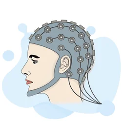
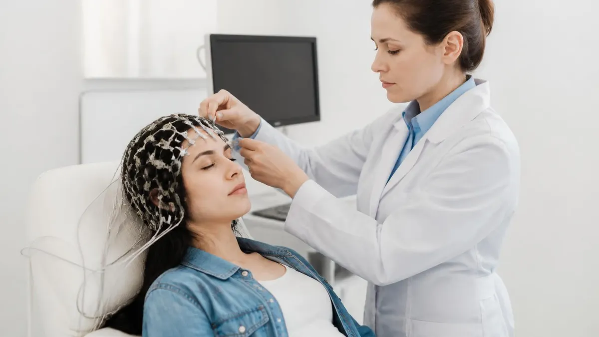
An EEG test — short for electroencephalogram — is the single most important diagnostic tool for evaluating seizures and epilepsy. When a neurologist suspects that a patient is experiencing seizure activity, the EEG test is almost always the first specialized study ordered. The test records the brain's electrical signals through small metal electrodes placed on the scalp, capturing wave patterns that can reveal abnormal discharges associated with seizure eeg activity. Understanding what this test involves helps patients prepare and helps clinicians interpret findings accurately.
The EEG medical test has been in clinical use for nearly a century, yet it remains irreplaceable in modern neurology. Unlike MRI or CT scans, which show the brain's structure, an EEG captures real-time function — specifically the millisecond-to-millisecond electrical communication between neurons. During a seizure, this communication becomes explosively synchronized in ways the EEG captures with remarkable precision. Even between seizures, the brain of someone with epilepsy often shows subtle spike-and-wave complexes that point directly to the underlying condition.
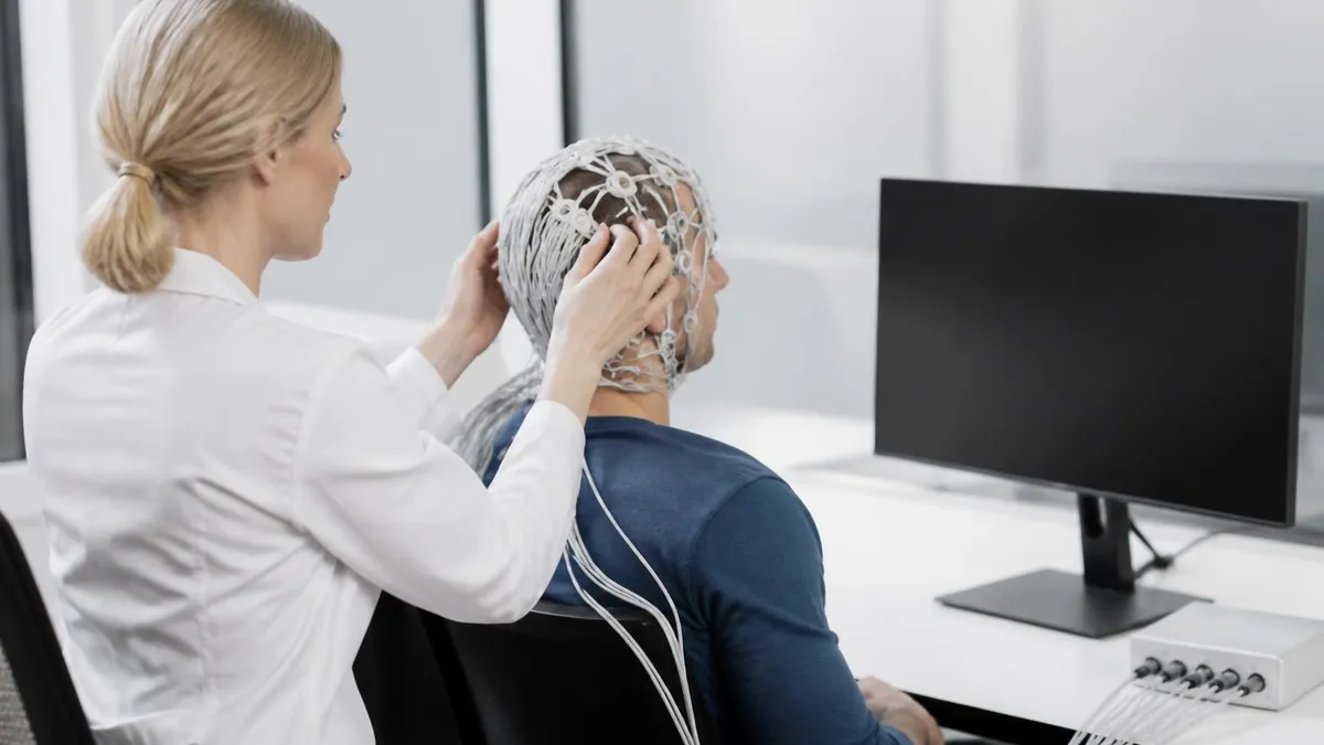
Many patients ask what is an EEG test and whether it is painful or dangerous. The short answer is that it is completely non-invasive and painless. Electrodes are attached to the scalp with a water-soluble gel or paste — nothing penetrates the skin. The machine simply listens to electrical signals your brain generates naturally. There are no electrical shocks, no radiation, and no sedating drugs required for standard recordings, though sleep-deprived studies and video-EEG monitoring may involve extended recording windows that require the patient to stay still for longer periods.
One of the most common misconceptions is that an EEG will always catch a seizure in the act. In reality, seizures are unpredictable events, and a routine 20–40 minute EEG may not record an ictal episode. However, neurologists are trained to recognize interictal abnormalities — spike discharges, sharp waves, and focal slowing that occur between seizures but still carry significant diagnostic weight. The location, frequency, and morphology of these abnormalities help classify the seizure type and guide treatment decisions about anti-seizure medications and surgical candidacy.
The EEG test cost in the United States varies depending on the facility, insurance coverage, and the type of recording performed. A routine outpatient EEG typically runs between $200 and $900 before insurance adjustments, while prolonged video-EEG monitoring in an epilepsy monitoring unit can cost several thousand dollars per day. Despite the expense, the EEG remains cost-effective because it directly informs treatment decisions that would otherwise require far more expensive trial-and-error medication adjustments or invasive evaluations.
For EEG technologists and students preparing for certification exams, mastering the patterns associated with seizure activity is a core competency. The R. EEG T. and CLTM examinations both include substantial content on epileptiform discharges, seizure classifications, and activation procedures designed to provoke abnormal activity. Recognizing the difference between a benign variant and a true epileptiform pattern is a skill that develops through systematic study and hands-on practice. Using an eeg test for seizure practice resource can significantly accelerate that learning curve.
This comprehensive guide covers everything patients and EEG professionals need to know about seizure EEG: the types of patterns seen, how the test is performed, how long it takes, what it costs, what side effects to expect, and how to interpret common findings. Whether you are a patient preparing for your first study or a technologist building clinical expertise, the information below will give you a solid foundation for understanding one of medicine's most valuable neurological tools.
EEG Test for Seizures by the Numbers

Types of EEG Tests Used to Evaluate Seizures
A 20–40 minute recording performed in an outpatient clinic or hospital department. Electrodes are applied using the 10–20 system. Activation procedures including hyperventilation and photic stimulation are typically performed. Best for initial evaluation of suspected seizure disorder.
The patient is asked to stay awake for part or all of the night before the test. Sleep deprivation lowers the seizure threshold and increases the likelihood of capturing epileptiform discharges, especially in patients with normal routine studies.
A portable recorder worn for 24–72 hours during normal daily activities. The patient keeps a diary of symptoms. Ideal for capturing events that occur infrequently or only during specific activities. Reviewed remotely by a neurophysiologist.
Inpatient admission to an epilepsy monitoring unit where continuous video and EEG are recorded simultaneously. Medications are often reduced to provoke seizures. Gold standard for seizure classification and pre-surgical evaluation. Duration ranges from days to weeks.
High-density surface EEG uses 128–256 electrodes for precise source localization. Intracranial EEG (SEEG, ECoG) places electrodes directly on or in the brain for surgical planning. Reserved for complex, medically refractory epilepsy cases.
Understanding what a seizure EEG actually looks like requires familiarity with the brain's normal electrical rhythms. At rest with eyes closed, a healthy adult brain produces alpha waves running at 8–13 Hz over the posterior regions. When alert or performing mental tasks, faster beta activity at 13–30 Hz predominates frontally. Drowsiness introduces theta waves at 4–7 Hz, and deep slow-wave sleep produces high-amplitude delta activity below 4 Hz. Any trained EEG reader begins by establishing this normal baseline before searching for abnormalities that could indicate epileptic activity.
The hallmark of epileptiform activity on an EEG is the spike-and-wave complex. A spike is a sharply contoured waveform lasting less than 70 milliseconds with an amplitude that clearly stands out from the background. It is typically followed by a slow wave of opposite polarity. The combination creates the classic spike-and-wave morphology that neurologists associate with epileptic discharges. Generalized 3 Hz spike-and-wave complexes — seen symmetrically across both hemispheres — are the defining pattern of childhood absence epilepsy, one of the most recognizable findings in all of clinical neurophysiology.
Focal epileptiform discharges tell a different story. When spikes or sharp waves appear predominantly over one region — say, the left temporal lobe — they suggest a focal seizure onset zone. Temporal lobe epilepsy, the most common form of medically refractory focal epilepsy, frequently shows anterior temporal spikes or sharp waves, sometimes with regional theta slowing in the same area. Frontal lobe epilepsy produces discharges that can spread rapidly and bilaterally, making localization more challenging. The specific topography of these discharges directly influences surgical planning when medications fail.
During an actual seizure — the ictal period — the EEG undergoes dramatic changes. A focal motor seizure might begin with low-amplitude, high-frequency activity building in one region before spreading. A generalized tonic-clonic seizure produces a characteristic sequence: the tonic phase shows high-frequency, high-amplitude recruiting rhythm, while the clonic phase shows repetitive spike-and-wave bursts that slow progressively until the seizure ends. The postictal period that follows is marked by diffuse slowing and suppression as the brain recovers from the metabolic demands of the seizure event.
Not all sharp-looking waveforms on an EEG represent true epileptiform activity. Benign variants — sometimes called normal variants with alarming morphology — can mimic epileptiform discharges and must be recognized to avoid misdiagnosis. Wicket spikes are temporal sharp transients seen in adults that resemble sharp waves but carry no pathological significance.
Small sharp spikes (SSS) appear during drowsiness as brief biphasic waveforms. The breach rhythm, seen over skull defects from prior surgery, produces high-amplitude sharp activity that reflects altered conductance rather than abnormal brain firing. Distinguishing these from true spikes requires experience and is a major focus of EEG technologist training.
Activation procedures are performed during routine EEG studies specifically to increase the yield of capturing epileptiform activity. Hyperventilation for three minutes causes cerebral vasoconstriction and can trigger absence seizures in susceptible individuals — a finding that is essentially diagnostic. Photic stimulation using a strobe light at varying frequencies tests for photoparoxysmal responses, which appear as generalized spike-and-wave bursts time-locked to the flash rate and are associated with photosensitive epilepsy syndromes. Sleep, whether natural or induced, is another powerful activator because the transition between sleep stages is particularly likely to unmask interictal discharges.
For EEG technologists studying for board certification, the ability to recognize and accurately document seizure patterns is tested extensively. Questions on the R. EEG T. examination cover ictal morphology, postictal changes, interictal abnormalities, and the classification of epilepsy syndromes based on EEG findings. Practicing with targeted question banks that focus on these patterns builds both the visual recognition skills and the clinical reasoning needed to perform well on examination day and in real clinical settings where accurate artifact rejection and pattern identification directly affect patient care decisions.
How Long Is an EEG Test? Duration by Study Type
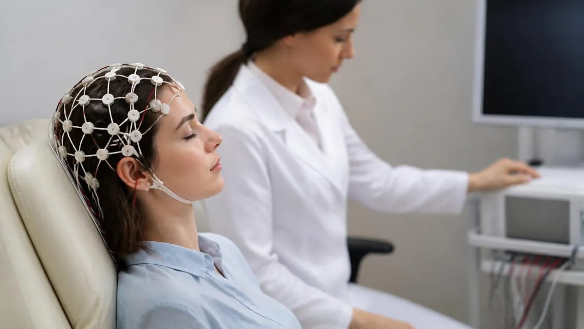
A standard outpatient EEG test typically lasts between 20 and 40 minutes from the time the technologist begins applying electrodes to the moment recording ends. Setup — cleaning electrode sites, applying conductive gel, and securing electrodes — takes an additional 20 to 30 minutes. Patients should plan for a total appointment time of approximately 60 to 90 minutes. The actual recording includes rest with eyes open and closed, hyperventilation, and photic stimulation.
Routine EEG is best suited for initial evaluations and follow-up monitoring in patients with a known epilepsy diagnosis. The short recording window means there is a meaningful chance of a normal result even in someone who has frequent seizures, simply because seizures did not happen to occur during that time window. A normal routine EEG never rules out epilepsy — it only means no abnormality was captured during the recording period.

Pros and Cons of EEG Testing for Seizure Evaluation
- +Non-invasive and painless — no needles, no radiation, no sedation required for standard studies
- +Real-time functional imaging that captures millisecond electrical events MRI and CT cannot show
- +Can detect interictal abnormalities between seizures, not just during seizure events
- +Multiple study types (routine, ambulatory, video-EEG) allow diagnostic workup to be scaled to clinical need
- +Directly guides medication selection and dosing by classifying epilepsy syndrome and seizure type
- +Essential for surgical planning — localizes the seizure onset zone before resective surgery
- −Routine EEG has a high false-negative rate — a single normal study does not rule out epilepsy
- −Cannot image structural lesions — must be combined with MRI for complete evaluation
- −Ambulatory and video-EEG monitoring are expensive and may not be fully covered by insurance
- −Prolonged studies require significant patient time commitment, including inpatient admissions
- −Scalp EEG has limited spatial resolution and may miss deep or small seizure onset zones
- −Artifact identification requires skilled technologists — muscle, movement, and electrode artifact can obscure true findings
How to Prepare for an EEG Test for Seizure Evaluation
- ✓Wash your hair the night before with regular shampoo and leave it completely free of conditioner, gel, hairspray, or oil products
- ✓Avoid caffeine for at least 8 hours before the test, as it can alter brain wave activity and affect results
- ✓Take all prescribed medications as usual unless your neurologist has specifically instructed otherwise
- ✓Eat a normal meal before arriving — low blood sugar from fasting can produce EEG artifacts and affect the recording
- ✓If ordered as a sleep-deprived study, follow your doctor's specific instructions about how long to stay awake beforehand
- ✓Arrive wearing comfortable, loose-fitting clothing and avoid wearing earrings or other jewelry that could interfere with electrode placement
- ✓Plan to have a driver take you home if you were instructed to sleep-deprive — drowsiness makes driving dangerous after a sleepless night
- ✓Bring your complete list of current medications including doses, as the reviewing neurologist will need this information for accurate interpretation
- ✓Inform the technologist at the start of the study about any recent seizures, changes in medication, or illnesses that could affect the recording
- ✓Plan to remain as still as possible during the recording — muscle movement and talking create artifacts that can obscure brain wave patterns
A Normal EEG Does Not Mean You Don't Have Epilepsy
Up to 50% of patients with confirmed epilepsy have a normal first routine EEG. The brain's epileptiform activity may simply not occur during the brief recording window. If your EEG result is normal but your symptoms are convincing, your neurologist will likely order a sleep-deprived EEG or ambulatory study to increase diagnostic yield. Never interpret a single normal EEG as definitive proof that seizures are absent.
The EEG test cost in the United States is one of the most common questions patients ask before scheduling their study. For a routine outpatient EEG, the technical and professional fee combined typically ranges from $200 to $900 depending on the geographic location, the facility type (hospital outpatient department versus independent clinic), and the specific billing codes used. Most major insurance plans, including Medicare and Medicaid, cover EEG when medically indicated for seizure evaluation. However, prior authorization may be required for extended studies such as ambulatory or video-EEG monitoring.
Ambulatory EEG typically costs between $500 and $1,500 for the professional interpretation fee, plus equipment rental or purchase costs that some facilities pass on to the patient. When performed in a hospital outpatient setting, the facility fee can push the total bill significantly higher. Patients with high-deductible health plans should contact their insurance provider before scheduling to understand what portion of the cost they will be responsible for paying out of pocket before their deductible is met.
Video-EEG monitoring in an epilepsy monitoring unit is the most expensive form of seizure evaluation, with costs ranging from $5,000 to over $40,000 for a multi-day admission. This includes inpatient room and board, continuous nursing care, technical EEG recording fees, and neurologist interpretation. Insurance coverage for EMU admissions is generally robust when documentation clearly supports medical necessity — specifically, when there is diagnostic uncertainty about seizure classification or when the patient is being evaluated for surgical candidacy after failing two or more appropriate anti-seizure medications.
For patients without insurance or those facing high out-of-pocket costs, several options exist. Community health centers and federally qualified health centers offer reduced-fee neurological services on a sliding scale. Some academic medical centers have financial assistance programs for patients who qualify based on income. The Epilepsy Foundation also maintains resources to help patients navigate the financial aspects of epilepsy care, including medication assistance programs and guidance on appealing insurance denials for medically necessary EEG studies.
The EEG test price also varies internationally. In Canada and the United Kingdom, where healthcare is publicly funded, routine EEG is covered entirely by provincial health insurance or the NHS, though wait times can be longer than in the US private system. In Mexico and parts of Central America, routine EEG costs as little as $30–$80 USD, making medical tourism a consideration for uninsured American patients who can coordinate care across borders. However, the quality of equipment and interpreter expertise must be verified carefully before pursuing this option.
From the perspective of healthcare economics, the EEG is extraordinarily cost-effective given its diagnostic power. Correctly classifying a seizure type on the first EEG can prevent years of inappropriate medication trials. Focal epileptiform discharges pointing to a resectable lesion can lead to surgical cure, eliminating lifetime medication costs and disability. Conversely, failing to obtain a proper EEG and empirically treating all seizures the same way leads to unnecessary drug toxicity, treatment failures, and the downstream costs of uncontrolled epilepsy including emergency department visits, hospitalizations, and lost productivity.
EEG technologists and students should understand the economics of the studies they perform because it directly affects workflow in clinical settings. Understanding billing codes, documentation requirements, and the distinction between routine, extended routine, and prolonged EEG services matters for ensuring studies are coded accurately and that the facility receives appropriate reimbursement. For certification candidates, this knowledge is also tested on the business and regulatory portions of EEG credentialing examinations, where understanding of CMS guidelines and coding principles is increasingly expected of credentialed professionals.

Status epilepticus — a seizure lasting more than 5 minutes or repeated seizures without recovery — is a neurological emergency requiring immediate EEG monitoring in the ICU setting. Non-convulsive status epilepticus (NCSE) can present as unexplained altered consciousness or coma with minimal outward signs, making urgent EEG the only way to diagnose the condition. If you or someone you care for experiences a prolonged seizure or unexplained unresponsiveness, call 911 immediately rather than waiting for an outpatient EEG appointment.
EEG test side effects are minimal compared to almost any other diagnostic procedure in neurology, which is one reason the test is so widely used across all age groups from premature neonates to elderly patients in the ICU. The most commonly reported experience after a routine EEG is mild scalp irritation or redness at electrode sites where the conductive paste was applied. This typically resolves within a few hours of washing the hair and scalp thoroughly with warm water and regular shampoo. No special skin care is needed in the vast majority of cases.
Some patients experience mild fatigue following a routine EEG, particularly if a sleep-deprived study was performed. Deliberately staying awake for 24 hours before a study causes the kind of tiredness that anyone would feel after a sleepless night, and this is not a side effect of the EEG itself. Patients who undergo sleep-deprived studies are strongly advised to arrange for a driver to take them home rather than driving while severely sleep-deprived. The EEG recording itself does not cause or worsen fatigue — the electrode application and the testing environment are entirely passive from the patient's perspective.
The hyperventilation activation procedure deserves special mention because it can produce sensations that alarm unprepared patients. Breathing rapidly and deeply for three minutes causes a fall in blood CO2 levels that leads to cerebral vasoconstriction. Most patients feel lightheaded, tingling in the hands and feet, or a sense of disorientation during this brief period. These sensations are expected, transient, and resolve within seconds of stopping hyperventilation. The procedure is contraindicated in patients with severe pulmonary disease, recent stroke, or sickle cell disease, and the technologist should always screen for these conditions before beginning.
Photic stimulation — flashing a strobe light at the patient at frequencies ranging from 1 to 60 Hz — can theoretically trigger a seizure in patients with photosensitive epilepsy. This is actually the intended purpose of the procedure in diagnostic context, but it means the technologist must be prepared to stop stimulation immediately if an abnormal response is observed.
The risk of triggering a clinically significant, prolonged seizure during photic stimulation is very low in a monitored medical setting, and the diagnostic value of identifying photosensitivity typically outweighs the small risk. Patients with known photosensitivity are usually evaluated by their neurologist before activation procedures are performed.
There are no long-term side effects associated with EEG testing. The procedure uses no ionizing radiation, no magnetic fields, no contrast agents, and no implanted devices in routine studies. Patients can return to normal activities immediately after a routine EEG — there is no recovery period, no dietary restrictions, and no activity limitations. The only preparation-related restriction is the recommendation to avoid caffeine before the test, and this restriction ends as soon as the recording is complete. Many patients drink coffee in the car on the way home from their appointment without any consequences.
In the context of invasive EEG monitoring — intracranial electrodes placed through burr holes or craniotomy for presurgical evaluation — the risk profile is entirely different. These procedures carry real surgical risks including bleeding, infection, and neurological deficits, and they are performed only in specialized epilepsy surgery centers under the direction of epileptologists and neurosurgeons. However, the EEG test that most patients and most healthcare discussions refer to is the non-invasive scalp recording, which remains one of the safest diagnostic procedures available in clinical medicine with essentially no meaningful side effects when performed by a trained technologist.
Understanding the safety profile of EEG is also important for EEG technologists who are frequently asked by anxious patients about risks before the study begins. Being able to explain clearly and confidently that the test is painless, safe, and non-invasive is part of patient communication competency tested on credentialing examinations. The ability to screen for contraindications to activation procedures, modify protocols appropriately for fragile patients, and recognize when an unexpected event during recording requires intervention are all clinical judgment skills that separate competent technologists from excellent ones — skills developed through education, certification preparation, and supervised clinical practice.
For EEG technologists and students preparing for the R. EEG T. examination or the CLTM credential, developing a systematic approach to reading seizure EEG patterns is as important as memorizing individual waveform morphologies. The best technologists approach every recording the same way: establish the background, characterize the dominant rhythm, assess symmetry between hemispheres, identify any regional abnormalities, and then review activation procedures and any clinical events. This methodical framework prevents errors of omission that occur when an examiner jumps directly to the most dramatic part of the tracing without understanding the baseline context.
Calibration and technical quality are prerequisites for accurate seizure EEG interpretation that are sometimes underemphasized in self-study resources. A study recorded at the wrong sensitivity setting will make normal activity appear abnormally high or low in amplitude. An inappropriate filter setting can create false-looking sharp activity from muscle artifact or attenuate real high-frequency oscillations.
Time base selection affects how waveform morphology appears — a 30 mm/sec standard paper speed is appropriate for most adult EEG, while neonatal EEG is typically read at 15 mm/sec. Technologists who understand these technical parameters produce better recordings and catch calibration errors before they compromise interpretation.
Electrode impedance and the 10–20 International System of electrode placement are foundational knowledge areas tested heavily on EEG certification examinations. The 10–20 system places electrodes at standardized anatomical landmarks — 10% and 20% of measured head distances — ensuring reproducible coverage of frontal, temporal, central, parietal, and occipital regions bilaterally.
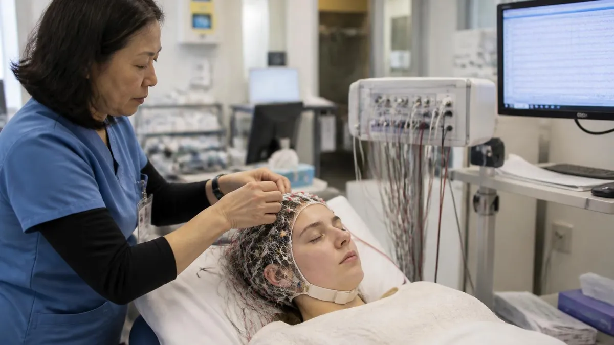
When electrodes are misplaced or have high impedance due to poor contact, focal-looking abnormalities can appear that are actually artifacts of poor recording rather than genuine brain pathology. The technologist's responsibility to achieve impedances below 5 kilohms at every electrode is not just a technical formality — it directly affects the clinical value of the study.
Montage selection is another area where technologist expertise affects seizure localization. A referential montage compares each electrode to a common reference and is useful for determining the absolute polarity and amplitude of discharges.
A bipolar longitudinal (double banana) montage compares adjacent electrode pairs along anterior-posterior chains and is excellent for localizing the field of a discharge by identifying the phase reversal — the point where the chain of deflections reverses direction, indicating the maximum of the electrical field. Experienced readers typically review recordings in multiple montages before rendering a final interpretation, and technologists who understand why montages matter can better anticipate what the interpreting physician needs.
Neonatal and pediatric EEG introduces additional complexity because normal developmental patterns change dramatically with age. A pattern that would be abnormal in an adult — such as burst-suppression or discontinuous background — is normal in a premature neonate at 24 weeks gestational age. Pediatric EEG requires age-corrected interpretation standards, and technologists working in pediatric settings must master these developmental norms to perform competent studies. Seizures in neonates are particularly subtle and may appear as rhythmic alpha or theta activity over a single region without the dramatic generalized discharges associated with adult tonic-clonic seizures.
Critical care EEG monitoring represents a growing subspecialty that focuses on detecting non-convulsive seizures and status epilepticus in ICU patients who cannot report symptoms. Continuous EEG monitoring (cEEG) in neurological intensive care units requires technologists to maintain electrode integrity over days to weeks, review incoming data in near-real-time for alarms, and communicate findings urgently to the clinical team when seizures are detected.
The American Clinical Neurophysiology Society has published guidelines standardizing the terminology used in critical care EEG reporting, and familiarity with these standards — including the ACNS Standardized Critical Care EEG Terminology — is increasingly expected of technologists working in hospital settings.
Building the clinical knowledge and technical skills required for excellence in seizure EEG work takes time, mentorship, and deliberate practice. Board-certified EEG technologists consistently report that regular practice with examination-style questions — especially questions focused on abnormal patterns, artifact recognition, and clinical correlation — is the most effective supplement to hands-on clinical experience. The combination of supervised patient contact and systematic cognitive review through practice testing creates the dual-pathway learning that produces technologists capable of contributing meaningfully to seizure diagnosis and epilepsy management from their first day of independent practice.
EEG Questions and Answers
EEG Electroencephalography Practice Test PDF (Free Printable 2026)
What Does an EEG Measure? Brain Waves, Electrical Activity, and What Your Results Reveal
EEG vs ECG vs EKG: Brain and Heart Test Differences
How to Read an EEG Test: A Complete Guide to Brain Wave Patterns and Results
EEG Waves Explained: Delta, Theta, Alpha, Beta, and Gamma Brain Rhythms
About the Author

Educational Psychologist & Academic Test Preparation Expert
Columbia University Teachers CollegeDr. Lisa Patel holds a Doctorate in Education from Columbia University Teachers College and has spent 17 years researching standardized test design and academic assessment. She has developed preparation programs for SAT, ACT, GRE, LSAT, UCAT, and numerous professional licensing exams, helping students of all backgrounds achieve their target scores.