EEG Sleep Stages: How Brain Waves Change During Sleep and What the Test Reveals
Learn how an EEG test maps sleep stages through brain waves. Understand NREM, REM, costs, and what abnormal results mean. ✅ Full guide inside.

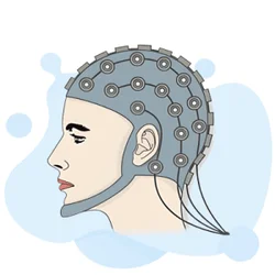
An EEG test — short for electroencephalogram — is one of the most powerful tools available for studying how the brain behaves during sleep. Unlike a standard waking EEG medical test that captures a snapshot of electrical activity over a few minutes, a sleep EEG tracks the brain through multiple cycles of distinct stages, each defined by a unique signature of brain waves. Understanding eeg sleep stages is essential not just for clinicians, but for anyone curious about what happens neurologically when the lights go out and consciousness fades.
During a typical night, a healthy adult cycles through four to five complete sleep cycles, each lasting roughly 90 minutes. Every cycle includes lighter non-REM stages, deep slow-wave sleep, and a period of rapid eye movement (REM) sleep. The EEG test records the electrical signals that mark each transition with remarkable precision. Neurologists and sleep technologists use these wave patterns to identify disorders ranging from insomnia and sleep apnea to narcolepsy and REM sleep behavior disorder.
Many patients wonder what is an EEG test before scheduling one. In simple terms, electrodes are placed on the scalp using a conductive gel, and a machine amplifies the tiny electrical signals produced by millions of neurons firing in synchrony. The resulting graph — a squiggly line of peaks and troughs — tells a complete story about brain state. During sleep, those peaks and troughs slow dramatically in some stages and accelerate in others, giving clinicians a biological timestamp of where in the sleep cycle you are at any given moment.
The clinical importance of sleep-stage EEG data cannot be overstated. Sleep disorders affect more than 70 million Americans, and misdiagnosis is common when clinicians rely on symptoms alone. An EEG medical test conducted overnight — called a polysomnogram — adds continuous brainwave monitoring to oxygen saturation, heart rate, and muscle movement data, creating a comprehensive biological portrait of your night. The combination helps separate, for example, a nightmare disorder from a nocturnal seizure, two conditions that look identical from the outside but require completely different treatments.
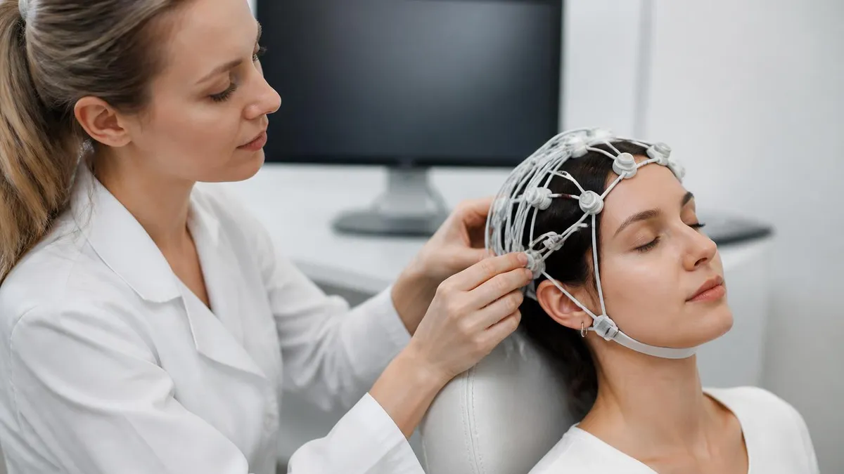
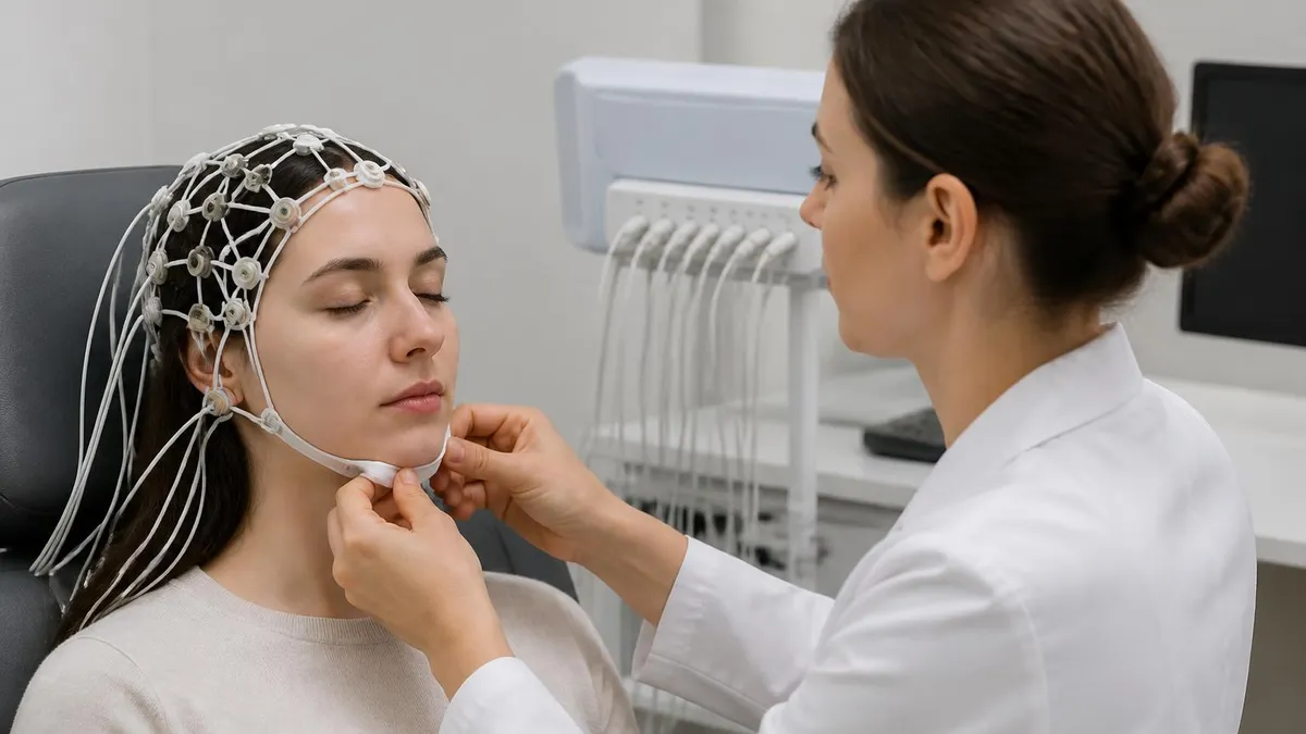
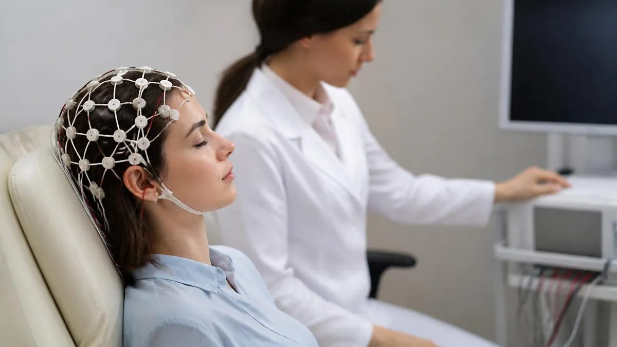
One frequently asked question is how long is an EEG test designed for sleep evaluation. A full polysomnographic study typically runs 7 to 8 hours in a sleep lab, though abbreviated home-based versions exist for specific indications like obstructive sleep apnea. The setup alone — applying up to 22 electrodes plus respiratory belts and leg sensors — takes 45 to 60 minutes before recording begins. Patients are encouraged to sleep as naturally as possible, and technologists monitor the recording remotely throughout the night to ensure signal quality and document any events.
If you are preparing for a certification exam that covers neurophysiology, a solid grasp of eeg sleep stages will appear in multiple question domains including normal patterns, artifacts, and clinical correlations. The R&K (Rechtschaffen and Kales) scoring system and the updated AASM (American Academy of Sleep Medicine) manual both define precise EEG criteria for each stage, and exam writers draw heavily from both frameworks. Knowing not just the wave names but their exact frequency ranges, amplitudes, and clinical meanings gives candidates a decisive advantage.
Beyond certification prep, this knowledge directly improves patient care. A technologist who can recognize a K-complex from an artifact in real time produces cleaner data, fewer misscored epochs, and ultimately a more accurate diagnosis. Sleep medicine is one of the fastest-growing subspecialties in the United States, and EEG-trained professionals who understand sleep architecture are in high demand across hospital sleep labs, outpatient neurology clinics, and telemedicine platforms offering remote monitoring services.
EEG Sleep Testing by the Numbers

The Four Sleep Stages: What the EEG Shows
The transition from wakefulness. The EEG shows low-amplitude, mixed-frequency activity with theta waves (4–7 Hz) replacing the alpha waves of relaxed wakefulness. Vertex sharp waves may appear. This stage lasts 1–7 minutes and accounts for roughly 5% of total sleep time.
Defined by two hallmark EEG features: sleep spindles (12–15 Hz bursts lasting 0.5–2 seconds) and K-complexes (sharp negative deflection followed by a positive wave). N2 comprises about 45–55% of total sleep in healthy adults and is critical for memory consolidation.
High-amplitude delta waves (0.5–2 Hz, >75 µV) dominate at least 20% of the epoch. Also called slow-wave sleep (SWS), N3 is the most restorative stage, supporting immune function, tissue repair, and growth hormone release. Parasomnias like sleepwalking originate here.
The EEG paradoxically resembles wakefulness: low-amplitude, mixed-frequency activity with sawtooth waves (2–6 Hz). Muscle atonia prevents acting out dreams. REM sleep supports emotional memory processing and occupies 20–25% of total sleep time, increasing in later cycles.
Brain wave frequencies are the language of the EEG, and fluency in this language separates a skilled neurophysiology technologist from someone who can merely operate the machine. Every frequency band has a name, a precise hertz range, a characteristic amplitude, and a set of clinical contexts in which it normally appears. During sleep, the brain moves through these bands in a predictable sequence, and any deviation from that sequence carries diagnostic weight. The AASM scoring manual, now the gold standard across US sleep labs, defines epoch-by-epoch criteria that map each 30-second window of the recording to a specific sleep stage.
Delta waves (0.5–4 Hz) are the slowest and highest-amplitude waves the brain routinely produces. They dominate deep NREM sleep, particularly Stage N3, and their presence signals that the brain is engaged in its most profound restorative processes. In adults younger than 30, N3 may comprise 20–25% of total sleep time.
That proportion declines with age — by age 60, many healthy adults show little or no delta activity, which partially explains why older individuals report feeling less refreshed after a full night in bed. On the EEG tracing, delta waves appear as large, slow undulations that are unmistakable even to a trainee.
Theta waves (4–8 Hz) define the drowsy-to-light-sleep transition and appear prominently in Stage N1. They also appear during REM sleep mixed with other frequencies, contributing to the low-amplitude, mixed-frequency pattern that characterizes that stage. In waking EEG recordings, excess theta in an alert individual can indicate encephalopathy or focal dysfunction, so context is everything. Sleep technologists learn to distinguish physiological drowsiness theta from pathological theta through a combination of electrode location, patient state documentation, and accompanying clinical features in the recording.
Alpha waves (8–13 Hz) are the relaxed wakefulness signature. A person lying quietly with eyes closed will typically show a prominent posterior alpha rhythm that disappears — a phenomenon called alpha blocking — the moment they open their eyes or engage in mental activity. During the transition into sleep, alpha slows and fragments before giving way to theta. Some patients show alpha intrusion into N2 or N3 sleep, a pattern associated with non-restorative sleep complaints and conditions like fibromyalgia. Recognizing this pattern on the EEG is a clinical skill with direct therapeutic implications.
Sleep spindles are bursts of sigma-range activity (12–15 Hz) generated by thalamo-cortical circuits and are among the most striking features in an N2 epoch. They typically last 0.5–2 seconds and are distributed broadly across central and frontal electrode sites. Research has linked spindle density to overnight memory consolidation — subjects who show more spindles on post-learning sleep EEGs tend to perform better on declarative memory tests the following morning. Abnormally low spindle counts have been observed in patients with schizophrenia and certain genetic developmental disorders, making spindle analysis an active area of translational neuroscience research.
K-complexes are large, biphasic waveforms — a sharp negative peak followed immediately by a slower positive deflection — that also define Stage N2. They can occur spontaneously or in response to external stimuli such as a door closing or a technologist entering the room. Their amplitude must exceed 75 µV by definition and they must have a duration of at least 0.5 seconds. Scoring rules specify that a K-complex followed within 3 seconds by a sleep spindle constitutes the most prototypical N2 pattern. Clinicians use K-complex reactivity to assess depth of sedation in ICU patients and anesthesia depth monitoring contexts.
REM sleep's sawtooth waves — notched, triangular waves appearing at 2–6 Hz in frontocentral channels — are the least well-known EEG hallmark but equally important for accurate staging. They often immediately precede bursts of rapid eye movements recorded by the EOG channels. In a polysomnogram, confirming REM requires three simultaneous features: low-amplitude mixed-frequency EEG, rapid eye movements on EOG, and reduced chin muscle tone on EMG.
When all three criteria align, the technologist scores the epoch as REM. Missing one feature — particularly in ambiguous transitional periods — requires careful application of AASM tie-breaking rules that experienced technologists internalize over thousands of scored records.
What Is an EEG Test — Preparation, Procedure, and Results
Preparing properly for an EEG medical test improves signal quality and reduces the need for repeat recordings. Patients are typically asked to wash their hair the night before using only shampoo — no conditioner, oils, or styling products — because residue on the scalp raises electrode impedance and introduces artifact. Depending on whether the ordering physician wants a sleep-deprived study, patients may be asked to stay awake all or part of the night before. This intentional sleep deprivation increases the likelihood of falling asleep naturally during a daytime recording, which allows the technologist to capture transitional and sleep-stage EEG patterns without sedation.
Medications can profoundly affect the EEG and must be disclosed to the ordering physician before the test. Benzodiazepines increase beta activity and can mask epileptiform discharges; anticonvulsants may suppress abnormalities that would otherwise confirm a seizure diagnosis. Some neurologists request a brief medication taper before diagnostic studies, though this must always be balanced against seizure risk. Caffeine should be avoided on the day of the study if possible, as it promotes wakefulness and can prevent the patient from achieving the sleep stages the clinician wants to capture. Patients should arrive on time because setup alone takes 45–60 minutes.

Advantages and Limitations of Sleep EEG Testing
- +Non-invasive with no radiation exposure — electrodes sit on the scalp surface and cause no tissue damage
- +Provides real-time, continuous brain wave data through all sleep stages simultaneously
- +Can diagnose nocturnal seizures that would be invisible on a daytime recording alone
- +Differentiates sleep disorders with similar symptoms (e.g., REM behavior disorder vs. sleepwalking)
- +Combined polysomnography adds respiratory and movement data for comprehensive sleep profiling
- +Results are permanent digital records that can be re-reviewed as clinical questions evolve
- −In-lab studies require sleeping away from home in an unfamiliar environment, which can alter natural sleep patterns
- −Setup time (45–60 minutes) and electrode gel in hair can be uncomfortable for some patients
- −EEG test cost ranges from $1,000–$3,500 for full polysomnography, creating access barriers without insurance
- −A single normal EEG does not rule out epilepsy; repeat or prolonged monitoring may be needed
- −Interpretation requires specialized training — misreading benign variants as pathological is common
- −Home sleep tests capture respiratory data but omit full EEG staging, limiting their diagnostic scope
Sleep EEG Prep Checklist: 10 Steps Before Your Test
- ✓Wash hair the night before with shampoo only — skip conditioner, oils, and all styling products.
- ✓Confirm with your doctor whether you need a sleep-deprived study and exactly how long to stay awake.
- ✓List all medications, supplements, and caffeine intake and discuss any required adjustments with your physician.
- ✓Avoid caffeine on the day of the study to maximize your ability to fall asleep during recording.
- ✓Arrive at the facility 15 minutes early — electrode setup takes 45–60 minutes before recording starts.
- ✓Bring comfortable sleepwear and any sleep accessories (pillow, white noise app) that help you fall asleep.
- ✓Inform the technologist of any scalp conditions, allergies to adhesives, or sensory sensitivities before setup.
- ✓Ask the technologist to explain each step during setup so anxiety does not prevent natural sleep onset.
- ✓After the study, verify whether your insurance requires a pre-authorization and confirm expected EEG test cost.
- ✓Follow up with the ordering physician within one week if you have not received your written results report.
The Hypnogram: Your Brain's Sleep Report Card
A hypnogram compresses an entire night of EEG data into a single graph showing how you cycled through N1, N2, N3, and REM sleep. A healthy adult's hypnogram shows deep N3 dominating the first half of the night and REM lengthening in the second half. A disrupted or abnormal hypnogram — with fragmented cycles, absent N3, or suppressed REM — points directly to specific disorders and guides treatment selection more precisely than any questionnaire or self-report.
Sleep disorders diagnosed through EEG monitoring span a remarkably wide clinical spectrum, from common conditions affecting millions of Americans to rare neurological syndromes that require subspecialty expertise. Understanding what an EEG test reveals in each disorder helps patients and providers make informed decisions about whether overnight monitoring is warranted and what findings to anticipate. Epilepsy is the most obvious indication, but the sleep EEG's diagnostic reach extends far beyond seizure detection into the realm of behavioral neurology, psychiatry, and movement disorders.
Nocturnal epilepsy is perhaps the most compelling indication for sleep EEG because seizures during sleep are frequently unwitnessed and may manifest only as unexplained morning fatigue, urinary incontinence, or tongue biting. Certain epilepsy syndromes are specifically sleep-related — autosomal dominant nocturnal frontal lobe epilepsy (ADNFLE) and benign childhood epilepsy with centrotemporal spikes (BECTS) both produce their characteristic discharges predominantly or exclusively during NREM sleep. In these cases, a daytime EEG may be entirely normal while a sleep recording is richly abnormal. Early diagnosis prevents years of misclassification as parasomnias or psychiatric disorders.
REM sleep behavior disorder (RBD) is a condition in which the normal muscle paralysis of REM sleep fails, allowing individuals to physically act out their dreams — punching, kicking, shouting — sometimes injuring themselves or a bed partner. EEG combined with EMG monitoring during a polysomnogram reveals sustained or intermittent muscle tone during REM epochs that should be atonic. RBD carries significant clinical urgency because it is a prodromal marker for alpha-synuclein neurodegenerative diseases including Parkinson's disease, Lewy body dementia, and multiple system atrophy. Diagnosing RBD through EEG monitoring can prompt neuroprotective discussions years before motor symptoms appear.
Narcolepsy with cataplexy is diagnosed in part through a multiple sleep latency test (MSLT), a standardized daytime nap protocol in which the patient is given five 20-minute opportunities to sleep at 2-hour intervals.
The EEG records how quickly sleep onset occurs and whether REM sleep appears within the nap — a finding called a sleep-onset REM period (SOREMP). Two or more SOREMPs on the MSLT, combined with a mean sleep latency under 8 minutes, meets diagnostic criteria for narcolepsy type 1. This EEG-based diagnostic standard has transformed narcolepsy from an underdiagnosed curiosity to a well-characterized condition with targeted pharmacological treatment options.
Periodic limb movement disorder (PLMD) involves repetitive, stereotyped leg movements during sleep that fragment the EEG record with arousal signatures. The EEG captures cortical arousals — brief accelerations of the dominant sleep frequency lasting 3–15 seconds — time-locked to each limb movement detected on the leg EMG channels. A periodic limb movement index above 15 events per hour in adults is considered clinically significant, particularly when accompanied by EEG-documented arousals that prevent reaching or sustaining deeper sleep stages. Treatment with dopaminergic medications often dramatically reduces both movement frequency and EEG arousal burden, restoring normal sleep architecture.
Non-REM parasomnias — sleepwalking, sleep terrors, and confusional arousals — arise from incomplete awakenings out of N3 sleep and are characterized on EEG by a distinctive pattern called slow-wave activity (SWA) intrusion into arousal.
During an episode, the EEG shows a mixture of delta activity (reflecting residual deep sleep) and faster frequencies (reflecting the partial arousal). This mixed pattern distinguishes NREM parasomnias from nocturnal seizures on EEG, though overlap cases with frontal lobe epilepsy require detailed video-EEG analysis and sometimes scalp-sphenoidal recordings to resolve. The availability of high-definition video synchronized with EEG in modern sleep labs has dramatically improved accuracy in these difficult cases.
Sleep-disordered breathing, including obstructive sleep apnea (OSA), produces characteristic EEG signatures even though the primary pathology is in the airway. Each apneic event ends with an arousal that the EEG captures as an abrupt frequency shift from deep slow-wave activity to alpha or theta frequencies.
Counting EEG-documented arousals per hour — the arousal index — provides a brainwave-based measure of sleep fragmentation that complements the apnea-hypopnea index derived from respiratory channels. Patients with high arousal indices but relatively low AHI may still experience profound daytime sleepiness, and EEG evidence of sleep fragmentation helps clinicians justify CPAP or other interventions even when respiratory criteria alone fall short of threshold.

EEG reports contain technical terminology that is easy to misread without specialized training. Terms like 'mild generalized slowing' or 'occasional sharp transients' have specific clinical meanings that depend heavily on the patient's age, medication status, and clinical context. Benign normal variants are frequently alarming to patients who look up terminology online. Always discuss your EEG results directly with the ordering neurologist or sleep physician before drawing any conclusions about your diagnosis or prognosis.
One of the most common practical questions about EEG testing involves cost, insurance coverage, and what patients can realistically expect to pay out of pocket. The answer depends on the type of study ordered, the facility performing it, geographic location, and whether the patient has insurance that covers the procedure. Understanding the financial landscape before scheduling helps patients plan appropriately and avoid unexpected bills that can create barriers to follow-up care. An EEG test cost for a standard outpatient study is substantially different from that of an in-lab polysomnogram or a prolonged video-EEG hospitalization.
A routine outpatient EEG test typically costs between $200 and $700 before insurance adjustments, depending on facility type. Hospital-based epilepsy monitoring units that conduct prolonged recordings lasting 3–7 days command significantly higher prices — often $3,000–$10,000 or more when room, board, nursing, and technical monitoring fees are included. An in-lab polysomnogram for sleep disorders generally falls between $1,000 and $3,500 at accredited sleep centers in the United States. EEG test price at academic medical centers may appear higher on the bill but often reflects comprehensive services including immediate technical review and next-day physician interpretation.
Most major health insurance plans, including Medicare and Medicaid, cover medically necessary EEG testing when ordered for appropriate indications. Prior authorization may be required, particularly for advanced studies like MSLT or prolonged video-EEG. Patients with high-deductible plans may still owe a substantial portion even with insurance, because sleep studies often occur early in the plan year before the deductible is met. It is worth calling the insurance company's pre-authorization line before scheduling to understand your specific cost-sharing obligations and to confirm that the performing facility is in-network.
For patients without insurance or those facing high out-of-pocket costs, several strategies can reduce the financial burden of an EEG medical test. Community health centers and federally qualified health centers (FQHCs) offer EEG services on a sliding-fee scale based on income. Teaching hospitals often provide discounted rates for patients who agree to participate in training programs. Some neurologists offer payment plans or can direct patients to hospital charity care programs for qualifying income levels. The emergence of home-based EEG monitoring services has also introduced lower-cost options for certain indications, though their diagnostic scope remains more limited than full in-lab polysomnography.
For professionals preparing for the ABRET (American Board of Registration of Electroencephalographic and Evoked Potential Technologists) examinations, the financial aspects of EEG practice — including CPT coding, billing compliance, and documentation requirements — represent a distinct knowledge domain. The R.EEG.T. and CLTM examinations include questions about regulatory and operational standards, and candidates who understand the full workflow from patient registration to insurance billing have a more complete professional preparation. Financial literacy about EEG test cost and insurance also helps technologists communicate with patients effectively, which is increasingly recognized as a core competency in patient-centered care models.
Travel EEG technologists — mobile professionals who staff sleep labs and neurology units on short-term contracts across multiple states — often encounter wide variation in facility billing practices, equipment, and documentation systems. A technologist who understands how an EEG medical test is coded, documented, and billed is a more valuable team member and is better positioned for advancement into supervisory or management roles. Many travel contracts offer significant premium pay — often $40–$70 per hour plus housing stipends — precisely because experienced EEG professionals who can function independently from day one are scarce in a tight labor market.
If you want to take your understanding of EEG sleep stages from theoretical to exam-ready, structured practice with realistic questions is the most efficient path. Reviewing scored hypnograms, identifying wave characteristics from descriptions, and working through clinical vignettes that test your ability to distinguish normal from pathological patterns will build the pattern recognition that exam questions demand. Consistent daily practice over 8–12 weeks, combined with review of the AASM scoring manual and ABRET content outlines, represents the evidence-based approach that most successful candidates report using in post-exam surveys.
Mastering EEG sleep stages for clinical practice or certification exams requires more than memorizing frequency ranges — it demands an integrated understanding of how brain wave patterns connect to patient physiology, clinical presentations, and real-world artifact challenges. The most effective study strategies combine visual learning from actual EEG tracings, active recall through practice questions, and spaced repetition to reinforce the wave characteristics that appear most frequently on ABRET and allied health examinations. Building this knowledge systematically over weeks rather than cramming before the test produces the durable recall that high-stakes exams measure.
Start with the fundamentals of the AASM scoring manual, which defines the criteria every accredited sleep technologist in the United States is expected to apply. Download the free summary rules from the AASM website and create a one-page reference card for each stage: the dominant EEG frequency, amplitude criteria, any required landmarks like spindles or K-complexes, and the minimum percentage of the epoch that must display those features.
This card approach forces active synthesis rather than passive reading and gives you a portable review tool during clinical shifts. Many successful candidates report reviewing these cards during slow periods in the EEG lab between patients.
Practice with real EEG examples whenever possible. Many textbooks now include digital supplementary materials with sample EEG segments and self-scoring exercises. Online repositories such as those maintained by professional societies allow registered members to access de-identified polysomnographic records. Working through 10–20 epochs daily — scoring them yourself before checking the answer key — builds the pattern recognition speed that exam conditions require. EEG experts describe this skill as analogous to reading music: at first you decode each note laboriously, but with enough practice the patterns become instantly recognizable gestalts.
Pay particular attention to the transitional periods between stages because that is where both clinical uncertainty and exam question difficulty concentrate. The N1-to-N2 transition requires identifying the first K-complex or spindle after predominantly theta activity. The N3-to-N2 transition requires careful epoch-by-epoch tracking of whether delta activity remains above or below the 20% threshold. The NREM-to-REM transition demands simultaneous attention to EEG, EOG, and chin EMG channels, and the rules for handling ambiguous epochs are among the most tested topics on advanced EEG certification examinations. Create specific practice sets focused on these transition zones rather than distributing practice uniformly across stages.
Artifact recognition is a competency that separates good sleep technologists from exceptional ones, and it appears prominently in both clinical practice and examination content. The most common artifacts encountered during sleep EEG include 60-Hz interference from poorly grounded equipment, slow rolling eye movements that contaminate frontal channels in early N1, electrode pop artifacts from loose contacts, and ECG artifact that creates a rhythmic waveform easily mistaken for a pathological pattern.
Each artifact has a characteristic morphology, a predictable electrode distribution, and a standard correction technique. Learning to recognize artifacts reduces misscoring and prevents the embarrassing situation of reporting epileptiform activity that is actually a loose F7 electrode.
Understanding the physiological basis of each sleep stage EEG pattern — not just its appearance but why the brain generates it — accelerates learning and improves long-term retention. N2 sleep spindles, for example, are generated by thalamic reticular nucleus neurons that rhythmically inhibit thalamocortical relay cells, producing synchronized oscillations that the cortex reflects in the spindle waveform.
K-complexes are thought to represent a cortical response to sensory stimuli that simultaneously promotes arousal threshold maintenance, effectively protecting sleep while momentarily acknowledging the environment. Understanding these mechanisms transforms isolated facts into a coherent framework that makes it easier to reason through novel questions you have never seen before.
Finally, build your exam stamina alongside your content knowledge. The ABRET R.EEG.T. examination consists of 225 questions administered over approximately 3.5 hours, and cognitive fatigue in the final third of the exam costs candidates points they earned in the first two-thirds. Regular timed practice sessions of 50–75 questions under exam-like conditions — no notes, no breaks, strict time limits — train the mental endurance needed to maintain accuracy through a full examination.
Combine this with adequate sleep the week before your exam date: the irony of poor sleep before a sleep EEG certification exam is not lost on anyone in the field, and the cognitive performance costs of sleep deprivation are precisely what all those EEG hours have taught you to recognize.
EEG Questions and Answers
EEG Electroencephalography Practice Test PDF (Free Printable 2026)
Sleep EEG: How Overnight Brain Wave Testing Works and What Your Results Mean
EEG Test Cost in 2026: Prices, Insurance Coverage, and How to Save
EEG vs ECG vs EKG: Brain and Heart Test Differences

Travel EEG Tech Jobs: The Complete Guide to Working as a Mobile Electroencephalography Technologist
About the Author
Educational Psychologist & Academic Test Preparation Expert
Columbia University Teachers CollegeDr. Lisa Patel holds a Doctorate in Education from Columbia University Teachers College and has spent 17 years researching standardized test design and academic assessment. She has developed preparation programs for SAT, ACT, GRE, LSAT, UCAT, and numerous professional licensing exams, helping students of all backgrounds achieve their target scores.