EEG Electroencephalography Practice Test PDF (Free Printable 2026 July)
✅ Pass the EEG Electroencephalography exam with confidence. Practice questions with detailed explanations and instant feedback on every answer.
The ABRET (American Board of Registration of Electroencephalographic and Evoked Potential Technologists) Registered EEG Technologist (R. EEG T.) certification exam tests your mastery of electrode placement, waveform identification, artifact recognition, and clinical EEG procedures. A free EEG practice test PDF gives you a portable, printable study resource you can work through away from a screen — ideal for reviewing the International 10-20 System diagrams, waveform frequency bands, and epileptiform patterns that appear repeatedly on the exam.
Whether you're preparing for the R. EEG T. credential or the Continuous Long-Term Monitoring (CLTM) examination, consistent offline practice with realistic question formats builds the pattern-recognition speed the exam demands.
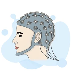
EEG Electrode Placement: The International 10-20 System
The 10-20 System is the foundation of every EEG technologist exam. Electrode positions are named by lobe prefix and number: Fp1/Fp2 (frontopolar), F3/F4/F7/F8/Fz (frontal), C3/C4/Cz (central), P3/P4/Pz (parietal), O1/O2 (occipital), and T3/T4/T5/T6 (temporal). Odd numbers always indicate left-hemisphere electrodes; even numbers indicate right; midline electrodes carry the suffix z.
Measurements are taken as 10% or 20% of the total nasion-to-inion or pre-auricular distances — hence the name. Accurate placement is critical because misplaced electrodes change the morphology of recorded waveforms and can mimic or mask pathology.
Normal and Abnormal EEG Waveforms
Alpha rhythm (8–13 Hz) is the dominant posterior rhythm in an awake, relaxed adult with eyes closed — it attenuates with eye opening. Beta activity (>13 Hz) predominates frontally during active thinking and with benzodiazepine use. Theta (4–7 Hz) is normal in drowsiness and children but abnormal in the waking adult EEG over focal regions. Delta (<4 Hz) is normal in deep NREM sleep but indicates severe dysfunction when present in an awake patient.
Key abnormal patterns: generalized 3 Hz spike-and-wave is the hallmark of absence (petit mal) epilepsy. Focal spikes or sharp waves suggest a localized seizure focus. Burst suppression indicates profound cortical dysfunction (anesthesia, hypoxia, or severe encephalopathy).
- ✓Download and print the free EEG practice test PDF above
- ✓Draw the 10-20 electrode map from memory and label all 21 standard positions
- ✓Memorize frequency bands: delta <4 Hz, theta 4-7 Hz, alpha 8-13 Hz, beta >13 Hz
- ✓Study sleep stage EEG correlates: vertex waves (N1), K-complexes and sleep spindles (N2), delta (N3)
- ✓Review generalized vs. focal epileptiform patterns and their clinical significance
- ✓Practice identifying muscle, eye movement, electrode pop, and 60 Hz artifacts on sample tracings
- ✓Study activation procedure protocols: hyperventilation duration, photic stimulation frequencies
- ✓Review impedance standards, inter-electrode distance requirements, and montage types
- ✓Complete at least two full-length timed practice exams before your test date
- ✓Review ABRET exam content outline and confirm all domain areas are covered in your study plan
EEG Artifacts and Activation Procedures
Artifact recognition is a heavily tested domain. Muscle (EMG) artifact produces high-frequency, irregular activity that can obscure underlying brain signals — reduce by instructing the patient to relax facial and neck muscles. Eye movement artifacts (lateral = square wave at F7/F8; blink = slow wave at Fp1/Fp2) are corrected by asking the patient to fixate or close their eyes. Electrode pop appears as a sudden high-amplitude spike at a single channel, caused by poor contact or dried gel. 60 Hz (powerline) interference is a regular, sinusoidal artifact corrected by improving electrode impedance or using a notch filter.
Activation procedures are used to provoke latent epileptiform activity. Hyperventilation (3–4 minutes of deep breathing) causes hypocapnia and vasoconstriction, which can elicit absence seizure discharges. Photic stimulation uses a strobe light at frequencies from 1 to 30 Hz to provoke photoparoxysmal responses in photosensitive patients. Sleep deprivation increases the likelihood of recording spontaneous seizure activity during drowsiness and light sleep. The PDF includes practice questions covering each procedure's indication, protocol, and expected EEG findings.
- +Industry-recognized credential boosts your resume
- +Higher earning potential (10-20% salary increase on average)
- +Demonstrates commitment to professional development
- +Opens doors to advanced career opportunities
- −Exam preparation requires significant time investment (4-8 weeks)
- −Certification fees can be $100-$400+
- −May require continuing education to maintain
- −Some employers may not require certification