What Is an EEG? Electroencephalography Complete Guide 2026 July
Prepare for the What Is an EEG? Electroencephalography certification. 📗 Practice questions with answer explanations covering all exam domains.

How EEG Works
The brain contains billions of neurons that communicate through electrical signals. When groups of neurons fire synchronously, their combined electrical activity produces measurable voltage differences that can be detected on the scalp surface. The EEG amplifies these tiny signals (measured in microvolts — millionths of a volt) and records them over time as a continuous waveform called the electroencephalogram.
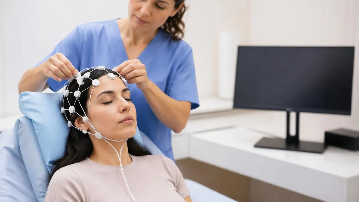
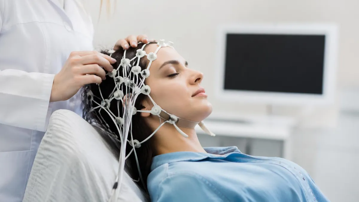
The electrodes used in standard EEG are small metal discs (usually silver/silver chloride or gold) attached to the scalp with conductive gel or paste. They do not emit any electricity — they are passive detectors. The standard clinical EEG uses the International 10-20 Electrode Placement System, which positions 19–21 electrodes at standardized anatomical landmarks measured as percentages of skull circumference. Each electrode is named by brain region (F=frontal, C=central, P=parietal, T=temporal, O=occipital) and numbered odd (left) or even (right).
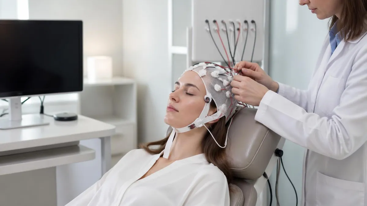
The recorded signals are amplified, filtered, and displayed as a series of channels — one channel per electrode pair — scrolling continuously across the monitor or recording system. Standard recordings run at 10 mm/sec paper speed with 30 mm/mV sensitivity. An EEG technologist monitors the recording in real time, marking events (patient movement, artifacts, clinical observations) and applying activation procedures.
Importantly, EEG records electrical activity at the brain's surface — it cannot image brain structure, detect tumor masses, or identify individual cellular pathology. It measures function (brain electrical rhythms) rather than anatomy.
EEG Overview
Delta Waves (0.5–4 Hz)
Delta waves are the slowest EEG frequency band. They are normal in infants, in deep (stage 3) non-REM sleep in adults, and under general anesthesia. Abnormal delta activity in a waking adult suggests encephalopathy, severe brain injury, deep coma, or structural lesions (tumors, strokes). Focal delta activity (slowing in one region) points to a focal lesion in that brain region. Generalized delta in a waking adult is a serious neurological finding requiring immediate investigation.
Types of EEG Studies
Standard clinical EEG has evolved into several specialized recording formats designed to capture different types of brain activity and specific clinical presentations.
Routine EEG (20–40 minutes): The standard diagnostic EEG performed in an outpatient setting or at bedside. Includes hyperventilation (3 minutes of deep breathing that can activate absence seizures) and photic stimulation (a strobe light that activates photosensitive epilepsy). A single routine EEG captures a random 20–40 minute window of brain activity — and since most people with epilepsy don't have a seizure during those minutes, a normal routine EEG does NOT rule out epilepsy.
Sleep-deprived EEG: The patient is kept awake for 24 hours (or significantly sleep-limited) before the EEG. Sleep deprivation increases seizure likelihood and activates epileptiform discharges that may not appear in a routine waking EEG. The patient typically falls asleep during the recording, capturing both waking and sleep EEG states.
Ambulatory EEG (24–72+ hours): Electrodes are applied and connected to a small portable recording device the patient wears at home or work during normal daily activities. This extended recording dramatically increases the chance of capturing seizures and documenting the patient's typical events. Ambulatory EEG is particularly useful for infrequent events that are unlikely to occur during a brief in-lab recording.
Video-EEG monitoring (VEEG): The gold standard for epilepsy evaluation prior to surgery. The patient is admitted to an epilepsy monitoring unit (EMU) and has simultaneous video and EEG recording for several days. Seizures are captured with both electrical (EEG) and behavioral (video) data, allowing precise localization of the seizure onset zone. Anti-seizure medications may be reduced during admission to induce seizures.
Intraoperative EEG and monitoring: EEG is monitored in real time during brain or spinal surgery to detect ischemia (brain tissue being damaged from insufficient blood flow) and to guide resection margins in epilepsy surgery. Specialized EEG technologists and neurophysiologists staff intraoperative neuromonitoring (IONM) teams.
Neonatal EEG: Specialized protocols for premature infants and newborns, with different electrode placement patterns and distinct normal and abnormal patterns. Neonatal seizures frequently have no visible clinical signs (electrographic-only seizures), making EEG essential for neonatal intensive care unit (NICU) seizure detection. For more detail, see our guide on the EEG test process and clinical applications.
What Conditions EEG Can Detect
EEG is most valuable for conditions that affect brain electrical rhythms — it is not a useful test for structural lesions (tumors, bleeding) where MRI or CT are preferred.
Epilepsy and seizure disorders: The primary clinical indication for EEG. Epileptiform discharges — spikes, sharp waves, and spike-wave complexes — are the electrical signature of epileptic activity. The EEG helps classify seizure type (focal vs. generalized), identify epilepsy syndrome, guide medication selection, and locate seizure onset zone for surgical evaluation. The EEG abnormal epileptiform patterns practice tests cover the specific waveform types and their clinical significance.
Sleep disorders: EEG is a component of polysomnography (PSG, or sleep study), which records brain activity along with eye movements, muscle tone, respiratory effort, oxygen saturation, and heart rate during sleep. EEG staging of sleep (N1, N2, N3, and REM) is essential for diagnosing insomnia, sleep apnea, narcolepsy, REM sleep behavior disorder, and parasomnias.
Encephalopathy: Diffuse brain dysfunction from metabolic causes (liver failure, kidney failure, electrolyte abnormalities, infection), toxic causes (medication toxicity, drug overdose), or structural causes (widespread brain injury) produces characteristic EEG slowing, abnormal patterns (triphasic waves in hepatic encephalopathy), or periodic discharges.
Brain death determination: Electrocerebral silence (ECS) — the absence of any brain electrical activity above 2 microvolts — is one component of brain death determination protocols. EEG confirmation of ECS requires strict technical standards for electrode placement, gain settings, and artifact exclusion.
Status epilepticus (SE): Prolonged seizure activity that is a neurological emergency. Non-convulsive status epilepticus (NCSE) — continuous seizure activity without visible convulsions — can only be diagnosed by EEG, making continuous EEG (cEEG) monitoring essential in ICU patients with altered consciousness of unknown cause.
EEG Study Tips
What's the best study strategy for EEG?
Focus on weak areas first. Use practice tests to identify gaps, then study those topics intensively.
How far in advance should I start studying?
Most successful candidates begin 4-8 weeks before the exam. Create a structured study schedule.
Should I retake practice tests?
Yes! Take each practice test 2-3 times. Focus on understanding why answers are correct, not memorizing.
What should I do on exam day?
Arrive 30 min early, bring required ID, read questions carefully, flag difficult ones, and review before submitting.
EEG Checklist
- ✓Review the 10-20 electrode placement system: all electrode names, landmarks, and measurements
- ✓Study normal EEG patterns by age: newborn, infant, child, adult waking and sleep stages
- ✓Memorize the five EEG frequency bands and their normal vs. abnormal significance
- ✓Learn epileptiform discharge patterns: spikes, sharp waves, spike-wave, GPFA, burst suppression
- ✓Study activation procedures: hyperventilation response, photic driving, drowsiness/sleep changes
- ✓Practice artifact recognition: muscle, movement, eye movement, electrode, 60 Hz, cardiac
- ✓Review EEG montages: referential vs. bipolar, longitudinal bipolar (double banana), transverse bipolar
- ✓Study ABRET R.EEG.T exam blueprint content domains: knowledge, instrumentation, patient care
- ✓Complete at least 3 full-length practice exams
- ✓Review HIPAA, patient safety protocols, and electrode application techniques
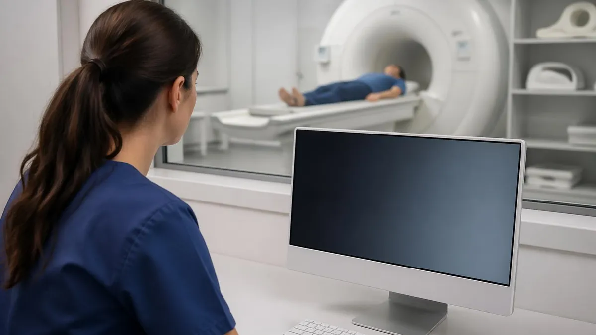
EEG vs MRI vs CT: When Each Is Used
EEG, MRI, and CT are complementary neurological diagnostic tools, not alternatives. Each answers different clinical questions and is chosen based on what information is needed.
EEG measures brain function — electrical activity patterns. It is the test of choice when the clinical question involves seizure activity, encephalopathy severity, sleep disorders, or ongoing neurological status in ICU patients. EEG provides real-time dynamic information about brain state but gives no structural information.
MRI reveals brain structure in exquisite soft-tissue detail. For epilepsy evaluation, brain MRI (particularly high-resolution sequences at 3T) identifies structural causes: hippocampal sclerosis, cortical dysplasia, tumors, cavernous malformations, and vascular lesions. MRI and EEG together provide complementary functional and structural data for comprehensive epilepsy evaluation.
CT is fast and widely available, making it the first-line neurological imaging in emergencies (acute stroke, head trauma, intracranial hemorrhage). CT does not provide the soft-tissue detail of MRI and cannot identify the subtle structural lesions that MRI detects in epilepsy evaluation. CT uses ionizing radiation; MRI does not.
In most epilepsy workups, all three tests contribute: EEG characterizes seizure activity and localizes the epileptic focus electrically; MRI identifies (or rules out) a structural cause; CT may be obtained acutely if an emergency presentation makes rapid imaging necessary before MRI is available.
EEG Technologist Career 2026
An EEG technologist (also called a neurodiagnostic technologist) applies electrodes, operates EEG equipment, performs the recording, monitors for artifacts and clinical events, and prepares the recording for neurologist interpretation. The role requires a blend of technical instrumentation skill, patient interaction ability, and foundational neurophysiology knowledge.
Most EEG technologists work in hospital neurology departments, epilepsy monitoring units, neurology outpatient clinics, sleep labs, and neurodiagnostic testing companies. Intraoperative neuromonitoring (IONM) technologists work in operating rooms and may travel to multiple surgery sites.
Certification: The American Board of Registration of Electroencephalographic and Evoked Potential Technologists (ABRET) offers the R.EEG.T (Registered EEG Technologist) credential. Most employers prefer or require ABRET certification. Eligibility requires: completing a formal neurodiagnostic technology program OR equivalent supervised work experience, plus passing the written knowledge examination and a practical skills assessment for some pathways.
Salary and outlook: The Bureau of Labor Statistics reports a median annual salary of $58,760 for electroneurodiagnostic technologists (2023), with job growth projected at approximately 10% from 2022–2032 — faster than average. The growing aging population increases demand for neurological diagnostic services, and EEG technologists in specialized IONM and long-term monitoring roles can earn premium compensation.
EEG Practice Test Questions
Prepare for the EEG - Electroencephalography exam with our free practice test modules. Each quiz covers key topics to help you pass on your first try.
EEG Abnormal Epileptiform Patterns
EEG Exam Questions covering Abnormal Epileptiform Patterns. Master EEG Test concepts for certification prep.
EEG Activation Procedures
Free EEG Practice Test featuring Activation Procedures. Improve your EEG Exam score with mock test prep.
EEG Artifact Identification
EEG Test Prep for Artifact Identification. Practice EEG Quiz questions and boost your score.
EEG Electrode Placement and Montages
EEG Questions and Answers on Electrode Placement and Montages. Free EEG practice for exam readiness.
EEG - Electroencephalography Abnormal Epil...
EEG Mock Test covering - Electroencephalography Abnormal Epileptiform Patterns. Online EEG Test practice with instant feedback.
EEG - Electroencephalography Activation Pr...
Free EEG Quiz on - Electroencephalography Activation Procedures. EEG Exam prep questions with detailed explanations.
EEG - Electroencephalography Artifact Iden...
EEG Practice Questions for - Electroencephalography Artifact Identification. Build confidence for your EEG certification exam.
EEG - Electroencephalography Electrode Pla...
EEG Test Online for - Electroencephalography Electrode Placement and Montages. Free practice with instant results and feedback.
EEG - Electroencephalography Instrumentati...
EEG Study Material on - Electroencephalography Instrumentation and Calibration. Prepare effectively with real exam-style questions.
EEG - Electroencephalography Neuroanatomy ...
Free EEG Test covering - Electroencephalography Neuroanatomy and Physiology. Practice and track your EEG exam readiness.
EEG - Electroencephalography Non-Epileptif...
EEG Exam Questions covering - Electroencephalography Non-Epileptiform Abnormalities. Master EEG Test concepts for certification prep.
EEG - Electroencephalography Normal EEG Wa...
Free EEG Practice Test featuring - Electroencephalography Normal EEG Waveforms. Improve your EEG Exam score with mock test prep.
EEG - Electroencephalography Patient Safet...
EEG Mock Exam on - Electroencephalography Patient Safety and Ethics. EEG Study Guide questions to pass on your first try.
EEG - Electroencephalography Pediatric and...
EEG Test Prep for - Electroencephalography Pediatric and Neonatal EEG. Practice EEG Quiz questions and boost your score.
EEG ICU and Continuous EEG Monitoring
EEG Questions and Answers on ICU and Continuous EEG Monitoring. Free EEG practice for exam readiness.
EEG Instrumentation and Calibration
EEG Mock Test covering Instrumentation and Calibration. Online EEG Test practice with instant feedback.
EEG Neuroanatomy and Physiology
Free EEG Quiz on Neuroanatomy and Physiology. EEG Exam prep questions with detailed explanations.
EEG Non-Epileptiform Abnormalities
EEG Practice Questions for Non-Epileptiform Abnormalities. Build confidence for your EEG certification exam.
EEG Normal EEG Waveforms
EEG Test Online for Normal EEG Waveforms. Free practice with instant results and feedback.
EEG Patient Safety and Ethics
EEG Study Material on Patient Safety and Ethics. Prepare effectively with real exam-style questions.
EEG Pros and Cons
- +EEG has a defined, publicly available content blueprint — candidates know exactly what to prepare for
- +Multiple preparation pathways (self-study, courses, coaching) accommodate different learning styles and schedules
- +A growing ecosystem of study resources means candidates at any budget level can access quality preparation materials
- +Clear score reporting allows candidates to identify specific strengths and weaknesses for targeted remediation
- +Professional recognition associated with strong performance provides tangible career and academic benefits
- −The scope of tested content requires substantial preparation time that competes with existing professional or academic commitments
- −No single resource covers the full content scope — candidates typically need multiple study tools for comprehensive preparation
- −Test anxiety and exam-day performance variability mean preparation effort does not always translate linearly to scores
- −Registration, preparation, and potential retake costs accumulate into a significant financial investment
- −Content and format can change between exam versions, making older preparation materials less reliable
EEG Questions and Answers


About the Author

Educational Psychologist & Academic Test Preparation Expert
Columbia University Teachers CollegeDr. Lisa Patel holds a Doctorate in Education from Columbia University Teachers College and has spent 17 years researching standardized test design and academic assessment. She has developed preparation programs for SAT, ACT, GRE, LSAT, UCAT, and numerous professional licensing exams, helping students of all backgrounds achieve their target scores.