What Does an EEG Measure? Brain Waves, Electrical Activity, and What Your Results Reveal
🆕 What does an EEG measure? Learn how the EEG test records brain electrical activity, frequencies, costs, side effects, and what abnormal results mean.
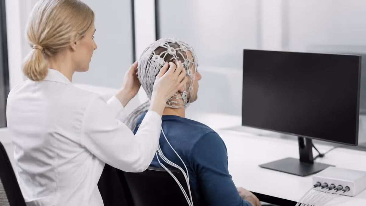
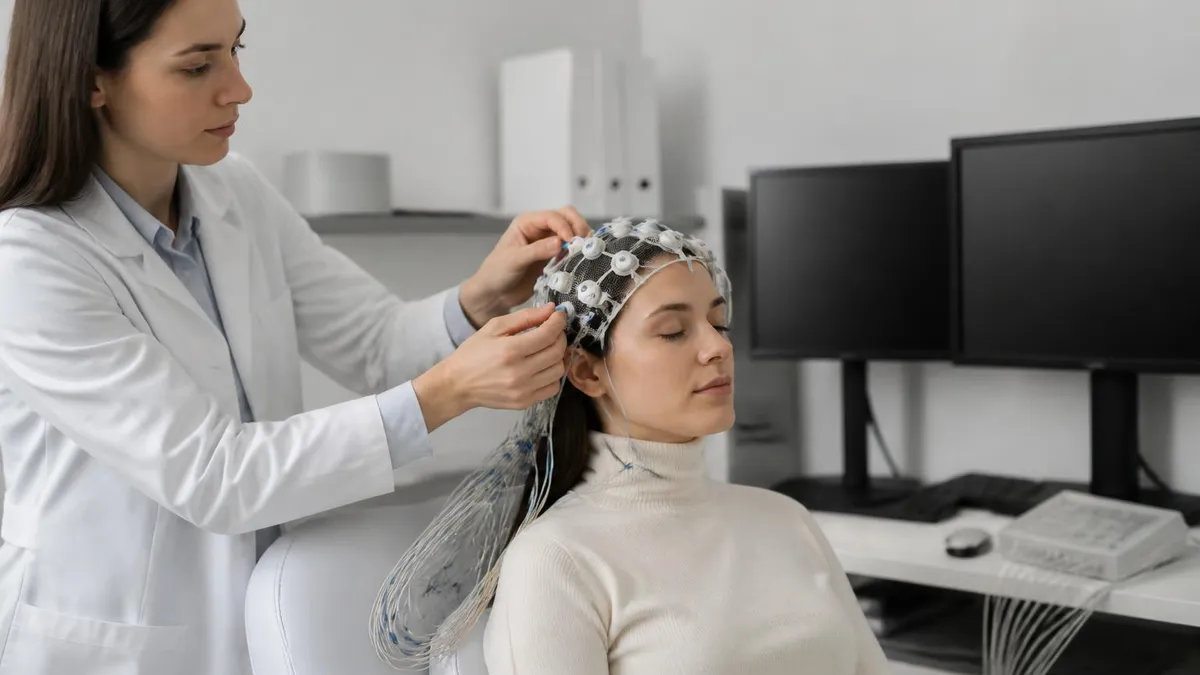
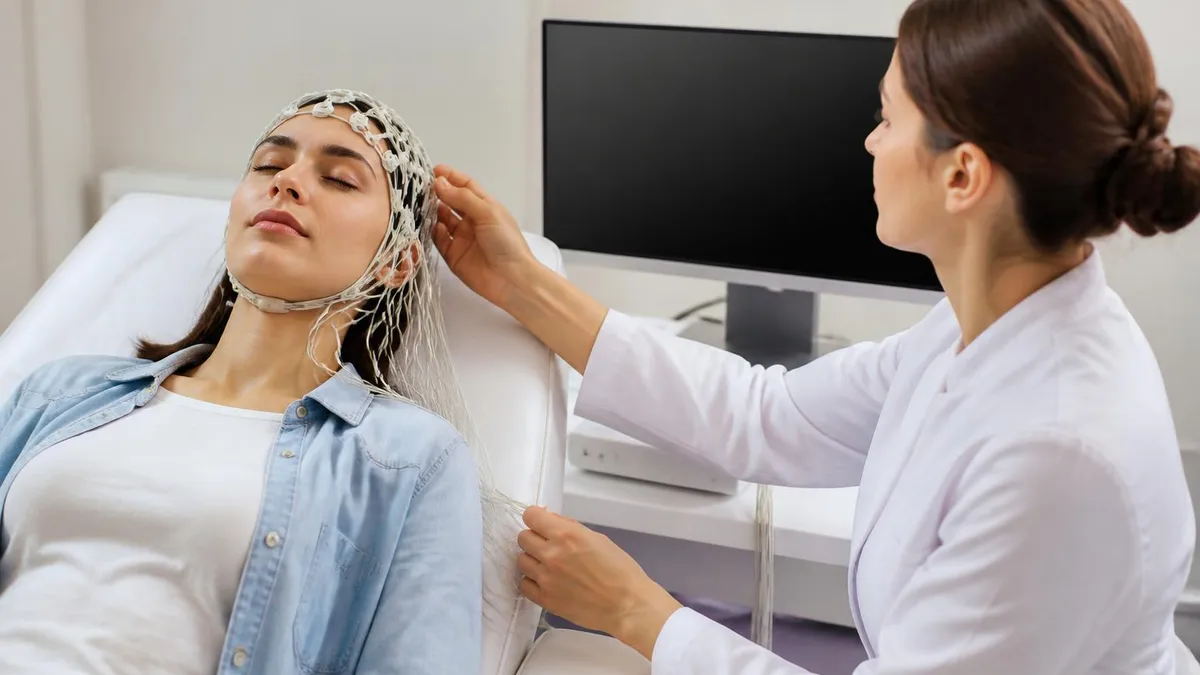
If you have ever wondered what does an EEG measure, the short answer is the tiny electrical voltages produced by the firing of billions of neurons in your brain. An EEG test, short for electroencephalogram, records these voltage fluctuations from the scalp using small metal discs called electrodes. The signals are amplified hundreds of thousands of times and displayed as wavy lines that neurologists read to look for seizures, sleep disorders, head injuries, encephalitis, and other neurological problems that purely structural scans like MRI cannot reveal.
Unlike imaging that shows brain anatomy, an EEG captures real-time function. It measures the summed postsynaptic potentials of pyramidal cells in the cerebral cortex, primarily those oriented perpendicular to the scalp. These rhythmic electrical changes are organized into recognizable frequency bands — delta, theta, alpha, beta, and gamma — each tied to specific states of consciousness, ages, and brain regions. When a doctor orders an eeg test price estimate, they are pricing this functional snapshot of your cortex.
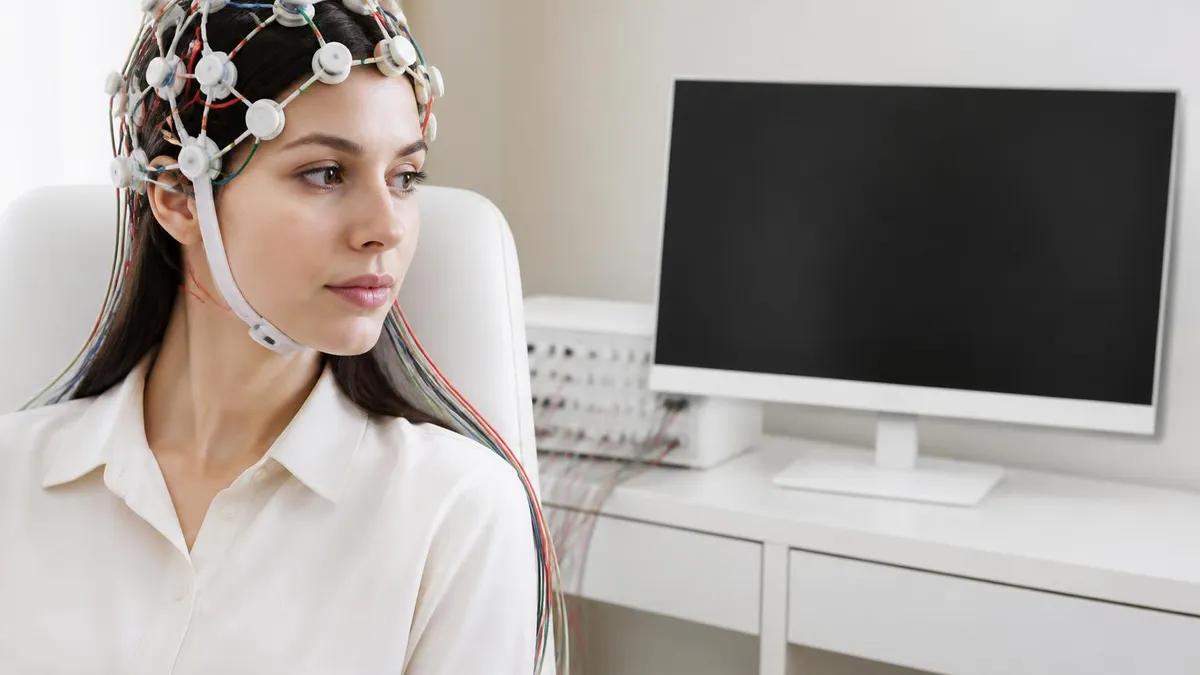
A standard EEG records 19 to 21 channels using the international 10–20 system, with electrodes labeled by hemisphere and lobe (Fp1, F3, C4, O2, and so on). Each channel shows the voltage difference between two electrodes, which is why technologists configure montages such as longitudinal bipolar, transverse bipolar, and average reference. The resulting traces tell the reader where activity originates, how symmetric the two hemispheres are, and whether any focal slowing, spikes, or sharp waves are present.
Voltages on a routine scalp EEG are extremely small — usually between 10 and 100 microvolts, roughly one-millionth of a typical AA battery. Because the skull attenuates signals dramatically, the EEG mostly sees activity from gyri close to the surface. Deep structures like the hippocampus rarely contribute directly to scalp recordings, though their downstream effects on cortex often do. That is why some patients eventually need long-term monitoring, ambulatory EEG, or intracranial recordings to localize a seizure focus.
EEG is one of the oldest neurodiagnostic tools — Hans Berger recorded the first human EEG in 1924 — yet it remains indispensable because it has millisecond time resolution that no other clinical test matches. Functional MRI captures activity changes over seconds; EEG captures them in milliseconds, which is the timescale of actual neural communication. That speed is exactly what is needed to catch epileptiform discharges, characterize sleep stages, monitor anesthesia depth, and confirm or rule out brain death.
Common reasons doctors order an EEG include first-time seizures, episodes of staring or confusion, unexplained loss of consciousness, suspected encephalopathy, evaluation of coma, sleep disturbances, and pre-surgical mapping for epilepsy. The test is painless, non-invasive, and uses no radiation. Most outpatient routines last about 20 to 40 minutes of recording, plus setup, though longer sleep-deprived, ambulatory, and inpatient video-EEG studies can run from several hours to many days when more data is needed.
This guide explains in plain language what does an EEG measure, how the recording works, which brain wave frequencies matter, what abnormal patterns suggest, what the test costs in the United States, how long it takes, what side effects to expect, and how to prepare so your results are as clean and useful as possible.
What an EEG Measures by the Numbers
EEG Frequencies and What They Mean
Slowest and highest amplitude waves. Normal during deep, dreamless sleep in all ages and dominant in infants. When seen while awake in adults, focal delta suggests a structural lesion and diffuse delta points to encephalopathy.
Seen in drowsiness, light sleep, and during meditation or memory tasks. Theta is normal in children but excessive theta in awake adults can indicate mild encephalopathy, medication effect, or focal cortical dysfunction.
The classic posterior dominant rhythm of relaxed wakefulness with eyes closed. Alpha attenuates when you open your eyes or concentrate. Asymmetry or slowing of the alpha rhythm is one of the earliest clues to focal pathology.
Low-amplitude fast activity associated with active thinking, anxiety, and benzodiazepine or barbiturate use. Excessive frontal beta is one of the most reliable EEG signatures of sedative medication on a patient's report.
High-frequency oscillations linked to attention, sensory binding, and cognitive processing. Gamma is challenging to record cleanly on scalp EEG because of muscle artifact, but it is increasingly studied in research and neurosurgical mapping.
To understand what an EEG measures at a physical level, picture a cortical pyramidal neuron as a tiny battery. When thousands of these cells fire together, their summed extracellular currents create a voltage field that travels through brain tissue, cerebrospinal fluid, skull, and scalp before reaching the electrode. The skull alone attenuates the signal by roughly a factor of ten and blurs it spatially, which is why scalp EEG sees averaged populations of neurons rather than individual cells.
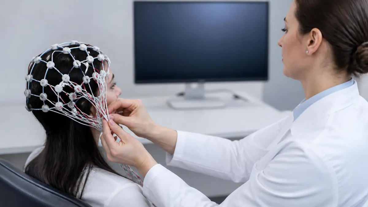
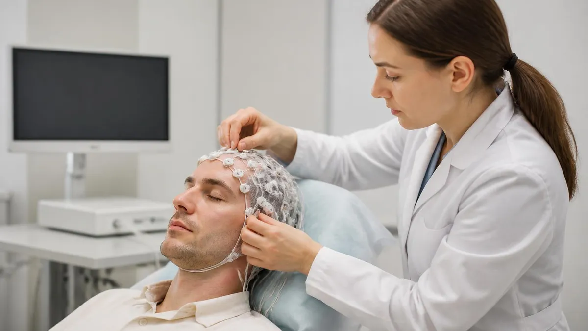
The electrode itself is usually a small silver/silver-chloride disc attached with conductive paste, collodion glue, or a snug cap. A technologist measures your head, marks landmarks at the nasion, inion, and preauricular points, and places electrodes at standardized distances. Good impedance — typically below 5 kΩ — is critical, because poor electrode contact introduces noise that can mimic real brain activity. A skilled tech spends as much time on prep as on recording. The eeg medical test career pays for exactly this expertise.
Each channel on the recording is the voltage difference between two electrodes. In a bipolar montage, adjacent electrodes are paired (Fp1-F3, F3-C3, C3-P3) and a phase reversal between channels helps localize a discharge. In a referential montage, every electrode is compared to a common reference like the ear, mastoid, or an average. Modern digital EEG lets the reader re-montage the same recording instantly, viewing it from multiple perspectives without re-acquiring data.
Activation procedures add diagnostic value by stressing the brain. Hyperventilation for three to five minutes induces hypocapnia and can bring out generalized spike-wave activity in absence epilepsy. Photic stimulation at 1–30 Hz tests for photoparoxysmal responses. Sleep — especially after sleep deprivation — unmasks epileptiform discharges that are silent during wakefulness in roughly 30% of patients with epilepsy whose routine EEG looked normal.
Artifacts are the constant enemy of clean EEG. Eye blinks produce frontal slow waves, lateral eye movement creates lateral rectus spikes, swallowing generates muscle bursts, sweat causes slow undulating baselines, and a single loose electrode can produce 60 Hz line noise across the entire trace. Technologists are trained to spot, label, and minimize these patterns; readers are trained to ignore them. Misreading artifact as pathology is one of the most common EEG errors and a frequent reason for second opinions.
Time resolution is where EEG truly shines. A spike-and-wave discharge lasts about 200 milliseconds; a seizure-onset rhythmic theta can be seen within a second of clinical symptoms; sleep spindles last 0.5–1.5 seconds. Capturing these features requires a sampling rate of at least 200 Hz, and modern systems sample 500–1000 Hz to avoid aliasing. The trade-off is file size and storage, which is why prolonged video-EEG produces multi-gigabyte datasets.
Spatial resolution, by contrast, is EEG's weakness. Sources within a couple of centimeters of each other can be indistinguishable on the scalp. High-density EEG (64, 128, or 256 channels) plus source-localization software improves this, but for routine clinical use, 19–21 channels remain the standard. When precise localization is needed for epilepsy surgery, intracranial electrodes still outperform any non-invasive method.
EEG Practice Test Questions
Prepare for the EEG - Electroencephalography exam with our free practice test modules. Each quiz covers key topics to help you pass on your first try.
EEG Abnormal Epileptiform Patterns
EEG Exam Questions covering Abnormal Epileptiform Patterns. Master EEG Test concepts for certification prep.
EEG Activation Procedures
Free EEG Practice Test featuring Activation Procedures. Improve your EEG Exam score with mock test prep.
EEG Artifact Identification
EEG Test Prep for Artifact Identification. Practice EEG Quiz questions and boost your score.
EEG Electrode Placement and Montages
EEG Questions and Answers on Electrode Placement and Montages. Free EEG practice for exam readiness.
EEG - Electroencephalography Abnormal Epil...
EEG Mock Test covering - Electroencephalography Abnormal Epileptiform Patterns. Online EEG Test practice with instant feedback.
EEG - Electroencephalography Activation Pr...
Free EEG Quiz on - Electroencephalography Activation Procedures. EEG Exam prep questions with detailed explanations.
EEG - Electroencephalography Artifact Iden...
EEG Practice Questions for - Electroencephalography Artifact Identification. Build confidence for your EEG certification exam.
EEG - Electroencephalography Electrode Pla...
EEG Test Online for - Electroencephalography Electrode Placement and Montages. Free practice with instant results and feedback.
EEG - Electroencephalography Instrumentati...
EEG Study Material on - Electroencephalography Instrumentation and Calibration. Prepare effectively with real exam-style questions.
EEG - Electroencephalography Neuroanatomy ...
Free EEG Test covering - Electroencephalography Neuroanatomy and Physiology. Practice and track your EEG exam readiness.
EEG - Electroencephalography Non-Epileptif...
EEG Exam Questions covering - Electroencephalography Non-Epileptiform Abnormalities. Master EEG Test concepts for certification prep.
EEG - Electroencephalography Normal EEG Wa...
Free EEG Practice Test featuring - Electroencephalography Normal EEG Waveforms. Improve your EEG Exam score with mock test prep.
EEG - Electroencephalography Patient Safet...
EEG Mock Exam on - Electroencephalography Patient Safety and Ethics. EEG Study Guide questions to pass on your first try.
EEG - Electroencephalography Pediatric and...
EEG Test Prep for - Electroencephalography Pediatric and Neonatal EEG. Practice EEG Quiz questions and boost your score.
EEG ICU and Continuous EEG Monitoring
EEG Questions and Answers on ICU and Continuous EEG Monitoring. Free EEG practice for exam readiness.
EEG Instrumentation and Calibration
EEG Mock Test covering Instrumentation and Calibration. Online EEG Test practice with instant feedback.
EEG Neuroanatomy and Physiology
Free EEG Quiz on Neuroanatomy and Physiology. EEG Exam prep questions with detailed explanations.
EEG Non-Epileptiform Abnormalities
EEG Practice Questions for Non-Epileptiform Abnormalities. Build confidence for your EEG certification exam.
EEG Normal EEG Waveforms
EEG Test Online for Normal EEG Waveforms. Free practice with instant results and feedback.
EEG Patient Safety and Ethics
EEG Study Material on Patient Safety and Ethics. Prepare effectively with real exam-style questions.
What Is an EEG Test? Common Types Explained
A routine EEG is the standard outpatient test most patients receive first. It lasts 20 to 40 minutes of actual recording with a 15 to 30 minute setup. The technologist places 19 to 21 electrodes, asks you to relax with eyes closed, then runs activation procedures: hyperventilation and photic stimulation. The goal is a high-quality baseline view of waking activity plus, ideally, a few minutes of drowsiness.
Routine EEGs are excellent screens for ongoing brain dysfunction, encephalopathy, and many seizure disorders, but a single normal routine does not rule out epilepsy. Sensitivity for catching interictal discharges in known epilepsy is only about 30 to 50 percent on the first study, which is why neurologists often order a repeat sleep-deprived EEG or move to ambulatory or video-EEG monitoring when suspicion remains high.
EEG Test: Strengths and Limitations
- +Captures brain activity in real time with millisecond resolution
- +Non-invasive, painless, and uses no radiation or contrast
- +Relatively inexpensive compared with MRI or PET imaging
- +Safe for pregnant patients, infants, and the critically ill
- +Detects epileptiform activity that imaging cannot show
- +Useful for sleep disorders, encephalopathy, and coma assessment
- +Can be performed at the bedside in the ICU or emergency room
- −Poor spatial resolution compared with MRI or fMRI
- −Cannot directly see deep brain structures like hippocampus
- −A normal routine EEG does not rule out epilepsy
- −Highly sensitive to muscle, eye, and electrical artifact
- −Requires skilled technologists and experienced readers
- −Long studies are uncomfortable and limit patient movement
- −Conductive paste and glue can be messy to wash out of hair

How to Prepare for Your EEG Test
- ✓Wash your hair the night before with regular shampoo only — no conditioner, oils, gels, sprays, or leave-in treatments
- ✓Avoid caffeine, energy drinks, and chocolate for at least eight hours before the test
- ✓Eat a normal meal beforehand so low blood sugar does not mimic abnormal slowing
- ✓Take all prescribed medications unless your doctor specifically instructs otherwise
- ✓If a sleep-deprived study is ordered, sleep only 4 hours the night before for adults
- ✓Wear a comfortable button-down shirt so electrodes can be removed without disturbing your scalp
- ✓Bring a list of current medications, supplements, and recent seizure or symptom diary
- ✓Arrange a ride home if you are sleep-deprived or expect drowsiness afterward
- ✓Tell the technologist about any scalp conditions, recent hair treatments, or implanted devices
- ✓Bring a snack and water for after the test, especially for ambulatory studies
- ✓Plan to wash your hair thoroughly after the test to remove conductive paste or collodion
- ✓For children, bring favorite books, a tablet, or a comfort item to help them stay calm during setup
A single clean EEG cannot rule out epilepsy
Roughly half of patients with confirmed epilepsy will have a normal first routine EEG. Sensitivity rises with repeated studies, sleep-deprived recordings, and prolonged ambulatory or inpatient video-EEG monitoring. If your doctor still suspects seizures after a normal test, a longer or more provocative study is usually the next step — not a reassurance that nothing is wrong.
When a neurologist reads your EEG, they look for several categories of abnormal findings. The most clinically important is epileptiform activity: spikes, sharp waves, polyspikes, and spike-and-wave complexes. A spike is a sharp transient lasting less than 70 milliseconds; a sharp wave lasts 70 to 200 milliseconds. Both have a pointed peak and stand out from background. Generalized 3 Hz spike-and-wave discharges are the classic signature of absence epilepsy, while focal spikes localize to a particular cortical region.
Slowing is the next major category. Focal slowing — say, a region of delta or theta over one temporal lobe — suggests an underlying structural lesion such as a tumor, stroke, or contusion. Generalized slowing reflects diffuse brain dysfunction from metabolic encephalopathy, hypoxia, sedating medications, or progressive neurodegeneration. The frequency, amplitude, reactivity, and distribution of slowing tell the reader how severe and reversible the underlying problem likely is.
Periodic patterns are particularly concerning. Periodic lateralized epileptiform discharges (PLEDs, now called LPDs) often appear in acute structural lesions like herpes encephalitis, large strokes, or recent post-ictal states. Generalized periodic discharges (GPDs) are seen in severe metabolic encephalopathy and Creutzfeldt-Jakob disease. Triphasic waves classically suggest hepatic or renal encephalopathy. Burst-suppression — high-voltage bursts alternating with flat periods — indicates very deep anesthesia, severe hypoxic injury, or pediatric epileptic encephalopathies.
Background features matter too. The posterior dominant rhythm should be 8–13 Hz alpha by age 8, symmetric within 0.5 Hz between hemispheres, and reactive to eye opening. A slowed, asymmetric, or non-reactive background is abnormal regardless of whether spikes are present. Sleep architecture — spindles, vertex waves, K-complexes — should be present and symmetric on a sleep recording. Loss of these normal markers is itself a finding. Studying real-world tracings, like those in our how long is an eeg test visual guide, builds pattern recognition fast.
Reactivity testing — checking whether the EEG changes in response to stimulation — is essential in comatose patients. A background that reacts to noise, pain, or eye-opening signals a better prognosis than an unreactive one. In the ICU, continuous EEG is increasingly standard for patients with altered consciousness because non-convulsive status epilepticus is common, easily missed clinically, and treatable when detected.
Specific syndromes have characteristic EEG signatures. Juvenile myoclonic epilepsy shows generalized 4–6 Hz polyspike-wave. Benign rolandic epilepsy of childhood shows centrotemporal spikes that activate in sleep. Lennox-Gastaut syndrome shows slow spike-wave at 1.5–2.5 Hz. Subacute sclerosing panencephalitis produces periodic high-voltage complexes every 4–15 seconds. Recognizing these patterns can change diagnosis and treatment immediately, which is why EEG remains a core test even in the imaging era.
It is also important to know what an EEG does not show. Normal EEG does not exclude migraine, mild traumatic brain injury, ADHD, autism, depression, or dementia in early stages. Pseudoseizures (psychogenic non-epileptic events) often have a completely normal EEG during the event — a finding that is itself diagnostic when captured on video-EEG. Quantitative EEG marketing claims for diagnosing concussion, ADHD, or psychiatric illness remain controversial and are not standard of care.
Side effects from a routine EEG are rare and minor — mild scalp irritation from electrode paste, fatigue from sleep deprivation, or transient dizziness after hyperventilation. The most important safety consideration is photic stimulation, which can rarely trigger a seizure in people with photosensitive epilepsy. The test is performed by trained staff who can stop stimulation immediately and manage any event. Call your provider if you develop a new headache, persistent scalp redness, or any seizure after going home.
The price of an EEG in the United States varies widely by setting, length, and insurance. A routine 20–40 minute outpatient EEG typically bills between $200 and $1,000 before insurance, with self-pay cash prices often available for $150 to $400 at independent neurology clinics. Hospital outpatient departments are usually the most expensive, while freestanding diagnostic centers tend to be cheaper. Always ask whether the quote includes both the technical fee (the recording) and the professional fee (the neurologist's interpretation).
Ambulatory EEG studies running 24 to 72 hours generally cost $1,000 to $3,000 in the outpatient setting. Inpatient video-EEG monitoring in an epilepsy monitoring unit can exceed $20,000 for a multi-day stay because it includes the room, nursing care, continuous technologist support, and physician oversight in addition to the EEG itself. Insurance coverage is usually good when there is a clear clinical indication like a documented seizure or unexplained loss of consciousness, but pre-authorization is often required. The what is a eeg test career page covers the staffing side of these costs.
How long is an EEG test, in practice? A routine study takes about an hour of your day: 15 to 30 minutes for electrode setup, 20 to 40 minutes of recording, and 10 to 15 minutes for cleanup. Sleep-deprived studies last about the same on the machine but require the night-before preparation. Ambulatory studies clip on in 30 to 60 minutes; you then wear the recorder for the prescribed duration. Inpatient monitoring is measured in days, not minutes, and is reserved for difficult diagnostic cases.
Most insurance plans, including Medicare and Medicaid, cover medically necessary EEGs. Patients with high-deductible plans may want to shop around — prices for the same CPT code (95812, 95816, 95819, 95822, 95957, etc.) can differ by 5x between facilities in the same city. Hospital chargemaster prices are often dramatically higher than negotiated rates, so requesting an itemized estimate and asking for the cash-pay price are both reasonable steps.
For families without insurance, federally qualified health centers and academic medical centers sometimes offer sliding-scale fees, and many neurology departments have financial counselors who help patients apply for charity care. The American Epilepsy Society and the Epilepsy Foundation maintain referral lists for low-cost diagnostic services. If your doctor strongly suspects epilepsy and cost is a barrier, ask explicitly — a delayed diagnosis often costs far more in ER visits and missed work than the test itself.
Side effects of the EEG itself are minimal. There is no radiation, no contrast, and no needle (unless you are getting needle electrodes, which are rarely used outside the operating room). Hyperventilation can cause lightheadedness, tingling, and rarely a brief faint, all of which resolve within minutes. Photic stimulation can theoretically trigger a seizure in photosensitive patients, but trained technologists stop stimulation at the first sign of a photoparoxysmal response. Collodion glue, used to attach electrodes for long studies, has a strong smell during application and removal.
After the test, you can usually drive yourself home, return to work, and resume normal activities the same day. Exceptions are sleep-deprived studies (arrange a ride) and studies done specifically because of a recent seizure, where driving restrictions may already be in place by state law. Results typically come back within one to seven days, depending on whether the study is read locally or sent to a tertiary center for specialist interpretation.
To get the most from your EEG, treat the preparation as part of the test. Clean, product-free hair lets electrodes make solid contact and dramatically reduces artifact. Avoiding caffeine prevents excess beta activity that can obscure subtler findings. Eating a normal meal keeps your blood sugar stable so any slowing the reader sees reflects your brain, not your fasting state. Bringing a complete medication list — including supplements and as-needed drugs — lets the neurologist correctly interpret any drug-related patterns such as benzodiazepine beta.
If you have had previous EEGs, request the actual digital files or written reports and bring them to your appointment. Comparison across time is often more diagnostic than a single snapshot. A focal slowing that has resolved suggests a healed lesion; a new focal slowing in the same region suggests progression. Quantitative interictal spike counts can also be tracked across studies to assess response to antiseizure medications, which matters for treatment decisions.
Keep an event diary in the days leading up to and during the recording. Note the time, duration, triggers, and symptoms of any spells. For ambulatory and inpatient studies, push the event button on the recorder at the start of any unusual sensation — even if you are not sure it is a seizure. The reader can then jump directly to those timestamps. Many diagnoses hinge on a single 30-second captured event correlated with the patient's button-press.
Communicate openly with the technologist. Tell them if you have a tremor, an itchy spot, or anxiety about the test — these all show up on the trace, and the tech can annotate the recording so the reader does not mistake them for pathology. If you fall asleep, that is good news for diagnostic yield. If you cannot relax, ask for the lights to be dimmed or for a brief break before activation procedures. A calm patient produces a clean trace.
After the test, do not expect immediate feedback from the technologist. They are highly trained but are not credentialed to interpret findings, and giving preliminary impressions is outside their scope. Results go to the ordering physician, usually within a few business days. If you do not hear back within a week, call the office — reports sometimes get stuck in inboxes, and follow-up is your responsibility as much as theirs.
If the result is abnormal, ask specific questions: what pattern was seen, where it was localized, whether it is consistent with the clinical suspicion, and what the next step is. If the result is normal but symptoms persist, ask whether a sleep-deprived, ambulatory, or video-EEG study is appropriate, and whether other tests such as MRI, cardiac monitoring, or tilt-table testing should be added. A normal EEG in the presence of clear clinical events is a reason to extend the workup, not to stop it.
Finally, treat EEG as one tool in a broader picture. The most accurate diagnoses combine the clinical history (what the events look like, how long they last, what triggers them), examination findings, imaging when indicated, and EEG. Patients who keep good records, ask thoughtful questions, and stay engaged with their neurology team consistently get faster, more accurate answers than those who rely on a single test result to settle every question.
EEG Questions and Answers
EEG for Seizures: How the Test Diagnoses Epilepsy and What to Expect
EEG Tech Salary 2026: Pay by State, Experience, and Certification
EEG Images: A Visual Guide to Brain Wave Patterns, Recordings, and Test Results
EEG Tech Jobs in 2026: Career Paths, Salaries, and How to Get Hired
Reading EEG Patterns: A Practical Reference for Techs and Trainees
About the Author
Educational Psychologist & Academic Test Preparation Expert
Columbia University Teachers CollegeDr. Lisa Patel holds a Doctorate in Education from Columbia University Teachers College and has spent 17 years researching standardized test design and academic assessment. She has developed preparation programs for SAT, ACT, GRE, LSAT, UCAT, and numerous professional licensing exams, helping students of all backgrounds achieve their target scores.