
Does an EEG Test Show Brain Damage? What the EEG Medical Test Reveals
Does an EEG test show brain damage? Learn what the EEG medical test detects, how long it takes, costs, and its real limitations. 🧠

The EEG test — short for electroencephalogram — is one of the most commonly ordered neurological studies in the United States, and many patients and families wonder: does EEG show brain damage? The honest answer is nuanced.
An EEG measures the brain's electrical activity through small electrodes placed on the scalp, and while it cannot photograph structural injury the way an MRI can, it can reveal abnormal electrical patterns that strongly suggest underlying damage, dysfunction, or disease affecting the brain's neural networks. Understanding what this EEG medical test does and does not detect is essential for anyone navigating a neurological diagnosis.
When the brain sustains injury — from stroke, traumatic impact, oxygen deprivation, or toxic exposure — the affected neurons often fire abnormally or stop firing altogether. These changes show up on an EEG as slowing of background rhythms, loss of normal wave architecture, burst-suppression patterns, or focal asymmetries. A neurologist reading the tracing can frequently pinpoint which region of the brain is affected and how severely, even before imaging confirms the structural picture. This makes the EEG brain test a valuable early-warning tool in urgent clinical settings such as the ICU and emergency department.
However, it is equally important to know what an EEG cannot do. A completely normal EEG does not rule out brain damage. Certain types of injury — particularly small lesions in deep white matter, early concussions, or mild diffuse axonal injury — may not produce detectable changes in the surface electrical signal. In these cases, neurologists rely on MRI or CT scanning alongside the EEG rather than treating any single test as definitive. The EEG is most powerful when interpreted as part of a broader clinical picture that includes symptoms, imaging, and history.
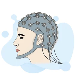
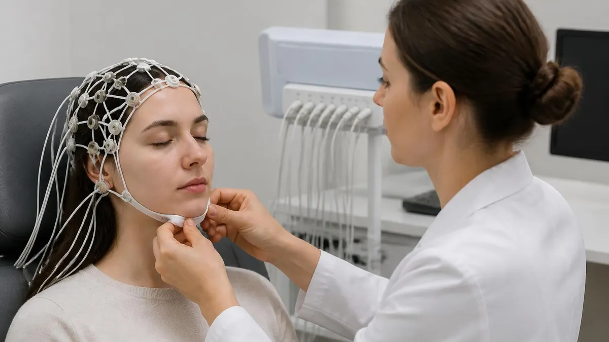
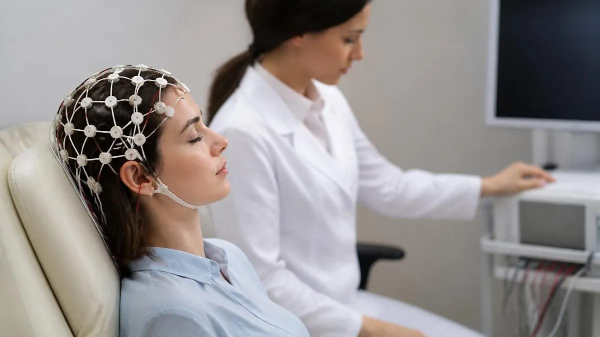
The question of what is an EEG test often arises when patients are referred for evaluation of seizures, altered consciousness, or unexplained cognitive decline. During the procedure, a technician applies 19 to 25 electrodes to the scalp using a conductive gel. These electrodes passively detect tiny voltage differences between regions of the brain and transmit them to an amplifier, where they appear as wavy lines representing different frequency bands: delta, theta, alpha, and beta waves.
Each band corresponds to a different mental state and physiological process, and deviations from age-appropriate norms can signal pathology. You can learn more about eeg brain test preparation and interpretation through practice resources designed for technicians and students.
One important consideration is how long an EEG test takes. A routine outpatient EEG typically runs 20 to 40 minutes of actual recording time, though the full appointment — including electrode placement and removal — usually lasts 60 to 90 minutes. Prolonged monitoring studies, which may be ordered for patients with suspected ongoing seizure activity or disorders of consciousness following brain injury, can last 24 hours, 72 hours, or even weeks in specialized epilepsy monitoring units. The duration ordered depends heavily on what the referring clinician is trying to detect and how intermittent the suspected abnormality may be.
Patients frequently search for information about EEG test side effects, and the good news is that the procedure is extremely safe. There is no radiation, no electrical current passed through the brain, and no sedation required for most adult studies. Some patients experience mild scalp irritation from the electrode gel, and the sleep deprivation sometimes requested before the test can cause fatigue. Rare patients with photosensitive epilepsy may experience symptoms during the photic stimulation portion of the test, which is why qualified technicians monitor patients carefully throughout. Overall, the EEG ranks among the safest diagnostic tools available in modern neurology.
Because the EEG is non-invasive, repeatable, and relatively affordable compared to advanced imaging, it remains a cornerstone of neurological evaluation decades after its introduction. Whether used to diagnose epilepsy, monitor coma depth, confirm brain death, or assess recovery from encephalopathy, this versatile study provides real-time windows into living brain function that no static imaging technique can replicate. The sections below explore each of these clinical applications in depth, covering what patterns appear in brain damage, how to interpret results, what the EEG test price looks like, and how professionals prepare to read these complex tracings.
EEG & Brain Damage: Key Numbers

How the EEG Detects Signs of Brain Damage
When one brain region is damaged, neurons in that area generate slower frequencies. A neurologist sees this as focal delta or theta waves appearing consistently over one hemisphere or lobe, indicating localized structural or functional injury such as a stroke or tumor.
This severe pattern alternates between brief bursts of high-amplitude activity and periods of near-electrical silence. It indicates profound global brain dysfunction and is commonly seen after cardiac arrest, severe anoxic injury, or deep anesthesia in ICU patients.
A healthy awake adult shows a dominant alpha rhythm of 8–13 Hz. Diffuse brain damage from encephalopathy, metabolic disturbance, or traumatic injury typically replaces this with slower delta or theta rhythms across the entire recording, reflecting widespread neuronal dysfunction.
Sharp waves, spikes, and spike-and-wave complexes indicate cortical irritability, often occurring at the edges of a damaged zone. These discharges reflect neurons that are hyper-excitable due to local injury, scar tissue, or disrupted inhibitory signaling in the surrounding tissue.
The complete absence of EEG activity above 2 microvolts, confirmed on two recordings at least six hours apart, is one criterion used to support a determination of brain death. This finding requires strict technical standards and must be interpreted alongside clinical exam findings.
Understanding the types of brain damage that an EEG can and cannot reveal helps clinicians order the right tests and helps patients set realistic expectations. Ischemic strokes produce characteristic focal slowing over the affected vascular territory within hours of onset. When a large stroke affects the middle cerebral artery, for example, the EEG typically shows prominent delta waves over the ipsilateral frontotemporal region. This focal asymmetry mirrors the clinical deficits — weakness, language problems, or neglect — and helps confirm the anatomical location of injury before MRI is available or when imaging is contraindicated by a pacemaker or severe claustrophobia.
Traumatic brain injury presents a more heterogeneous EEG picture because the damage is often diffuse and multifocal. Mild concussions may produce very subtle slowing that normalizes within days, while moderate-to-severe TBI produces persistent background slowing, loss of sleep architecture, and in some cases post-traumatic epileptiform discharges that mark the beginning of post-traumatic epilepsy. Research suggests that up to 25 percent of patients with moderate-to-severe TBI develop epilepsy within the first five years of injury, and the EEG plays a central role in detecting this complication early enough to initiate prophylactic treatment.
Hypoxic-ischemic encephalopathy — brain injury caused by oxygen deprivation during cardiac arrest, near-drowning, or neonatal birth complications — produces some of the most dramatic EEG changes seen in clinical practice. Continuous EEG monitoring in the ICU following cardiac arrest has become standard of care at major medical centers because the EEG provides prognostic information that clinical examination alone cannot offer. The presence of burst-suppression, periodic discharges, or absence of sleep-wake cycling within the first 24 to 72 hours correlates with poor neurological outcomes, while the early return of organized background activity and sleep spindles signals a more favorable recovery trajectory.
For patients with encephalopathy caused by metabolic disorders — including hepatic failure, uremic encephalopathy, and hypoglycemia — the EEG reflects the degree of systemic disruption rather than a fixed structural lesion. Triphasic waves, named for their distinctive three-phase morphology, were historically associated with hepatic encephalopathy but are now recognized as a non-specific marker of severe metabolic brain dysfunction.
As the underlying metabolic abnormality is corrected, the EEG typically normalizes in parallel, making serial EEG recordings a useful monitoring tool in this setting. To understand how EEG cost factors into long-term monitoring decisions, explore resources on eeg brain activity test pricing and insurance coverage.
Infectious encephalitides — including herpes simplex encephalitis, autoimmune encephalitis, and Creutzfeldt-Jakob disease — each produce recognizable EEG signatures that contribute to diagnosis. Herpes simplex encephalitis classically generates periodic lateralized epileptiform discharges arising from the temporal lobes, reflecting the virus's predilection for limbic structures.
In Creutzfeldt-Jakob disease, generalized periodic sharp wave complexes appearing at a rate of approximately one per second are so characteristic that their presence in the appropriate clinical context is considered a major diagnostic criterion. These disease-specific patterns illustrate how the EEG functions not just as a generic indicator of brain dysfunction but as a differential diagnostic tool in the hands of an experienced reader.
Neonatal brain injury deserves special mention because the immature brain produces a completely different EEG background than the adult brain. Premature infants display discontinuous tracings with long interburst intervals that are normal for gestational age. Pathological changes in neonates include abnormal sharp transients, depressed or excessively discontinuous backgrounds, and seizure patterns that may be purely electrographic with no visible clinical manifestations. Continuous EEG monitoring in neonatal ICUs has transformed the detection and treatment of neonatal seizures, many of which would otherwise go unrecognized and untreated, leading to additional brain injury from repeated uncontrolled electrical storms.
Autoimmune encephalitis has emerged in recent years as a critically important cause of brain dysfunction, particularly in young adults and children. Conditions such as anti-NMDA receptor encephalitis produce a distinctive EEG pattern called extreme delta brush — a pattern of delta waves superimposed with rhythmic beta activity — that is rarely seen in other conditions.
Recognizing this pattern on EEG can prompt the clinician to order the appropriate antibody panels, initiate immunotherapy, and search for an underlying teratoma before the patient deteriorates further. This example demonstrates how EEG pattern recognition is an active, evolving field that requires ongoing education and exposure to the latest literature.
What Is an EEG Medical Test? Procedure, Cost & Duration
A routine EEG begins with the technician measuring the patient's head to place electrodes at standardized positions defined by the International 10-20 System. Conductive gel is applied under each electrode to ensure good electrical contact. The patient lies still with eyes closed while the technician records resting brain activity, then performs activation procedures including hyperventilation for three minutes and photic stimulation using a strobe light to provoke latent abnormalities that may not appear at rest.
Most adult EEGs are performed without sedation. Patients are asked to remain relaxed but awake unless a sleep study is specifically requested, in which case they may be asked to arrive sleep-deprived the night before. The recording technician continuously monitors the tracing for movement artifacts from eye blinking, jaw clenching, or muscle tension, and may ask the patient to adjust position to obtain a clean signal. After recording, electrodes are removed and the gel is washed from the scalp. The raw data is then reviewed and reported by a board-certified clinical neurophysiologist or neurologist.

EEG for Brain Damage: Benefits and Limitations
- +Non-invasive and safe — no radiation or electrical current delivered to the brain
- +Provides real-time functional information that MRI and CT cannot offer
- +Can detect abnormalities within minutes to hours of brain injury onset
- +Useful prognostic tool in coma and post-cardiac arrest monitoring
- +Identifies non-convulsive seizures invisible to clinical observation alone
- +Repeatable and relatively affordable compared to advanced neuroimaging
- −Cannot show structural lesions such as contusions, hematomas, or tumors directly
- −Normal EEG does not rule out brain damage, especially mild or deep injuries
- −Highly sensitive to movement, muscle, and electrode artifacts requiring skilled technicians
- −Interpretation requires specialized training; errors by untrained readers can mislead clinicians
- −Routine 30-minute study may miss intermittent abnormalities that only appear during events
- −Limited spatial resolution compared to high-field MRI for precise localization of damage
Preparing for Your EEG Test: 10-Step Checklist
- ✓Wash your hair the night before with regular shampoo but skip conditioner, oil, or styling products.
- ✓Ask your ordering physician whether you should be sleep-deprived the night before the study.
- ✓Confirm with your neurologist whether to continue, reduce, or temporarily stop anti-seizure medications.
- ✓Eat a normal meal before the appointment — low blood sugar can alter EEG findings.
- ✓Avoid caffeine on the morning of the test if instructed by your technician.
- ✓Wear comfortable, loose-fitting clothing so you can recline comfortably during recording.
- ✓Arrive 15 minutes early to complete paperwork and allow time for electrode placement.
- ✓Inform the technician about all medications, supplements, and any recent sleep changes.
- ✓Remain as still as possible during recording to minimize muscle and movement artifacts.
- ✓Ask the technician how and when you will receive results, and arrange follow-up with your neurologist.
A Normal EEG Does Not Mean a Normal Brain
Up to 10 percent of patients with confirmed structural brain damage — including small strokes, mild traumatic brain injury, and early neurodegenerative disease — will have a completely normal routine EEG. Always combine EEG findings with neuroimaging, clinical history, and cognitive assessment for the most accurate picture of brain health.
The limitations of the EEG test are as important to understand as its strengths, particularly for patients and families who may expect a single test to answer all questions about brain injury. The spatial resolution of standard scalp EEG is constrained by the distance between the electrodes and the cortical surface, as well as the volume conduction properties of the skull and scalp tissues.
This means that deep brain structures — the hippocampus, thalamus, brainstem, and white matter tracts — generate signals that are severely attenuated by the time they reach scalp electrodes. Damage to these regions may therefore produce little or no change in the routine EEG, even when the clinical deficits are substantial.
High-density EEG systems with 64, 128, or 256 electrodes improve spatial resolution considerably by sampling more points across the scalp surface and applying sophisticated mathematical algorithms to reconstruct the likely cortical sources of recorded activity. These advanced systems are increasingly used in research settings and specialized epilepsy centers for source localization prior to surgical planning. However, they remain impractical for routine clinical use due to the time required for electrode application, the complexity of data analysis, and cost considerations that limit their availability to major academic medical centers.
Neuroimaging and EEG are complementary rather than competitive tools. MRI excels at revealing structural anatomy — showing exactly where a lesion is located, how large it is, and whether it involves gray matter, white matter, or both. The EEG excels at capturing dynamic functional information — showing how the brain is actually working at this moment, whether abnormal synchrony is occurring, and whether treatable electrical events are ongoing.
In the acute setting, both tests are often ordered simultaneously because each answers a different question. A patient with a subdural hematoma, for example, needs CT or MRI to define the blood collection, but also needs EEG to determine whether the pressure is provoking subclinical seizures that require treatment.
Functional MRI, positron emission tomography, and single-photon emission computed tomography provide additional layers of information about cerebral blood flow and metabolism that neither structural MRI nor EEG can provide. These modalities are particularly valuable in disorders of consciousness — vegetative state and minimally conscious state — where EEG may show organized background activity but the patient shows no reliable behavioral responses.
Research groups using high-density EEG combined with transcranial magnetic stimulation have demonstrated that some patients diagnosed as vegetative are actually processing information covertly, challenging clinicians and families to reconsider prognosis and care decisions. To explore how EEG patterns specifically relate to seizure activity associated with brain damage, read more about eeg test for brain evaluation and what seizure patterns reveal about the injured brain.
Quantitative EEG analysis represents a growing bridge between traditional visual EEG interpretation and objective, reproducible brain function metrics. By applying computerized algorithms to the raw EEG signal, quantitative tools can calculate power in each frequency band, asymmetry indices between hemispheres, connectivity measures, and event-related potentials with far greater precision than the human eye can achieve reviewing paper tracings. These metrics are increasingly used in sports medicine to quantify the effects of concussion, in anesthesiology to titrate sedation depth, and in psychiatry to guide medication selection using neurophysiological biomarkers.
The emerging field of EEG-based brain-computer interfaces takes the technology far beyond diagnostics. Patients with severe motor impairments from conditions such as amyotrophic lateral sclerosis or locked-in syndrome can learn to modulate their own brain rhythms to control external devices — spelling systems, robotic arms, or environmental controls — using only their neural signals. While this application is still largely in the research phase, it illustrates that EEG is not merely a passive window into a damaged brain but potentially a platform for rehabilitative interaction that exploits whatever residual function remains after injury.
For EEG technologists and neurologists in training, developing fluency with abnormal patterns associated with brain damage requires sustained practice with a wide variety of tracings. Recognizing the difference between a true focal slow wave and an electrode artifact, between an authentic sharp transient and a normal variant waveform, and between genuine burst-suppression and the pattern produced by poor electrode contact demands years of supervised experience combined with systematic self-study using validated question banks and annotated atlas resources.

If you or someone you know experiences sudden confusion, loss of consciousness, uncontrolled shaking, inability to speak, or one-sided weakness, call 911 immediately. These symptoms may indicate a stroke, severe seizure, or other acute brain injury requiring emergency neuroimaging and continuous EEG monitoring — delays in evaluation can significantly worsen outcomes.
Continuous EEG monitoring in the intensive care unit has transformed how clinicians manage critically ill patients with brain injury. Before continuous monitoring became widely available, non-convulsive seizures and non-convulsive status epilepticus were vastly underrecognized in the ICU population.
Studies using continuous EEG in ICUs at major academic centers have found that approximately 8 to 34 percent of comatose patients — depending on the underlying etiology — have ongoing electrographic seizures that produce no visible clinical signs and would therefore go entirely undetected without continuous monitoring. Each undetected seizure episode contributes additional metabolic stress to an already damaged brain, potentially worsening the ultimate degree of injury.
The 2021 American Clinical Neurophysiology Society guidelines recommend continuous EEG monitoring for all patients with unexplained altered consciousness, subarachnoid hemorrhage with poor neurological grade, moderate-to-severe traumatic brain injury, and post-cardiac arrest encephalopathy managed with targeted temperature therapy. These recommendations reflect the growing body of evidence that EEG-guided management — specifically, identifying and treating electrographic seizures — improves survival and functional outcomes in these critically ill populations. Implementation of these guidelines remains inconsistent across hospitals, however, because continuous EEG requires trained personnel to apply and maintain electrodes, specialized remote monitoring infrastructure, and qualified neurophysiologists available around the clock.
Prognostication after cardiac arrest is one of the most emotionally and medically complex challenges in critical care medicine. Families and medical teams must make decisions about continuation or withdrawal of life-sustaining treatment within a relatively short time window, often before the full extent of brain injury is known. The EEG provides objective data that complements the neurological examination and biomarkers such as serum neuron-specific enolase.
The presence of a continuous and reactive EEG background within 24 to 48 hours of resuscitation is associated with favorable neurological recovery in multiple large observational studies, while burst-suppression and electrocerebral silence at 72 hours — in the absence of sedative medication confounding — are associated with very poor prognosis.
Understanding EEG coherence — a measure of how synchronously different brain regions communicate with each other — adds an additional dimension to the assessment of brain damage. Normal brains maintain tightly regulated coherence patterns that vary with cognitive task and arousal state. In patients with diffuse axonal injury from TBI or hypoxic damage, coherence between frontal and posterior regions is often severely reduced, reflecting the disruption of the long-range white matter tracts that normally coordinate inter-regional communication.
Quantitative coherence analysis may eventually serve as a sensitive biomarker for subclinical connectivity damage not visible on conventional imaging. Learn more about how this metric is measured in our detailed guide to eeg for brain damage and connectivity analysis.
Neuroprognostication in traumatic brain injury also relies heavily on EEG data. The presence of sleep architecture — specifically, spindles and K-complexes — in a comatose TBI patient suggests that the thalamocortical circuits responsible for generating sleep rhythms are at least partially intact, which is generally a favorable prognostic sign.
Conversely, the complete absence of sleep features in a patient who has been observed for several days suggests severe disruption of these circuits and is associated with worse long-term outcomes. Serial EEG recordings that document the evolution of background activity over the first week after injury often provide the most useful prognostic trajectory rather than any single snapshot recording.
Pediatric applications of EEG for brain damage assessment have unique considerations because the developing brain produces age-specific background patterns that differ dramatically from adult norms. A two-month-old infant's EEG looks completely different from a two-year-old's, which in turn differs from a school-age child's recording. Neonatologists and pediatric neurologists interpreting these studies must have specialized training in developmental EEG norms to avoid misclassifying normal developmental patterns as pathological or, conversely, missing genuine abnormalities that fall within the range of normal for a given gestational age. This developmental variability makes neonatal and pediatric EEG interpretation a distinct subspecialty within clinical neurophysiology.
The integration of artificial intelligence and machine learning into EEG interpretation represents the frontier of this field. Several research groups and commercial companies have developed algorithms capable of detecting seizures, classifying sleep stages, and identifying patterns associated with specific diagnoses with accuracy approaching or in some cases exceeding that of trained human reviewers.
These AI tools are particularly promising for continuous ICU monitoring, where the sheer volume of data generated over days of recording exceeds what any individual can manually review with full attention. As these systems mature and receive regulatory clearance, they are likely to become standard components of the neurophysiology workflow, freeing clinicians to focus their expertise on ambiguous and complex cases.
For EEG technologists preparing for the ABRET credentialing examination, mastering the patterns associated with brain damage is not merely an academic exercise — it is a core clinical competency tested extensively on the exam and applied every working day. The Registered EEG Technologist (R. EEG T.) credential requires candidates to demonstrate knowledge of normal and abnormal patterns, artifact recognition, electrode application, and activation procedures. Questions on abnormal patterns in the context of structural and metabolic brain disease account for a substantial proportion of the exam blueprint, making this topic one of the highest-yield areas for focused study.
A systematic approach to studying EEG brain damage patterns involves first building a solid foundation in normal EEG for each age group, then methodically learning the deviations that characterize major pathological categories. Start with the most common and clinically urgent patterns: focal slowing, generalized slowing, and epileptiform discharges.
Then progress to the more specialized patterns: triphasic waves in metabolic encephalopathy, periodic lateralized epileptiform discharges in focal brain damage, burst-suppression in severe global injury, and the extreme delta brush of anti-NMDA encephalitis. Each pattern should be studied from multiple angles — its appearance on the page, the clinical conditions associated with it, and the technical factors that can mimic or mask it.
Practice questions and case-based learning are essential complements to textbook reading. Reading a description of burst-suppression in a textbook chapter conveys the concept intellectually, but actually identifying it in a practice tracing alongside a clinical vignette builds the pattern recognition skills that translate to real clinical performance. The ABRET examination emphasizes clinical application over rote memorization, so candidates who practice with realistic clinical scenarios consistently outperform those who study definitions alone. Timed practice under exam conditions also builds the mental stamina needed to sustain attention through a multi-hour credentialing examination.
Beyond credentialing preparation, the EEG field is evolving rapidly enough that even experienced practitioners benefit from continuing education. New pathological entities are being described — particularly in the autoimmune encephalitis spectrum — that require updating pattern recognition skills that were established during training. Journals such as the Journal of Clinical Neurophysiology, Epilepsia, and Neurocritical Care regularly publish case series and consensus statements that refine the criteria for specific EEG diagnoses. Subscribing to these resources and attending annual meetings of the American Epilepsy Society or the American Clinical Neurophysiology Society provides ongoing exposure to the latest developments in this dynamic field.
Self-assessment using structured question banks is one of the most efficient study strategies because it simultaneously exposes knowledge gaps, reinforces correct information through immediate feedback, and simulates the exam environment. When reviewing incorrect answers, the goal should not merely be to memorize the correct answer but to understand the underlying principle well enough to apply it to a novel clinical scenario. This transfer of learning from memorized facts to flexible problem-solving is the hallmark of genuine expertise and the level of competence that ABRET credentialing is designed to certify.
Time management during EEG preparation is itself a skill worth developing. Candidates who spread study over 12 to 16 weeks consistently perform better than those who attempt intensive cramming in the final two weeks before the examination.
Spacing repetitions across multiple sessions exploits the well-documented spacing effect in memory consolidation, whereby information reviewed at increasing intervals is retained far more durably than information reviewed repeatedly in a single sitting. A practical schedule might involve covering one major topic category per week, reviewing it again at two weeks, and then incorporating it into mixed-content practice tests during the final month before the examination.
Finally, understanding the clinical context behind each EEG finding transforms rote pattern recognition into meaningful clinical reasoning. When a technologist understands that the focal slowing they are recording over the left temporal region likely reflects ischemic injury to the speech cortex — and that the patient's aphasia reported in the clinical history confirms this localization — the EEG ceases to be an abstract pattern and becomes a window into a real person's neurological status.
This integration of technical skill and clinical understanding is what distinguishes excellent EEG professionals from merely competent ones, and it is the quality that the most demanding employers and most discerning patients ultimately rely upon.
EEG Questions and Answers
EEG Electroencephalography Practice Test PDF (Free Printable 2026)

Seizure EEG: What the EEG Test Shows, How It Works, and What to Expect
EEG Test Cost in 2026: Prices, Insurance Coverage, and How to Save

EEG Coherence: What It Is, How It Works, and What It Reveals About Brain Connectivity
EEG vs ECG vs EKG: What Each Test Measures, How They Differ, and What to Expect
About the Author

Educational Psychologist & Academic Test Preparation Expert
Columbia University Teachers CollegeDr. Lisa Patel holds a Doctorate in Education from Columbia University Teachers College and has spent 17 years researching standardized test design and academic assessment. She has developed preparation programs for SAT, ACT, GRE, LSAT, UCAT, and numerous professional licensing exams, helping students of all backgrounds achieve their target scores.