
EEG vs ECG vs EKG: What Each Test Measures, How They Differ, and What to Expect
Learn what an EEG test, ECG, and EKG each measure, how they differ, cost, side effects, and duration. 🧠 Clear guide for patients and students.

When a doctor orders an EEG test, many patients immediately wonder whether it is the same as an ECG or EKG they may have had before. The confusion is understandable because all three acronyms sound similar and all three involve attaching electrodes to the body to record electrical signals — but the organs they monitor, the conditions they diagnose, and the clinical workflows surrounding them are entirely different.
Understanding the distinction between ecg vs ekg vs eeg is essential whether you are a patient preparing for a procedure, a nursing student reviewing fundamentals, or a healthcare professional studying for a certification exam.
The EEG test — short for electroencephalography — measures the brain's electrical activity. Electrodes placed on the scalp pick up tiny voltage fluctuations produced by millions of neurons firing simultaneously. Neurologists use EEG results to identify seizure disorders, evaluate altered consciousness, investigate sleep disturbances, and even confirm brain death in the most serious clinical scenarios. The EEG medical test is the gold standard for epilepsy diagnosis and cannot be replaced by imaging studies like MRI or CT scans, which show structure rather than function.
The ECG and EKG, by contrast, record electrical activity in the heart rather than the brain. The two terms refer to the same test — ECG is an abbreviation of the English term electrocardiogram, while EKG comes from the German spelling Elektrokardiogramm. Cardiologists adopted EKG historically to distinguish the test from EEG in verbal communication and written orders, reducing the risk of one being confused for the other in a busy clinical environment. Both acronyms describe identical technology and produce the same waveform tracing of the cardiac cycle.
One of the most common questions patients ask before scheduling is what is an eeg test and how it compares to the heart test their cardiologist ordered. The procedural experience is superficially similar: a technician applies conductive gel and attaches electrodes, and the machine records signals passively without delivering any electricity into the body. However, standard EEG uses 19 to 256 electrodes arranged across the entire scalp according to the international 10–20 system, while a standard 12-lead ECG uses just 10 electrodes placed on the chest, wrists, and ankles. Preparation, duration, and interpretation are all distinct.
Knowing the difference between these tests also has real financial implications. The eeg test cost and the cost of a cardiac workup follow very different billing pathways, involve different specialist referrals, and are covered by insurance under separate diagnostic codes. A routine EEG typically runs between $200 and $700 at an outpatient clinic, while a standard ECG in the same facility may cost as little as $50 to $150. When a physician orders both in the same visit, patients should confirm their insurance covers each procedure separately to avoid unexpected out-of-pocket expenses.
This article walks through what each test measures, how the procedures compare in practice, how long each lasts, what side effects to anticipate, and how the results are interpreted. Whether you are preparing for an upcoming appointment or studying for the R. EEG T. board examination, the side-by-side comparison in the following sections will give you a clear, clinically accurate foundation. Readers who want to reinforce their understanding with practice questions can explore our full guide on eeg vs ecg vs ekg for downloadable study materials alongside this article.
By the end of this guide, you will understand not just what each acronym stands for but why the distinction matters clinically, what a technologist does during each procedure, what abnormal findings look like, and how patients can prepare to get the most accurate results from whichever test their physician has ordered. The information below reflects current US clinical practice standards and is written to be useful for both general audiences and allied health professionals seeking certification-level depth.
EEG vs ECG vs EKG by the Numbers

EEG, ECG, and EKG: The Core Differences at a Glance
Records electrical activity of the brain using scalp electrodes. Used to diagnose epilepsy, sleep disorders, encephalopathy, and brain death. Involves 19–256 electrodes and lasts 20 minutes to several days for ambulatory monitoring.
Records electrical activity of the heart using 10 electrodes on the chest, wrists, and ankles. Used to diagnose arrhythmias, heart attacks, and conduction abnormalities. A standard resting test takes under 10 minutes total.
ECG and EKG are identical procedures. EKG derives from the German Elektrokardiogramm and was adopted in English-speaking hospitals to prevent verbal mix-ups with EEG orders. Both generate the same cardiac waveform tracing.
Some neurological conditions mimic cardiac arrhythmias and vice versa. Syncope workups, for example, may include both an EEG and an ECG. Each is billed and interpreted separately by different specialist teams.
Understanding what is an eeg medical test requires a brief look at neurophysiology. The human brain contains approximately 86 billion neurons, and at any given moment billions of them are communicating via electrochemical signals.
When large groups of neurons fire in synchrony — as they do during normal brain states like sleep and wakeful rest — the cumulative electrical activity is strong enough to be detected through the skull and scalp by sensitive amplifiers. The EEG captures these signals in the form of oscillating waves categorized by frequency: delta (0.5–4 Hz), theta (4–8 Hz), alpha (8–13 Hz), beta (13–30 Hz), and gamma (above 30 Hz).
Each frequency band corresponds to a different brain state. Alpha waves are dominant when you close your eyes and relax; beta waves appear during focused cognitive activity; delta waves are hallmarks of deep, dreamless sleep. During a seizure, the normal background rhythms are disrupted by high-amplitude, high-frequency discharges that appear on the EEG tracing as sharp waves, spike-and-wave complexes, or generalized paroxysmal bursts depending on the seizure type. A trained electroencephalographer — typically a neurologist with subspecialty training — reads these patterns and produces a clinical interpretation report.
The ECG measures something fundamentally different: the depolarization and repolarization of cardiac muscle cells that drive the mechanical pumping of the heart. The classic ECG waveform shows a P wave (atrial depolarization), a QRS complex (ventricular depolarization), and a T wave (ventricular repolarization). Cardiologists analyze the shape, duration, and intervals of these components to identify conditions ranging from atrial fibrillation and ventricular tachycardia to ST-elevation myocardial infarction, bundle branch blocks, and prolonged QT syndrome. No brain activity is captured, and no neurological interpretation is performed.
A common misconception is that the EEG can somehow detect heart problems or vice versa. This is physiologically impossible: the electrode placements, amplifier sensitivity ranges, frequency filters, and signal processing pipelines of the two devices are optimized for entirely different signal sources. An EEG amplifier tuned to detect microvolt-level brain signals would be overwhelmed by the millivolt-scale cardiac signals if a chest electrode were accidentally included in the montage. Clinical technologists are trained to recognize and eliminate cardiac artifact — the ECG signal that sometimes bleeds into EEG recordings through the body — rather than interpret it diagnostically.
For patients asking what is a eeg test from a procedural standpoint, the experience begins with a technologist measuring the patient's head and marking electrode positions using a soft pencil and measuring tape. The scalp is lightly abraded at each marked site to reduce skin resistance, and electrodes filled with conductive paste are applied.
Once impedance values at all electrode sites fall below acceptable thresholds — typically under 5 kilohms — the recording begins. The patient is asked to rest quietly with eyes closed, then eyes open, and may be asked to breathe rapidly (hyperventilation) or view a flickering strobe light to provoke abnormal activity if epilepsy is suspected.
The ECG procedure, by comparison, takes only a few minutes. The patient lies flat, gel patches or sticky electrode stickers are placed on standardized positions on the limbs and chest, and the 10-second recording is captured automatically. No special preparation of the skin beyond cleaning is required, no scalp measurement occurs, and no provocative maneuvers are performed during a routine resting ECG. The speed and simplicity of the ECG make it one of the most commonly performed diagnostic tests in the world, used in emergency departments, primary care offices, and pre-operative screenings daily.
Students preparing for the R. EEG T. examination need to understand both normal and abnormal waveform patterns in detail. Practice resources such as the printable question sets linked from our eeg vs ecg vs ekg study page offer targeted review of waveform identification, electrode placement, and artifact recognition — exactly the type of knowledge tested on the ABRET certification exam. Mastery of how EEG differs from cardiac monitoring is a foundational competency that appears across multiple domains of the examination blueprint.
How Long Is an EEG Test? Duration by Type
A standard outpatient routine EEG typically takes between 20 and 40 minutes of actual recording time, though patients should budget 60 to 90 minutes for the full appointment including setup and electrode removal. The technologist spends approximately 20 to 30 minutes placing and verifying electrodes before recording begins. The actual tracing includes a baseline resting phase, eyes-open and eyes-closed epochs, hyperventilation for 3 minutes, and photic stimulation at varying flash frequencies from 1 to 25 Hz.
Patients often ask whether the test is uncomfortable. Electrode placement involves mild scalp abrasion and the application of a slightly cool gel, but no needles, no electricity, and no pain. After the recording, the paste is washed out with warm water. Most outpatient facilities complete the entire visit within one hour, and patients may drive themselves home and return to normal activities immediately unless sedation was used for the procedure.

EEG vs ECG/EKG: Benefits and Limitations of Each Test
- +EEG directly measures brain electrical function — no imaging equivalent can replicate this
- +ECG/EKG is extremely fast, taking under 10 minutes including setup and recording
- +EEG can record over days, capturing rare spontaneous seizures that brief tests miss
- +ECG is widely available in virtually every clinical setting including primary care offices
- +EEG is non-invasive and radiation-free, safe for all ages including newborns and pregnant women
- +ECG/EKG can identify life-threatening cardiac events like STEMI within seconds of tracing review
- −EEG setup takes 20–30 minutes and requires specialized technologist training and equipment
- −ECG cannot detect intermittent arrhythmias that don't occur during the brief recording window
- −EEG interpretation requires subspecialty neurologist training; misreading is a documented risk
- −ECG cannot diagnose structural heart disease — echocardiography or angiography are needed
- −EEG results can be obscured by muscle artifact, electrode pop, and patient movement
- −ECG/EKG does not provide any information about brain function or neurological conditions
EEG Test Preparation Checklist: What to Do Before Your Appointment
- ✓Wash your hair thoroughly the night before or morning of the test — do not apply conditioner, oil, or styling products
- ✓Get adequate sleep the night before unless your doctor specifically ordered a sleep-deprived EEG
- ✓Eat a full meal before arriving — low blood sugar can affect EEG findings and may cause symptoms during hyperventilation
- ✓Continue taking all prescribed medications unless your neurologist has explicitly instructed you to hold a specific drug
- ✓Avoid caffeine for at least 8 hours before the test, as stimulants can alter background EEG frequency patterns
- ✓Arrive 15 minutes early to complete paperwork and allow the technologist to begin setup without rushing
- ✓Wear comfortable, loose-fitting clothing — you will be reclining or lying down for the duration of the recording
- ✓Inform the technologist of all medications, supplements, and any recent illnesses or sleep changes
- ✓Remove earrings and other scalp jewelry before the appointment to avoid interference with electrode placement
- ✓Arrange transportation if sedation was ordered — most facilities will not allow self-driving after sedated EEG
A Normal EEG Does Not Rule Out Epilepsy
Up to 50% of people with confirmed epilepsy have a normal routine EEG on their first study. A single 30-minute recording captures only a brief snapshot of brain activity, and seizure discharges may simply not occur during that window. A normal result reduces suspicion but never eliminates it — prolonged or sleep-deprived studies are frequently needed when clinical suspicion remains high despite an initial normal tracing.
The eeg test cost in the United States varies considerably depending on the type of study ordered, the facility where it is performed, the geographic region, and whether the patient has insurance. A routine outpatient EEG at a hospital-affiliated neurology clinic typically costs between $200 and $700 before insurance adjustments.
Freestanding diagnostic imaging centers and private neurology practices may charge less — sometimes as low as $150 for a routine study — while academic medical centers in high-cost cities like New York or San Francisco can bill $900 or more for the same service. These prices cover the technical component only; a separate professional interpretation fee ranging from $100 to $300 is billed by the neurologist who reads the recording.
Insurance coverage for EEG tests is generally robust when a clear medical indication is documented. Commercial insurers, Medicare, and Medicaid all cover the EEG medical test when ordered for medically recognized indications including evaluation of epilepsy, assessment of altered mental status, monitoring of sedation in the ICU, or brain death determination. The key to avoiding claim denials is ensuring that the ordering physician documents the clinical indication explicitly in the referral. Routine EEG falls under CPT code 95816 (awake and drowsy) or 95819 (awake and asleep), while ambulatory studies use different code sets that carry higher reimbursement rates.
Patients without insurance or with high-deductible plans can negotiate the eeg test price directly with the facility. Most hospitals and large clinic systems have financial assistance programs, and the list price can often be reduced by 30 to 60 percent through self-pay discounts. Community health centers that operate under federally qualified health center (FQHC) status offer EEG services on a sliding fee scale based on household income. Patients should ask the scheduling department specifically about financial assistance before providing insurance information, as this can improve bargaining position when negotiating rates.
By comparison, the ECG/EKG is one of the most affordable diagnostic tests in medicine. A resting 12-lead ECG in an outpatient setting typically costs $50 to $200 before insurance, and many primary care practices include it as part of an annual wellness visit with no additional charge beyond the office visit copay. Emergency department ECGs are billed differently because they are interpreted in the context of an acute presentation, and facility fees can push the total charge significantly higher — sometimes $500 or more on the itemized hospital bill — though insurers typically pay a fraction of the chargemaster rate.
For healthcare students and professionals studying for certification examinations, understanding billing and procedure codes is not just academically interesting — it appears directly on the R. EEG T. exam as part of the professional responsibilities domain. The ABRET blueprint expects candidates to recognize appropriate CPT coding categories for various EEG study types, understand documentation requirements for medical necessity, and identify which clinical scenarios justify extended versus routine monitoring. These are practical competencies that affect how EEG labs operate financially and how technologists communicate with administrative teams about procedure scheduling and authorization.
Long-term video-EEG monitoring represents the most expensive end of the spectrum. Inpatient epilepsy monitoring unit stays that run 5 to 7 days with continuous video-EEG, nursing care, and daily neurologist review can generate hospital bills ranging from $10,000 to $40,000 depending on the facility and complication rate. These costs are almost always covered by insurance when pre-authorized, because the clinical value — definitive seizure classification enabling surgical candidacy evaluation — is well established and the alternative (continued medication trials for drug-resistant epilepsy) carries its own substantial cost and morbidity burden over time.
Patients who have been quoted a high eeg test cost should not delay or avoid necessary testing for financial reasons without first exploring all assistance options. The Epilepsy Foundation offers a helpline and local affiliate network that can connect patients with financial assistance resources, community grants, and charity care programs. Many neurologists' offices also have financial counselors who can submit prior authorization requests, appeal insurance denials, and identify patient assistance programs that significantly reduce out-of-pocket expenses for uninsured and underinsured patients nationwide.

Before scheduling an EEG test, ask the facility for their self-pay rate and request an itemized cost estimate that separates the technical fee from the professional interpretation fee. Many outpatient neurology centers offer 30–50% discounts for self-pay patients who pay at the time of service, and some academic medical centers waive interpretation fees for patients who qualify for financial assistance. Always get the quoted price in writing before your appointment date.
A question that appears frequently on patient intake forms and online forums is whether eeg test side effects are a concern. The straightforward answer is that a standard EEG is among the safest diagnostic procedures in medicine. The test records electrical signals passively — it does not deliver any electrical current to the brain or body — and the electrodes are held in place by paste rather than needles.
Skin irritation at electrode sites is the most commonly reported minor side effect, occurring when the skin abrasion used to reduce impedance is aggressive or when a patient has sensitive skin. This redness typically resolves within a few hours to a day.
The hyperventilation portion of the EEG carries a somewhat higher risk of producing transient symptoms. Breathing rapidly for 3 consecutive minutes causes hypocapnia — a drop in blood carbon dioxide — which produces cerebral vasoconstriction and can trigger tingling in the hands and feet, lightheadedness, and occasionally a sensation of faintness. These symptoms are expected, self-limited, and resolve within minutes of stopping hyperventilation.
In rare cases, hyperventilation can provoke an absence seizure in a patient with untreated childhood absence epilepsy — which is actually the point of the procedure, since provoking a brief absence during the recording confirms the diagnosis. Technologists are trained to monitor for and respond to seizures during the procedure.
Photic stimulation — the flashing strobe light used during the final phase of a routine EEG — carries a very small risk of triggering a generalized tonic-clonic seizure in patients with photosensitive epilepsy. Studies estimate that photosensitivity is present in approximately 3 percent of people with epilepsy, and among those, photic stimulation during EEG can occasionally provoke a seizure rather than simply subclinical epileptiform discharges. Technologists are required to have seizure first aid training and emergency protocols in place precisely because of this known risk, which is why EEG is performed in a supervised clinical setting rather than at home.
Sedated EEG, ordered for very young children or patients with intellectual disabilities who cannot cooperate with the standard procedure, carries the additional risks associated with the sedating medication used. Oral chloral hydrate, oral midazolam, or intranasal dexmedetomidine are common choices in pediatric EEG labs.
Each carries a small risk of respiratory depression, allergic reaction, or paradoxical agitation. Facilities that perform sedated EEG are required to have resuscitation equipment and trained personnel available, and the patient must be monitored for a recovery period after the recording before discharge — which is why these studies take longer and cost more than standard unsedated EEG.
The ECG and EKG are even lower risk than the EEG. Because no electrode abrasion is typically required and the recording lasts only 10 seconds, the only meaningful side effects are minor skin irritation from the adhesive electrode patches — particularly in patients with latex sensitivity if latex-containing leads are used — and the very rare vasovagal response in anxious patients who discomfort from the sticky electrode removal.
Exercise stress testing (the ECG variant performed during treadmill testing) carries higher cardiovascular risk, but a resting 12-lead ECG does not stress the heart in any way and poses essentially no physiological risk.
For patients who have been told they need both an EEG and an ECG in the same workup — common in syncope evaluations, for example — the two tests can often be scheduled on the same day at facilities that have both cardiology and neurology diagnostic services. The EEG should typically be performed first when both are on the schedule, as the electrode paste used in EEG can occasionally create artifact in ECG recordings if it drips onto the neck or chest.
A brief wash and dry of the face and neck between tests eliminates this potential source of interference. Patients curious about additional aspects of brain wave testing may find our article on eeg vs ecg vs ekg study materials particularly useful for connecting clinical concepts to examination-style questions.
Long-term ambulatory EEG monitoring introduces a different category of side effects: skin breakdown and pressure sores at electrode sites from days of continuous wear, scalp itching from prolonged contact with electrode paste, and the psychological stress of wearing a visible medical device in public. Modern disposable electrode arrays and hypoallergenic adhesives have substantially reduced skin-related complications, and EEG technologists perform daily skin checks during inpatient monitoring stays to identify and treat any early signs of irritation before they progress to more serious skin breakdown that would require premature termination of the study.
For students and technologists working toward ABRET certification, the comparison of EEG versus ECG versus EKG is more than academic trivia — it represents a foundational domain of professional competency. Examination candidates are expected to explain each test's clinical purpose, recognize the electrode systems used, understand artifact sources and how to eliminate them, and communicate clearly with patients and clinical teams about procedural details. The R. EEG T. examination blueprint explicitly tests knowledge of neurodiagnostic modalities in the context of related cardiac and neurological diagnostic procedures.
One of the most practically important skills for a working EEG technologist is recognizing cardiac artifact on a brain wave recording. Because the heart's electrical signals are many times stronger than brain signals, the QRS complex of the ECG can appear as a periodic spike artifact on EEG channels — particularly in recordings from patients with large hearts, arrhythmias, or unusual electrode placements near the temporal regions. Experienced technologists identify this artifact by its strict periodicity, its waveform shape matching the cardiac cycle, and its disappearance when electrode impedance and referential montages are optimized.
Understanding the clinical overlap between neurological and cardiac conditions is equally important for technologists who work in epilepsy monitoring units or cardiac ICU settings. Long QT syndrome, for example, can cause syncopal episodes that are clinically indistinguishable from generalized tonic-clonic seizures. Patients with this condition may be admitted to neurology services and undergo EEG monitoring before a cardiac etiology is identified through concurrent ECG review. Technologists who understand both modalities are better positioned to flag suspicious patterns to the reading physician and advocate for appropriate cross-specialty consultation.
Career pathways in neurodiagnostics continue to expand as hospitals recognize the value of specialized EEG monitoring beyond traditional epilepsy applications. Continuous EEG monitoring in the neurocritical care unit — where technologists manage long-term recordings in sedated, intubated patients with traumatic brain injury, stroke, or hypoxic-ischemic encephalopathy after cardiac arrest — requires deep technical competency and rapid artifact recognition in a high-stakes clinical environment. These positions command salaries at the upper end of the EEG technologist pay range, often $65,000 to $85,000 annually in major metropolitan areas, reflecting the specialized skill set and critical care environment.
The distinction between EEG and ECG also matters for patient education, which is a core responsibility of every EEG technologist. Patients who arrive expecting a quick 10-second test like an ECG — because a family member described their own cardiac test — will be surprised and potentially anxious to learn that an EEG involves 30 minutes of electrode placement followed by 40 minutes of recording.
Technologists who proactively explain the procedure, normalize the experience, and address patient concerns about electricity, pain, and what the test can and cannot detect are far more likely to obtain a high-quality, artifact-free recording from a relaxed, cooperative patient.
Telehealth and remote EEG interpretation services have expanded significantly in the United States over the past decade, allowing community hospitals and rural clinics to transmit EEG recordings securely to remote neurologist readers for same-day interpretation. This infrastructure has improved access to EEG medical test results in underserved regions where board-certified epileptologists were previously unavailable, reducing the time from recording to treatment decision from days or weeks to hours.
Technologists in these settings operate with greater autonomy, making the technical quality of the recording even more critical because there is no neurologist physically present to request repeat recording if artifact is identified after the patient leaves.
Looking ahead, artificial intelligence tools for automated EEG interpretation are entering clinical practice, with several FDA-cleared algorithms available to flag abnormal patterns for neurologist review. These tools do not replace physician interpretation but serve as a first-pass screening layer — similar to how AI algorithms now assist with ECG interpretation in primary care settings. Understanding the capabilities and limitations of automated analysis is becoming a new competency dimension for EEG technologists, who will increasingly work alongside AI-assisted systems while maintaining the technical expertise to verify, contextualize, and communicate results to clinical teams and patients.
EEG Questions and Answers
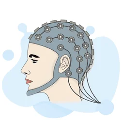
EEG Electroencephalography Practice Test PDF (Free Printable 2026)
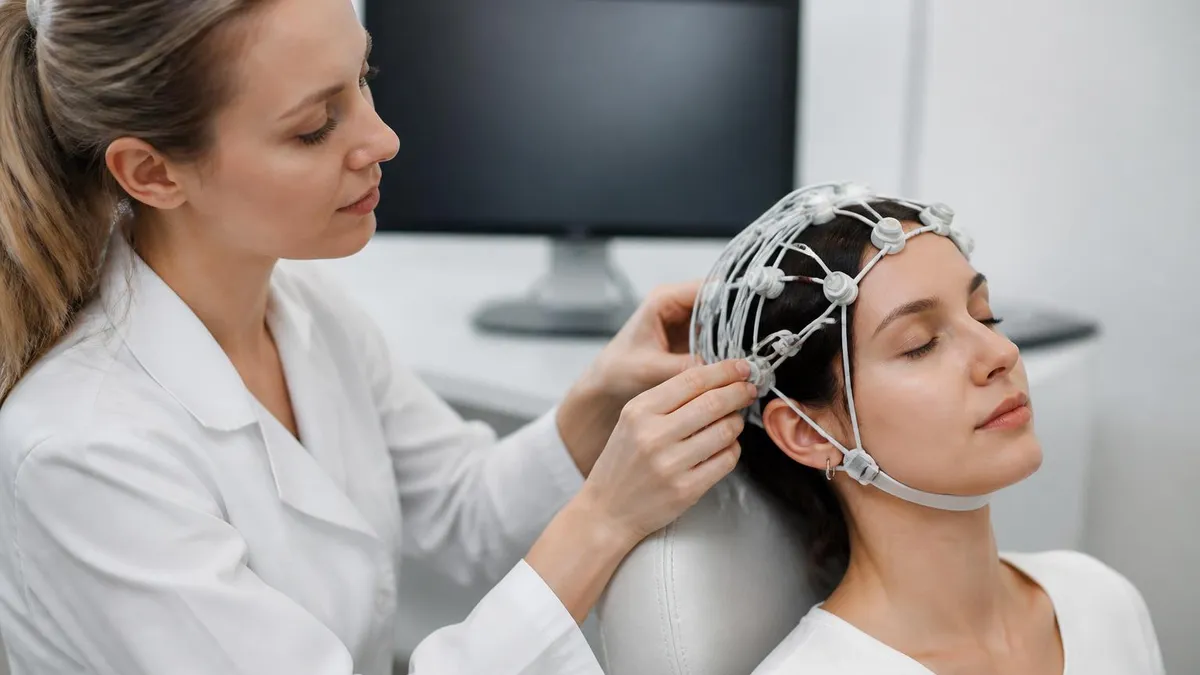
Sleep EEG: How Overnight Brain Wave Testing Works and What Your Results Mean
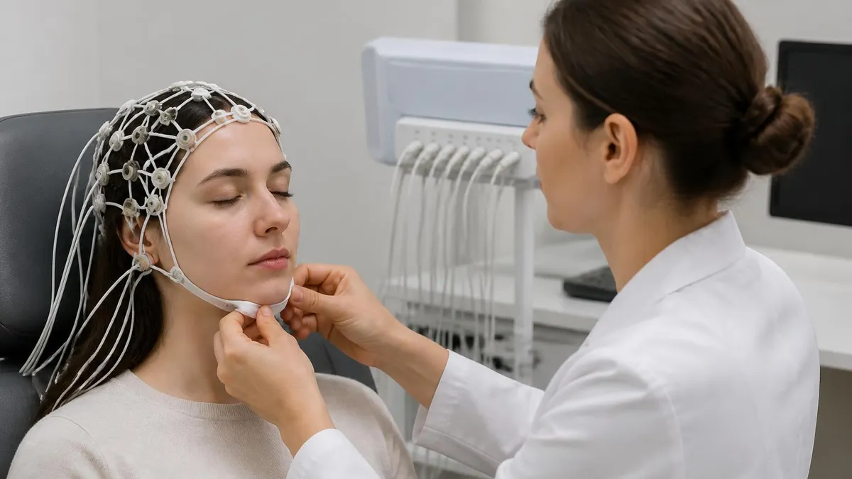
EEG Test Cost in 2026: Prices, Insurance Coverage, and How to Save

Travel EEG Tech Jobs: The Complete Guide to Working as a Mobile Electroencephalography Technologist

EEG Test: What Is Mapping EEG, How It Works, and What to Expect
About the Author

Educational Psychologist & Academic Test Preparation Expert
Columbia University Teachers CollegeDr. Lisa Patel holds a Doctorate in Education from Columbia University Teachers College and has spent 17 years researching standardized test design and academic assessment. She has developed preparation programs for SAT, ACT, GRE, LSAT, UCAT, and numerous professional licensing exams, helping students of all backgrounds achieve their target scores.