
EEG Test: What Is Mapping EEG, How It Works, and What to Expect 2026 October
Learn what an EEG test is, how mapping EEG works, what it costs, and what to expect. 🧠 Complete 2026 October guide for patients and EEG students.

An EEG test — short for electroencephalogram — is one of the most valuable diagnostic tools in modern neurology, and mapping EEG takes that capability several steps further by producing a detailed, color-coded picture of electrical activity across the entire brain. During a standard EEG test, technologists attach small metal electrodes to specific points on your scalp using a conductive paste, then record the tiny voltage fluctuations produced by millions of firing neurons beneath the skull. Mapping EEG processes those same raw signals through specialized software to generate a topographic brain map that clinicians can analyze region by region.
Understanding what an EEG medical test involves is important whether you are a patient preparing for the procedure or a student training to become a registered EEG technologist. The test itself is painless and non-invasive — no needles penetrate the skin, no radiation is used, and the electrodes only detect electrical signals rather than sending any current into the body. A routine diagnostic study typically takes between 45 minutes and two hours depending on the clinical indication, while extended or ambulatory recordings can run for 24 to 72 hours to capture infrequent events like seizures or sleep disturbances.
The eeg test price varies considerably depending on where the study is performed. Hospital outpatient departments typically charge between $800 and $3,000 for a standard routine recording, while free-standing neurology clinics and outpatient imaging centers often price the same study between $400 and $1,200. Quantitative EEG mapping — which adds computational analysis on top of the raw recording — generally adds $200 to $600 to whatever the base recording fee happens to be. Insurance coverage, geographic location, and whether you have met your deductible all influence your final out-of-pocket EEG test cost significantly.
From a clinical perspective, the eeg test is ordered for a wide range of indications. Epilepsy evaluation is by far the most common reason a neurologist requests the study, since the test can identify characteristic spike-and-wave discharges, focal slowing, and other abnormal epileptiform patterns that help confirm or rule out a seizure disorder. Beyond epilepsy, clinicians use EEG to evaluate altered states of consciousness, suspected encephalopathy, sleep disorders, head trauma sequelae, and certain types of dementia. Brain mapping adds a spatial dimension that helps localize abnormal activity to specific lobes or networks.
For those studying toward the R. EEG T. credential offered by the American Board of Registration of Electroencephalographic and Evoked Potential Technologists (ABRET), mastering the principles of eeg mapping is essential for both the written examination and clinical competency evaluations. The registration examination tests candidates on electrode placement according to the International 10-20 System, artifact recognition, normal and abnormal waveform identification, and the technical parameters that govern a high-quality recording. Understanding how mapping software interprets multichannel data prepares technologists to troubleshoot studies and communicate findings to supervising neurologists.
EEG test side effects are minimal because the procedure is entirely passive. Occasional patients report mild scalp irritation from the electrode paste, temporary redness at electrode sites, or difficulty washing the collodion adhesive out of their hair after the study. Patients undergoing activation procedures such as hyperventilation or photic stimulation may experience brief lightheadedness or visual sensations during those specific portions of the test, but these effects resolve within seconds of stopping the stimulus. Serious adverse events attributable directly to the EEG recording itself are exceedingly rare in routine clinical practice.
This comprehensive guide walks through everything you need to know about the EEG test — from the science behind mapping technology to practical cost-saving strategies, preparation tips, and the clinical significance of common findings. Whether you are a patient navigating an upcoming appointment or a technologist candidate reinforcing your examination knowledge, the sections that follow provide accurate, evidence-based information organized for clarity and depth.
EEG Test by the Numbers

How EEG Mapping Technology Works
Electrode Placement (10-20 System)
Signal Acquisition and Amplification
Reference and Montage Selection
Quantitative Analysis and Mapping
Neurologist Interpretation and Report
The term EEG test encompasses several distinct recording modalities, each tailored to different clinical questions. A routine outpatient EEG is the most commonly ordered type — it lasts 20 to 40 minutes of actual recording time (plus setup), captures the patient in a resting state with eyes open and closed, and includes standard activation procedures such as hyperventilation for three minutes and photic stimulation at frequencies ranging from 1 to 30 flashes per second.
This abbreviated study is sufficient to detect many epileptiform abnormalities and background rhythm disturbances, but it has an inherent limitation: if a patient's seizures are infrequent, the brief recording window may miss the event entirely.
Extended ambulatory EEG monitoring addresses this limitation by sending the patient home with a portable recording unit attached to their head for 24, 48, or 72 hours. The ambulatory device uses the same 10-20 electrode system as a routine study but stores data on a compact digital recorder the patient wears on a belt or carries in a small bag.
When a clinical event occurs, the patient or a companion presses an event button on the device, placing a time-stamp marker that helps the interpreting neurologist correlate the EEG at that moment with the reported symptoms. How long is an EEG test in this format? The setup appointment typically takes 30 to 45 minutes, and the patient returns the following day or two days later for lead removal and data download.
Video-EEG telemetry is the gold standard for pre-surgical epilepsy evaluation and for confirming or ruling out non-epileptic events. In this modality, the patient is admitted to a specialized epilepsy monitoring unit (EMU) and connected to a continuous EEG recording system that also captures synchronized video footage. Anti-epileptic medications may be tapered to provoke habitual events under controlled conditions. Neurologists can then review the behavioral manifestations of each clinical event alongside the simultaneous EEG changes, providing definitive seizure classification and localization data that guides surgical planning or medication adjustment.
High-density EEG mapping uses arrays of 64 to 256 electrodes rather than the standard 19 to achieve much greater spatial resolution. The denser electrode coverage reduces the distance between recording sites from roughly 6 centimeters to 2 centimeters or less, which allows source localization algorithms to more precisely estimate the cortical generators of abnormal electrical activity. Neurosurgical centers use high-density mapping as a non-invasive complement to intracranial electrode implantation when planning resective surgery for drug-resistant epilepsy. Research applications include mapping the neural correlates of cognitive processes, sleep stage transitions, and real-time brain-computer interface control signals.
Neonatal EEG is a specialized subspecialty with its own electrode placement conventions, normal maturational standards, and pattern recognition criteria. Premature infants as young as 23 weeks gestational age can be recorded, and the presence of specific patterns such as burst-suppression, trace alternant, or seizure activity carries profound prognostic implications for neurodevelopmental outcome. Neonatal EEG technologists must understand the dramatic changes in background activity that occur week by week as the immature brain matures, since a pattern that is completely normal at 28 weeks corrected age would be markedly abnormal at 40 weeks.
Intraoperative neurophysiological monitoring (IONM) represents another specialized application of EEG technology. During neurosurgical procedures, continuous EEG recording combined with evoked potential monitoring alerts the surgical team in real time to changes in cerebral perfusion or cortical function that may indicate impending neurological injury. EEG technologists working in the operating room must be highly proficient at rapid artifact identification and at distinguishing anesthetic-induced changes in background activity from true pathological changes, because a false alarm or a missed alert can have serious consequences for the patient on the table.
Sleep EEG studies — including standard polysomnography and split-night titration studies — incorporate EEG channels as part of a broader multi-channel recording that also captures eye movements, chin muscle tone, respiratory effort, oxygen saturation, and limb movements. The EEG channels are essential for staging sleep into the recognized AASM categories: N1, N2, N3, and REM sleep. Understanding sleep EEG scoring rules is increasingly relevant for EEG technologists, since many hospital-based departments handle both epilepsy monitoring and sleep laboratory functions using overlapping technical staff and shared equipment resources.
What Is an EEG Medical Test Used For?
Epilepsy is by far the most common indication for ordering an EEG medical test. The recording can reveal interictal epileptiform discharges — spikes, sharp waves, or spike-and-slow-wave complexes — that appear between actual seizures and confirm a diagnosis of epilepsy in the right clinical context. Focal discharges help localize the seizure onset zone to a specific brain region, which is critical information when evaluating patients for surgical candidacy or when choosing between focal and generalized anti-seizure medications.
Not every patient with epilepsy has an abnormal routine EEG, however. Studies show that a single routine recording is normal in roughly 50 percent of people with confirmed epilepsy. Repeat studies, sleep-deprived recordings, and prolonged ambulatory monitoring substantially increase the yield. The EEG is always interpreted alongside the patient's clinical history — a normal recording does not rule out epilepsy, just as an abnormal one must be interpreted cautiously in the context of low pre-test probability.

Advantages and Limitations of Mapping EEG
- +Non-invasive and painless — no needles, no radiation, no injected contrast agents required
- +High temporal resolution captures millisecond-by-millisecond brain electrical dynamics in real time
- +Widely available in hospital and outpatient settings across the United States
- +Relatively low EEG test cost compared to MRI or PET imaging studies
- +Quantitative mapping software adds spatial context that raw waveforms cannot provide alone
- +Can be performed continuously over days in ambulatory or ICU settings to capture rare events
- −Spatial resolution is inherently limited — even 256-electrode arrays cannot pinpoint generators with MRI-level precision
- −Highly sensitive to artifacts from eye movement, muscle activity, electrode movement, and ambient electrical interference
- −Normal EEG does not rule out epilepsy — up to 50 percent of first routine studies in confirmed epilepsy cases are normal
- −Quantitative mapping analysis requires specialized software and trained interpreters, limiting availability
- −EEG test side effects from activation procedures can temporarily produce symptoms in susceptible patients
- −Results are heavily dependent on technologist skill during acquisition and neurologist expertise during interpretation
Pros and Cons at a Glance
| Pros | Cons |
|---|---|
| Non-invasive and painless — no needles, no radiation, no injected contrast agents required | Highly sensitive to artifacts from eye movement, muscle activity, electrode movement, and ambient electrical interference |
| High temporal resolution captures millisecond-by-millisecond brain electrical dynamics in real time | Quantitative mapping analysis requires specialized software and trained interpreters, limiting availability |
| Widely available in hospital and outpatient settings across the United States | EEG test side effects from activation procedures can temporarily produce symptoms in susceptible patients |
| Relatively low EEG test cost compared to MRI or PET imaging studies | Results are heavily dependent on technologist skill during acquisition and neurologist expertise during interpretation |
Preparing for Your EEG Test: Complete Checklist
- ✓Wash your hair the night before or morning of the test — avoid using conditioner, oils, sprays, or styling products that increase scalp impedance.
- ✓Confirm with your ordering physician whether to take your regular medications on the test day, especially anti-seizure drugs.
- ✓Get a full night of sleep unless your physician has specifically ordered a sleep-deprived study to increase seizure likelihood.
- ✓Eat a normal meal before the appointment — arriving hungry or hypoglycemic can alter the EEG background and trigger symptoms during hyperventilation.
- ✓Avoid caffeine for at least 8 hours before the test, as caffeine can suppress normal drowsy and sleep patterns needed for a complete recording.
- ✓Bring a list of all current medications and supplements to the appointment so the technologist can document them in the study record.
- ✓Plan for the appointment to last 2 to 3 hours including setup, recording, and lead removal — arrange transportation if you feel uncomfortable driving after sedation.
- ✓Wear comfortable, loose-fitting clothing and avoid wearing earrings or other jewelry near the scalp that could interfere with electrode placement.
- ✓Inform the technologist about any skin conditions, open wounds, or areas of scalp tenderness before electrode placement begins.
- ✓Ask the ordering physician whether a routine study, sleep-deprived study, or ambulatory monitoring is most appropriate for your specific clinical question.
A Normal EEG Does Not Rule Out Epilepsy
Research consistently shows that a single routine EEG is normal in approximately 50 percent of patients with confirmed epilepsy. If your first EEG comes back normal but your neurologist still suspects a seizure disorder based on your history and symptoms, repeat testing, sleep-deprived protocols, or prolonged ambulatory monitoring significantly increase the diagnostic yield and may be the next appropriate step in your evaluation.
The EEG test cost in the United States depends on a complex interplay of facility type, geographic market, insurance status, and the specific type of recording ordered. For insured patients, the out-of-pocket expense after insurance processes the claim is typically far lower than the facility's billed charge, but it still varies enormously based on plan design.
A patient with a high-deductible health plan who has not yet met their annual deductible might pay the full allowed amount — often $400 to $900 for a routine outpatient study — while a patient with a low-copay plan might owe only $50 to $150 for the same procedure after a specialist visit copay applies.
Medicare covers routine and extended EEG testing when it is medically necessary, defining medical necessity primarily through the clinical documentation provided by the ordering neurologist. The Medicare allowed amount for a routine EEG (CPT code 95816) is approximately $150 to $250 depending on facility type and geographic locality, while a 24-hour ambulatory EEG (CPT code 95953) carries an allowed amount typically in the $350 to $600 range. Patients in traditional Medicare Part B generally owe 20 percent of the allowed amount after meeting their annual deductible, which translates to roughly $30 to $120 out of pocket for a routine study.
Medicaid programs cover EEG testing in all states, though prior authorization requirements and covered CPT codes vary by state plan. Patients covered by Medicaid typically owe little to nothing out of pocket for covered EEG studies, making cost a much lower barrier than for uninsured or underinsured patients. However, not all facilities accept Medicaid, so patients may need to call ahead to confirm participation before scheduling an appointment at a specific hospital or outpatient center.
Uninsured patients face the highest financial exposure and should proactively explore several cost-reduction strategies before assuming the study is unaffordable. Most hospital systems have charity care or financial assistance programs that can reduce or eliminate the bill for patients who meet income thresholds — typically 200 to 400 percent of the federal poverty level. Free-standing neurophysiology labs and academic medical center outpatient clinics frequently charge significantly less than hospital outpatient departments for identical studies, sometimes by a factor of three to five. Asking the scheduling department for the self-pay or cash-pay price before the appointment is always worthwhile.
Telehealth cannot replace the actual EEG recording — a technologist must physically place electrodes on the patient's scalp. However, the neurologist's interpretation of the recording can now be performed remotely by a telepresence neurophysiologist using cloud-based EEG review platforms. This model, called tele-EEG, has expanded access to expert interpretation in rural and underserved communities where board-certified clinical neurophysiologists are scarce. Tele-EEG interpretation services charge separately from the technical component of the study and are covered by most major insurers when performed by a credentialed physician.
For EEG technologist students, understanding the billing and reimbursement landscape is part of professional competency. Technologists often assist with prior authorization paperwork, insurance verification, and patient education about what to expect financially. Knowing the difference between the technical component (the recording itself, billed by the facility or tech) and the professional component (the physician interpretation) helps technologists explain why patients may receive two separate bills for what felt like a single appointment.
Academic medical centers affiliated with major neurology training programs sometimes offer EEG testing at reduced rates as part of research protocols or clinical training programs. Patients willing to have their study observed by neurology residents or EEG technology students — always under direct attending supervision — may access these discounted rates. This arrangement benefits both parties: patients receive expert care at lower cost, while trainees gain supervised clinical experience with real diagnostic studies that prepare them for board examinations and independent practice.

Many insurance plans require prior authorization for EEG testing, especially for ambulatory studies, video-EEG telemetry, and quantitative brain mapping. Scheduling and attending the appointment without obtaining required authorization first can result in the claim being denied entirely, leaving you responsible for the full facility charge. Call your insurance company's member services line at least five business days before your scheduled appointment to confirm coverage and authorization requirements.
For candidates preparing for the ABRET R. EEG T. examination, a thorough understanding of EEG mapping principles is not optional — it is tested directly. The examination blueprint published by ABRET identifies electrode placement, montage construction, artifact identification, normal and abnormal EEG patterns, and activation procedures as core competency domains. Each of these areas connects directly to the technical foundation of brain mapping: you cannot interpret a topographic map correctly if you do not understand where each electrode sits, what artifact looks like in various montages, and what constitutes a normal or abnormal finding in the underlying raw waveform data.
The International 10-20 System of electrode placement is the cornerstone of all clinical EEG work and the foundation of any mapping study. Candidates must know not only the letter-number codes for all 19 standard electrode positions but also the anatomical landmarks used to derive the measurements — the nasion, inion, and preauricular points — and the arithmetic that places electrodes at 10 and 20 percent intervals across the skull. High-density mapping systems extend this framework using additional electrodes interpolated between standard 10-20 positions, following nomenclature conventions defined by the American Clinical Neurophysiology Society (ACNS).
Artifact recognition is one of the most heavily tested skills on the R. EEG T. examination and one of the most consequential competencies in clinical practice. Movement artifacts, electrode pop artifacts, sweat artifacts, ECG artifacts, and 60-Hz electrical interference each have distinctive waveform morphologies and distribution patterns that allow a trained technologist to identify them quickly and take corrective action. On mapping studies, unrecognized artifacts can create spurious regions of apparent high-frequency activity on the topographic display that might be mistaken for pathological fast activity — a diagnostic error with potentially serious clinical consequences.
Normal EEG patterns across the lifespan represent a broad and nuanced body of knowledge that candidates must master. The posterior dominant alpha rhythm — typically 8 to 13 Hz in adults — is the most recognizable hallmark of a normal awake EEG, and candidates must know its expected frequency range at each stage of development from infancy through old age. Sleep architecture, drowsiness patterns, normal variants (such as wickets, SREDA, and 14-and-6 positive spikes), and the effects of common medications on background rhythms are all fair game on the examination.
Practice examinations are among the most effective tools available to R. EEG T. candidates, and targeted question banks organized by topic allow candidates to identify their weakest domains before sitting for the actual test.
The eeg mapping resources available through structured study programs provide realistic question formats, detailed answer explanations, and references to authoritative sources such as ACNS guidelines and the Niedermeyer and da Silva textbook that forms the backbone of most EEG training curricula. Spaced repetition of practice questions in the weeks leading up to the examination has been shown to improve retention and performance on high-stakes credentialing exams across multiple healthcare disciplines.
Clinical case-based learning is equally important alongside didactic study. Reviewing actual EEG recordings — whether from clinical rotations, online EEG libraries, or the archived studies available through academic neurology departments — develops the pattern recognition skills that written practice questions alone cannot fully replicate. Candidates should aim to review at least 50 to 100 complete EEG studies in the months before the examination, deliberately seeking out examples of rare but testable abnormalities such as hypsarrhythmia, NCSE patterns, Creutzfeldt-Jakob disease periodic complexes, and herpes encephalitis lateralized discharges.
Mentorship from experienced R. EEG T. technologists and supervising clinical neurophysiologists accelerates learning in ways that self-study cannot. Asking supervisors to narrate their thought process while reviewing a study, discussing the rationale for montage selection decisions, and requesting feedback on your own artifact management techniques all compound knowledge faster than passive reading. The EEG community is relatively small and collegial, and most experienced technologists are willing to mentor candidates who demonstrate genuine curiosity and professional commitment to the field.
Practical preparation for the EEG examination — whether you are the patient or the technologist candidate — benefits from a structured approach that addresses knowledge gaps systematically rather than hoping broad review will cover the highest-yield material. Patients preparing for an upcoming EEG test should focus primarily on the logistical preparation steps: hair washing, medication management, sleep and caffeine guidance, and understanding the general sequence of events during the appointment.
Anxiety about the procedure is common and understandable, but knowing that the test is painless, that the electrodes only listen and never transmit, and that side effects are minimal and transient can substantially reduce pre-appointment stress.
For technologist candidates, the weeks immediately preceding the examination are best used for active recall practice rather than passive re-reading of textbooks. Retrieval practice — the act of forcing yourself to recall information without looking at notes — produces far stronger long-term memory consolidation than simply re-reading the same material repeatedly. Writing out the 10-20 electrode positions from memory, sketching waveform morphologies of key abnormal patterns, and completing timed practice question sets under simulated examination conditions all engage active retrieval and build the test-taking stamina needed for a multi-hour credentialing examination.
Waveform recognition speed matters enormously in clinical EEG practice. Neurologists reviewing 24-hour ambulatory studies or long-term monitoring recordings often work at accelerated playback speeds of 30 to 60 times real time, relying on the technologist's preliminary review and flagging to guide their attention toward clinically significant segments. Developing the ability to quickly scan a page of EEG data — in the standard 10-second epoch display format — and immediately identify whether it contains artifact, normal background activity, or a potentially abnormal finding is a core clinical skill that separates proficient technologists from merely competent ones.
Understanding activation procedure protocols in depth is particularly valuable because these procedures carry both the highest yield for eliciting abnormal activity and the most important safety considerations. Hyperventilation is contraindicated in patients with recent stroke, severe pulmonary or cardiac disease, sickle cell disease, and moyamoya disease.
Photic stimulation protocols vary by laboratory but typically sweep through flash frequencies from 1 to 30 Hz in ascending and descending sequences, pausing at each frequency for 10 seconds. Photoparoxysmal responses — generalized spike-wave bursts triggered by the flickering light — are an important clinical finding that the technologist must recognize and document in real time.
Documentation skills are underappreciated in EEG technologist training but critically important in practice. A complete EEG record includes not only the digital waveform data but also a detailed technical description of the study conditions: the patient's level of arousal throughout the recording, any clinical events observed, the activation procedures performed and their results, artifacts encountered and the corrective measures taken, and all medications documented at the time of the study.
Thorough technical notes allow the interpreting neurologist to make sense of the recording even when reviewing the study hours or days after the actual acquisition, without access to the technologist's direct observations.
Continuing education after achieving R. EEG T. certification is both an ABRET requirement and a professional responsibility. Registered technologists must accumulate continuing education credits in defined categories over each certification cycle to maintain their credential.
The field of clinical neurophysiology evolves steadily — new guidelines for EEG terminology and pattern classification are periodically updated by the ACNS, new evidence emerges on the prognostic significance of various patterns in the ICU setting, and new technology platforms introduce capabilities that require technologists to expand their technical skills. Staying current with these developments positions certified technologists for career advancement into supervisory, educational, or specialized clinical roles.
The intersection of artificial intelligence and EEG analysis is one of the most exciting frontiers in clinical neurophysiology. Machine learning algorithms trained on large annotated EEG databases are now capable of detecting epileptiform discharges, classifying sleep stages, and screening for abnormal background patterns with sensitivity and specificity approaching that of expert human interpreters.
Rather than replacing EEG technologists, these tools are expected to augment clinical workflows — flagging segments of long recordings that warrant close human review, reducing the time neurologists spend on clearly normal studies, and potentially extending expert-level analysis to resource-limited settings. Technologists who understand the principles and limitations of AI-assisted EEG interpretation will be well positioned to work effectively alongside these emerging tools.
Sample EEG - Electroencephalography Practice Questions
Try these questions from our free EEG - Electroencephalography practice tests. The correct answer and an explanation follow each question.
The perisylvian language network includes Broca's area (IFG) and Wernicke's area connected by the arcuate fasciculus. EEG-based language mapping uses which technique to identify these regions before epilepsy surgery?
- A. Routine scalp EEG with hyperventilation
- B. Electrocorticography (ECoG) with cortical stimulation mapping
- C. Photic driving response analysis
- D. Z-score normalization of alpha power
Answer: B. Electrocorticography (ECoG) with cortical stimulation mapping
Electrocorticography with direct cortical stimulation (extraoperative or intraoperative) is the gold-standard EEG-based method to map language-eloquent cortex before resection.
A patient's ambulatory EEG diary notes going to sleep at 22:30. The technologist should expect to see which EEG pattern shortly after this time?
- A. Generalized paroxysmal fast activity
- B. Vertex sharp waves and sleep spindles
- C. Continuous 10 Hz alpha rhythm
- D. High-amplitude delta with sharp transients suggesting seizure
Answer: B. Vertex sharp waves and sleep spindles
Normal sleep onset is marked by vertex sharp waves (V waves) and sleep spindles (12–14 Hz) during NREM stage N2 sleep.
During hyperventilation, if the patient has an underlying brain tumor, you would most likely expect to see:
- A. Bilateral synchronous generalized slowing
- B. Focal or lateralized slow-wave abnormality that increases in prominence
- C. High-frequency gamma activity over the tumor site
- D. Suppression of all EEG activity
Answer: B. Focal or lateralized slow-wave abnormality that increases in prominence
Focal structural lesions such as tumors typically produce focal or lateralized slow-wave abnormalities that are accentuated by hyperventilation-induced hypocapnia.
During an EEG recording, a patient begins to have a generalized tonic-clonic seizure. What is the technologist's first priority?
- A. Continue recording and immediately stop all activation procedures
- B. Turn off the EEG machine to protect the equipment
- C. Restrain the patient to prevent movement artifact
- D. Remove all electrodes before calling for help
Answer: A. Continue recording and immediately stop all activation procedures
The technologist's first priority is to ensure patient safety while continuing the recording. This means stopping any activation procedures (hyperventilation, photic stimulation) and ensuring the patient is safe from injury, while keeping the EEG recording to capture the seizure.
EEG Questions and Answers
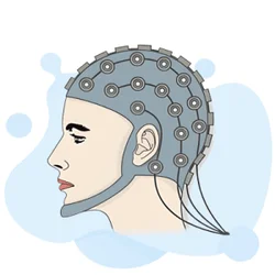
EEG Electroencephalography Practice Test PDF (Free Printable 2026)
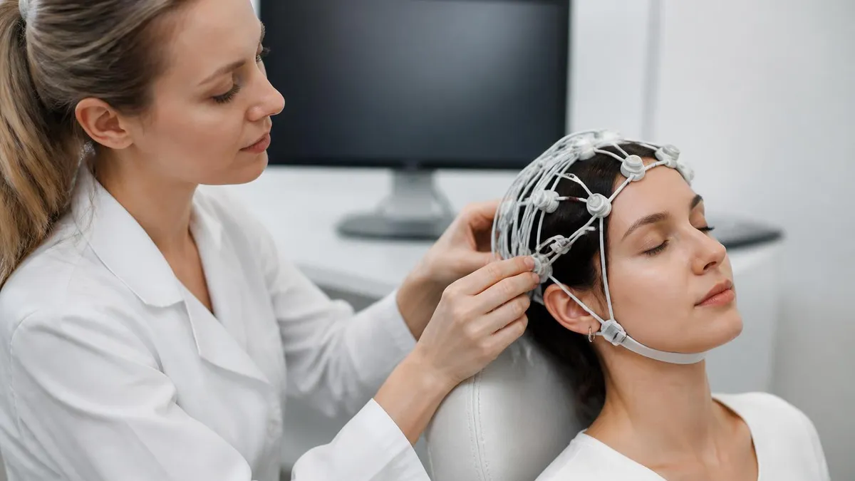
Sleep EEG: How Overnight Brain Wave Testing Works and What Your Results Mean
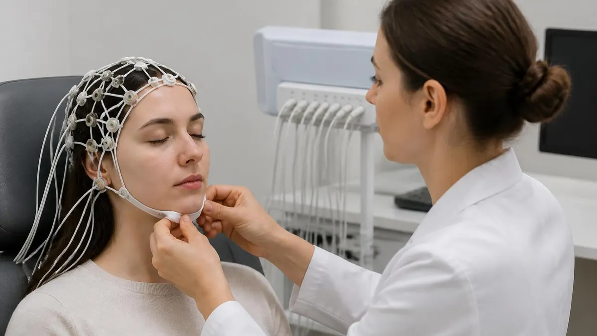
EEG Test Cost in 2026: Prices, Insurance Coverage, and How to Save

Travel EEG Tech Jobs: The Complete Guide to Working as a Mobile Electroencephalography Technologist
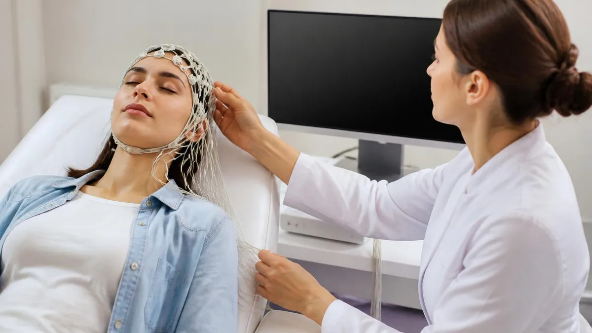
EEG Tech Salary 2026: Pay by State, Experience, and Certification
About the Author

Educational Psychologist & Academic Test Preparation Expert
Columbia University Teachers CollegeDr. Lisa Patel holds a Doctorate in Education from Columbia University Teachers College and has spent 17 years researching standardized test design and academic assessment. She has developed preparation programs for SAT, ACT, GRE, LSAT, UCAT, and numerous professional licensing exams, helping students of all backgrounds achieve their target scores.