Stereo EEG (sEEG): How Depth Electrode Brain Mapping Works, What It Costs, and What to Expect
Stereo EEG explained: 🎓 what an EEG test costs, how long it takes, side effects, and how sEEG depth electrodes map seizures before epilepsy surgery.
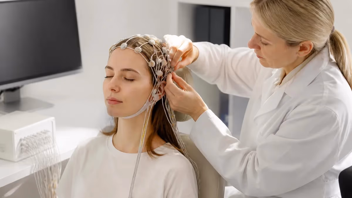
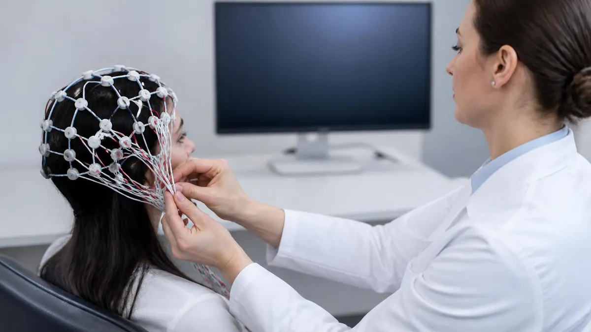
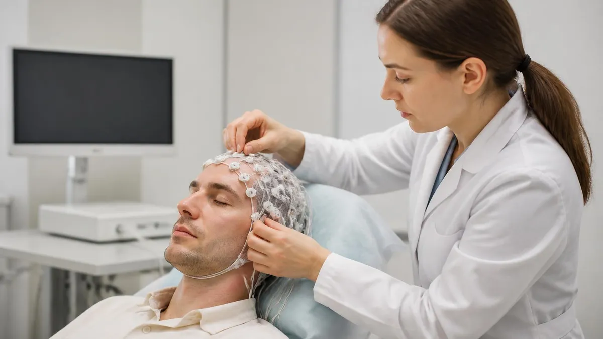
If your neurologist has mentioned a stereo eeg, you are almost certainly being evaluated for drug-resistant epilepsy, and you probably have a long list of questions about what an eeg test really involves, how long is an eeg test of this type, and what it will reveal. Stereo EEG, often abbreviated sEEG, is a specialized form of intracranial recording in which thin depth electrodes are placed through small holes in the skull to listen to electrical activity from inside the brain itself, far below the scalp surface that a routine EEG can reach.
A standard scalp eeg test records brain waves through electrodes glued to your hair and forehead. It is fast, painless, and gives a broad picture of cortical activity. Stereo EEG is fundamentally different. Each electrode is a slender wire roughly 0.8 millimeters wide carrying eight to eighteen tiny contacts, and a single patient typically receives between eight and fifteen of them. The result is a three-dimensional map of seizure activity that scalp recordings simply cannot produce.
The phrase eeg medical test gets used loosely in clinic notes, but sEEG sits at the most invasive end of that spectrum. It is performed only after non-invasive tests, video EEG monitoring, high-resolution MRI, PET, and sometimes magnetoencephalography have suggested a seizure focus but failed to define it precisely enough for surgery. For many candidates, the goal is to identify a small region of brain tissue that can be removed, ablated with laser, or treated with neuromodulation to stop seizures entirely.
People searching for an eeg test price for stereo EEG are often surprised by the range. A routine outpatient scalp study might cost a few hundred dollars, while a full sEEG admission can run into the tens of thousands because it involves a robotic surgical procedure, an intensive care recovery, and one to three weeks of inpatient monitoring. Insurance coverage is generally strong for medically necessary evaluations at comprehensive epilepsy centers.
This guide walks through every part of the experience: who qualifies, how the electrodes are planned and placed, what daily life on the epilepsy monitoring unit looks like, what eeg test side effects to expect, and how clinicians turn the recorded signals into a surgical plan. It is written for patients, families, and clinicians-in-training who want a deeper picture than the consent form provides.
You will also find statistics drawn from published epilepsy surgery outcomes, a comparison of sEEG against the older subdural grid approach, a practical checklist for the days before admission, and answers to the questions that come up most often during pre-operative counseling. Wherever possible, numbers reflect current US practice at level 4 epilepsy centers accredited by the National Association of Epilepsy Centers.
Stereo EEG is not the right tool for every seizure patient, and it is not a substitute for the screening tests that come before it. But when it is the right tool, it can change the entire trajectory of a person's life — turning intractable epilepsy into a problem with a clear surgical target and a realistic chance of long-term freedom from seizures.
Stereo EEG by the Numbers

The Stereo EEG Patient Journey
Phase I Non-Invasive Workup
Surgical Conference Review
Trajectory Planning
Implantation Surgery
EMU Monitoring
Electrode Removal and Plan
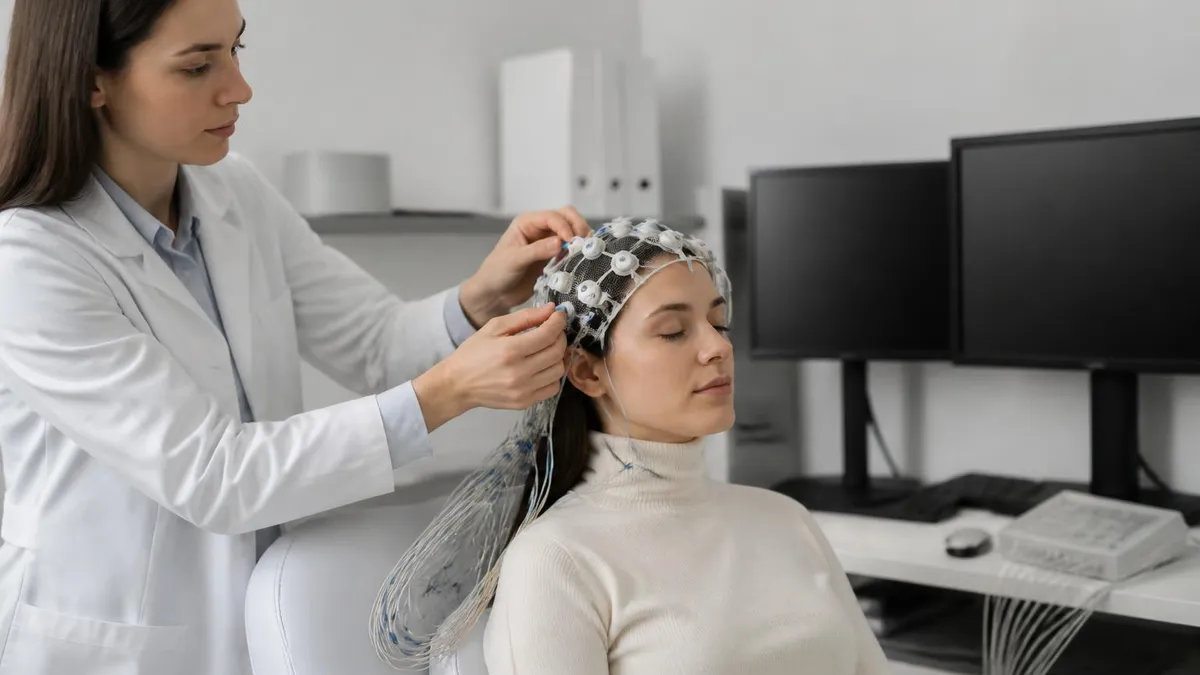
A scalp eeg medical test uses 19 or 25 silver-silver-chloride electrodes placed according to the international 10-20 or 10-10 system. The signal that reaches them has been heavily filtered by the cerebrospinal fluid, skull, and scalp, so deep structures such as the mesial temporal lobe, insula, and orbitofrontal cortex are essentially invisible. That filtering is why a person can have clearly focal seizures and still show only diffuse slowing on a routine recording.
Stereo EEG bypasses those layers entirely. Each depth electrode passes through the skull and brain in a straight line, recording from gray and white matter as it goes. Contacts on the same electrode are usually two millimeters apart, and bipolar montages between adjacent contacts produce very local measurements that can resolve millimeter-scale generators. This means clinicians can answer questions a scalp study cannot, such as whether seizures begin in the hippocampus or the amygdala on the same trajectory.
The other major intracranial approach, subdural grid recording, places sheets of electrodes directly on the cortical surface through a large craniotomy. Grids excel at mapping the lateral convexity in two dimensions, but they cannot sample deep structures and they carry higher rates of infection, hemorrhage, and CSF leak. Stereo EEG trades the dense surface coverage of a grid for the ability to sample many targets, including bilateral structures, through holes too small to require formal closure.
For patients, the practical differences are large. With grids, recovery from the implantation craniotomy is significant, hair must be shaved, and the second surgery to remove them is essentially another craniotomy. With sEEG, the implantation is performed through pinhole openings, hair is preserved, post-operative pain is usually controlled with mild analgesics, and electrode removal is a bedside procedure that takes minutes per wire and requires only suture closure and a small dressing.
Cost reflects this complexity. People searching how long is an eeg test for an outpatient study learn that 20-minute and 24-hour recordings are inexpensive screening tools. Stereo EEG is the opposite end of the price ladder, comparable to other neurosurgical admissions. Most commercial plans and Medicare cover the procedure when it is performed at an accredited level 4 epilepsy center for medically refractory focal epilepsy, but pre-authorization is almost always required.
Both approaches share a single goal: localize the seizure onset zone precisely enough that surgical treatment can offer a high chance of seizure freedom with minimal new neurological deficits. The choice between sEEG and grids is not about which technology is universally better. It is about which one best answers the specific question your team has formulated based on your scalp studies, imaging, and clinical history.
Most US centers have shifted dramatically toward sEEG over the last decade. National series suggest that more than 80% of new invasive evaluations at high-volume programs now use depth electrodes, while subdural grids are reserved for cases where dense mapping of a single lobe is essential. Hybrid implantations that combine a few strips with depth electrodes still occur, but they are increasingly the exception rather than the rule.
EEG Practice Test Questions
Prepare for the EEG - Electroencephalography exam with our free practice test modules. Each quiz covers key topics to help you pass on your first try.
EEG Abnormal Epileptiform Patterns
EEG Exam Questions covering Abnormal Epileptiform Patterns. Master EEG Test concepts for certification prep.
EEG Activation Procedures
Free EEG Practice Test featuring Activation Procedures. Improve your EEG Exam score with mock test prep.
EEG Artifact Identification
EEG Test Prep for Artifact Identification. Practice EEG Quiz questions and boost your score.
EEG Electrode Placement and Montages
EEG Questions and Answers on Electrode Placement and Montages. Free EEG practice for exam readiness.
EEG - Electroencephalography Abnormal Epil...
EEG Mock Test covering - Electroencephalography Abnormal Epileptiform Patterns. Online EEG Test practice with instant feedback.
EEG - Electroencephalography Activation Pr...
Free EEG Quiz on - Electroencephalography Activation Procedures. EEG Exam prep questions with detailed explanations.
EEG - Electroencephalography Artifact Iden...
EEG Practice Questions for - Electroencephalography Artifact Identification. Build confidence for your EEG certification exam.
EEG - Electroencephalography Electrode Pla...
EEG Test Online for - Electroencephalography Electrode Placement and Montages. Free practice with instant results and feedback.
EEG - Electroencephalography Instrumentati...
EEG Study Material on - Electroencephalography Instrumentation and Calibration. Prepare effectively with real exam-style questions.
EEG - Electroencephalography Neuroanatomy ...
Free EEG Test covering - Electroencephalography Neuroanatomy and Physiology. Practice and track your EEG exam readiness.
EEG - Electroencephalography Non-Epileptif...
EEG Exam Questions covering - Electroencephalography Non-Epileptiform Abnormalities. Master EEG Test concepts for certification prep.
EEG - Electroencephalography Normal EEG Wa...
Free EEG Practice Test featuring - Electroencephalography Normal EEG Waveforms. Improve your EEG Exam score with mock test prep.
EEG - Electroencephalography Patient Safet...
EEG Mock Exam on - Electroencephalography Patient Safety and Ethics. EEG Study Guide questions to pass on your first try.
EEG - Electroencephalography Pediatric and...
EEG Test Prep for - Electroencephalography Pediatric and Neonatal EEG. Practice EEG Quiz questions and boost your score.
EEG ICU and Continuous EEG Monitoring
EEG Questions and Answers on ICU and Continuous EEG Monitoring. Free EEG practice for exam readiness.
EEG Instrumentation and Calibration
EEG Mock Test covering Instrumentation and Calibration. Online EEG Test practice with instant feedback.
EEG Neuroanatomy and Physiology
Free EEG Quiz on Neuroanatomy and Physiology. EEG Exam prep questions with detailed explanations.
EEG Non-Epileptiform Abnormalities
EEG Practice Questions for Non-Epileptiform Abnormalities. Build confidence for your EEG certification exam.
EEG Normal EEG Waveforms
EEG Test Online for Normal EEG Waveforms. Free practice with instant results and feedback.
EEG Patient Safety and Ethics
EEG Study Material on Patient Safety and Ethics. Prepare effectively with real exam-style questions.
What Is an EEG Test Like When It Is a Stereo EEG?
You arrive at the hospital fasting and meet your neurosurgeon, anesthesiologist, and epileptologist. After IV placement and a final imaging check, you are taken to the operating room where general anesthesia is induced. A stereotactic frame or frameless system registers your head to the pre-planned MRI, and the robot positions itself for the first trajectory.
The surgeon makes a small skin incision, the robot drills a 2.5 millimeter hole, and the depth electrode is advanced to its target. Each trajectory typically takes ten to fifteen minutes, and most implantations involve eight to fifteen electrodes. You wake up in the recovery area with a soft turban dressing protecting the connector pigtails and can usually eat dinner the same evening.

Stereo EEG vs Subdural Grids: Pros and Cons
- +Samples deep structures such as hippocampus, insula, and cingulate that grids cannot reach
- +Allows bilateral and multi-lobar sampling through small skull holes in a single anesthetic
- +Lower rates of infection, CSF leak, and symptomatic hemorrhage in modern series
- +No hair shaving and minimal post-operative pain compared with a craniotomy
- +Bedside removal avoids a second major surgery and shortens total hospital time
- +Three-dimensional coverage supports laser interstitial thermal therapy planning
- +Robotic platforms achieve sub-millimeter accuracy and reproducible trajectories
- −Lower spatial density than a grid over a single gyrus or lobe
- −Cannot perform large-area functional mapping in the way a dense grid can
- −Requires sophisticated pre-implant hypothesis — random sampling is rarely useful
- −Inpatient EMU stay of one to two weeks is required to capture seizures
- −Procedure cost and need for accredited level 4 epilepsy center limit access
- −Medication tapering on the EMU can occasionally trigger status epilepticus
- −Not every payer recognizes sEEG codes the same way as grid recordings
Stereo EEG Pre-Admission Checklist
- ✓Confirm pre-authorization is approved and obtain a written cost estimate from the hospital
- ✓Bring a complete, dated medication list including doses and timing
- ✓Pack comfortable button-front pajamas that open easily over the head dressing
- ✓Bring chargers, headphones, books, and entertainment for a two-week inpatient stay
- ✓Arrange childcare, pet care, and time off work for at least three weeks total
- ✓Designate a primary contact who can attend daily rounds and the post-stay conference
- ✓Stop blood thinners only as directed by the neurosurgical team
- ✓Wash hair thoroughly the night before with the antimicrobial soap provided
- ✓Remove all hair products, extensions, and jewelry before arrival
- ✓Review your seizure semiology video with family so they can describe events accurately
- ✓Identify trigger questions you want the epileptologist to answer before discharge
- ✓Plan a quiet, low-stress home environment for the first week after removal
Stereo EEG only works when the hypothesis is right
Depth electrodes record only from where you put them. If the pre-implant hypothesis is wrong, even a perfectly executed sEEG can miss the seizure onset zone. That is why level 4 epilepsy centers spend weeks on Phase I evaluation and multidisciplinary review before a single electrode is planned. A successful sEEG is decided in the conference room as much as in the operating room.
Reading a stereo EEG is fundamentally different from reading a scalp study, and trainees who already understand what is a eeg test at the scalp level still face a steep learning curve. Each electrode produces 8 to 18 channels, so a typical implantation generates between 100 and 250 simultaneous traces. Modern review software lets the epileptologist switch between referential, bipolar, and Laplacian montages, color-code anatomical regions, and align the recording with high-resolution post-implant CT or MRI co-registration.
The first task is mapping each contact to anatomy. Co-registration software fuses the post-implant CT, which shows the exact electrode positions, with the pre-implant MRI, which shows the underlying anatomy. Contacts are labeled by structure — for example, hippocampal head, hippocampal body, entorhinal cortex, amygdala, temporal pole — so that any abnormal activity can be traced to a specific brain region rather than to a generic electrode number.
Interictal review then characterizes background activity, spikes, sharp waves, fast ripples, and pathological high-frequency oscillations. Fast ripples above 250 Hz are particularly important because they appear to mark epileptogenic tissue more specifically than ordinary spikes. Quantitative tools that calculate spike rates, connectivity, and time-frequency content support the visual review but rarely replace it. Experienced epileptologists still rely heavily on pattern recognition built up over thousands of cases.
Ictal analysis is the heart of the study. The team identifies the very first electrographic change at seizure onset, often a low-voltage fast activity in the gamma or ripple range, and tracks how it spreads to neighboring contacts and then to remote networks. The seizure onset zone is defined as the contacts showing the earliest, sustained, and reproducible changes across multiple seizures, ideally without contamination from artifact or medication-related background slowing.
Electrical stimulation mapping adds the functional dimension. Brief trains of current are delivered between adjacent contacts at progressively higher amplitudes while the patient performs language, motor, sensory, and memory tasks. Stimulation can also reproduce habitual auras or seizures, confirming that a candidate region is part of the epileptogenic network. Mapping eloquent cortex this way is essential when the seizure onset zone sits near language or motor areas slated for resection or ablation.
All of this information is synthesized into a single map that overlays the seizure onset zone, the irritative zone, and eloquent cortex on the patient's MRI. The map becomes the basis for the surgical recommendation. Sometimes the answer is a focal resection. Sometimes it is laser interstitial thermal therapy through a small probe. Sometimes it is responsive neurostimulation, deep brain stimulation, or — when seizures arise from multiple essential areas — continued medical management.
Patients and families are usually invited to a final conference where the team walks through the recording, the proposed plan, the expected probability of seizure freedom, and the realistic risks. Hearing the rationale directly from the people who reviewed your data is often the moment when an abstract evaluation becomes a concrete decision about your future, and it is the moment for which the entire sEEG admission was designed.

Reducing antiseizure medications on the EMU is what allows your usual seizures to be captured, but it can also provoke clusters or status epilepticus. The unit is staffed exactly for that reason, with continuous monitoring, rescue medications at the bedside, and protocols for rapid intervention. Tell your team immediately about new auras, unusual symptoms, or any change in your typical seizure pattern.
The most common eeg test side effects from a scalp recording are scalp irritation and tangled hair. The risks of stereo EEG are larger but, in experienced hands, still small. Multicenter series consistently report a symptomatic hemorrhage rate below 1% per patient, an infection rate of about 1%, and a permanent neurological deficit rate well under 0.5%. Asymptomatic small hemorrhages along electrode tracts are visible on routine post-implant imaging in up to 5% of cases and almost never require intervention.
Headache is the most common complaint after implantation and usually responds to acetaminophen, scheduled doses of a long-acting non-opioid, and elevation of the head of the bed. Mild scalp tenderness around entry sites can persist for one to two weeks. The skin holes themselves heal quickly because they are smaller than three millimeters, and most patients have only faint marks visible through the hair after a few months.
Status epilepticus during medication taper is uncommon but possible, and is one of the reasons sEEG is performed only at centers with dedicated epilepsy monitoring units. Unit nurses are trained to recognize prolonged or clustered seizures, abort them with intravenous benzodiazepines or rescue antiseizure drugs, and call the on-call epileptologist. Rapid escalation protocols are in place 24 hours a day, and emergency MRI and CT are available within the same building.
Functional outcomes after sEEG-guided surgery are where the procedure earns its place. In well-selected adults with mesial temporal lobe epilepsy, two-year seizure freedom rates of 65 to 75% are typical. In extra-temporal cases, which are harder to localize, rates range from roughly 40 to 60%. These are dramatic improvements compared with the natural history of drug-resistant epilepsy, where each additional medication adds only a small chance of seizure freedom.
Beyond seizures, patients often see improvements in mood, cognition, driving privileges, and employment after successful surgery. Even when complete seizure freedom is not achieved, a meaningful reduction in seizure frequency can transform daily life. Long-term studies show that early surgical evaluation, ideally within two years of failing two appropriate medications, leads to better outcomes than waiting a decade or more, although reading EEG patterns remains a lifelong skill for the treating team.
Patients who do not become seizure-free still benefit from a clear diagnosis. Knowing that seizures arise from a network that cannot be safely resected opens the door to responsive neurostimulation, deep brain stimulation, dietary therapy, or careful medication combinations chosen to match the network. Stereo EEG often answers the question that years of empirical drug trials could not: where exactly are these seizures coming from, and what should we do about them?
Finally, sEEG evaluations contribute to research that improves care for future patients. De-identified recordings are routinely shared with multicenter consortia studying high-frequency oscillations, network biomarkers, and machine learning classifiers for seizure prediction. Many patients say that knowing their experience supports the next generation of epilepsy surgery is one of the more meaningful parts of going through the procedure.
Practical preparation matters more than people expect. A typical sEEG admission lasts one to three weeks, and that is a long time to spend in a hospital room. Pack as if you were going on a long camping trip in a small cabin: comfortable button-front clothing that goes around your head wrap, slip-on shoes, multiple chargers, a long phone cable, noise-canceling headphones, and a small fan if the room runs warm. Most EMUs have free Wi-Fi but limited streaming bandwidth, so download shows and audiobooks in advance.
Bring a small notebook or use a notes app to track daily events. Write down when you feel auras, when staff press the seizure alarm, what medications change, and what the team tells you on rounds. Families who keep a structured log routinely catch details that turn out to be clinically important, and they are far less overwhelmed at the final conference because they have a clear record of the stay.
Talk to your employer and school early. Federal Family and Medical Leave Act protections cover most sEEG admissions, and many epilepsy centers have a social worker who can help with paperwork, short-term disability claims, and lodging assistance for out-of-town families. Ask whether your hospital has a Ronald McDonald House, Hope Lodge, or partnership with nearby hotels that offer medical rates, particularly if travel is involved.
Mental preparation is just as important as logistics. The first night with the head dressing in place can feel strange, and the slow pace of waiting for seizures can be emotionally tough. Set small daily goals: a phone call, a chapter of a book, a short walk in the room. Ask the unit whether music, gentle yoga, or guided meditation are allowed during recording; many EMUs encourage them as long as they are documented in the log.
Once you are home, plan for a quiet first week. Avoid heavy lifting and contact sports for at least two weeks, follow the wound care instructions exactly, and watch for fever, drainage, severe headache, or new neurological symptoms. Schedule the post-discharge surgical conference before you leave the hospital so you have a clear date for the next major decision rather than waiting passively for a call.
If the team recommends resective surgery, laser ablation, or neuromodulation, take time to read the operative consent carefully and ask for a written summary of expected benefits and risks. Most centers welcome a second opinion, and a strong sEEG dataset travels well between programs because it includes anatomy, semiology, and recordings that any qualified epileptologist can review. The data you generated belongs to your care, and you should feel empowered to use it.
Finally, remember that stereo EEG is a means to an end. It is one of the most sophisticated diagnostic tools in modern medicine, but its value is measured entirely by what happens afterward — by the seizures that do not happen, the driving license that is restored, the job that becomes possible, the years of life that are reclaimed. Going in with realistic expectations, an organized plan, and a supportive team is the best way to make sure the procedure delivers on its potential.
EEG Questions and Answers
EEG for Seizures: How the Test Diagnoses Epilepsy and What to Expect
EEG Tech Salary 2026: Pay by State, Experience, and Certification
EEG Images: A Visual Guide to Brain Wave Patterns, Recordings, and Test Results
EEG Tech Jobs in 2026: Career Paths, Salaries, and How to Get Hired
Reading EEG Patterns: A Practical Reference for Techs and Trainees
About the Author
Educational Psychologist & Academic Test Preparation Expert
Columbia University Teachers CollegeDr. Lisa Patel holds a Doctorate in Education from Columbia University Teachers College and has spent 17 years researching standardized test design and academic assessment. She has developed preparation programs for SAT, ACT, GRE, LSAT, UCAT, and numerous professional licensing exams, helping students of all backgrounds achieve their target scores.