
EEG Monitoring: What Is an EEG Test, How It Works, Cost, and What to Expect
Learn what an EEG test is, how EEG monitoring works, ✅ how long it takes, what it costs, and what to expect before, during, and after.
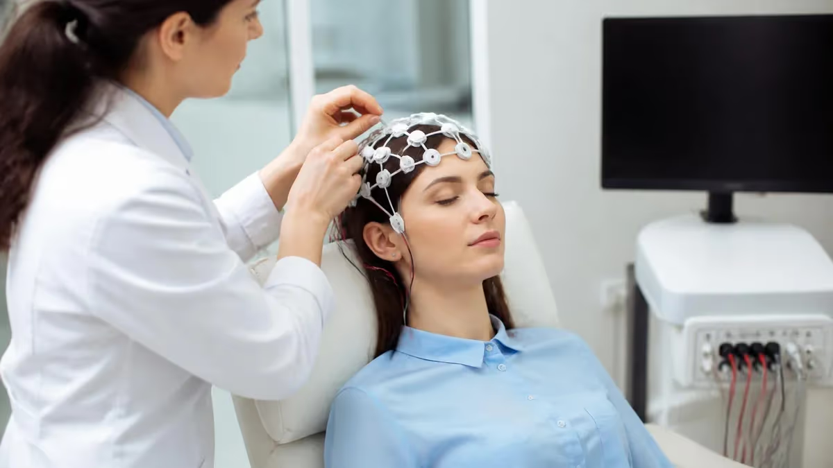
An EEG test — short for electroencephalogram — is one of the most important diagnostic tools in modern neurology. EEG monitoring records the electrical activity of the brain through small metal electrodes placed on the scalp, producing a continuous readout of brainwave patterns that clinicians use to detect seizure disorders, sleep conditions, metabolic encephalopathies, and a wide range of other neurological problems. If your doctor has ordered this study, understanding what an EEG medical test involves can ease anxiety and help you prepare effectively.
The history of the EEG dates back to 1924, when Hans Berger first recorded human brain electrical activity. Since then, the technology has evolved dramatically — from single-channel paper tracings to high-density digital systems capable of recording 256 channels simultaneously. Despite these advances, the core principle remains unchanged: neurons communicate through electrical impulses, and those impulses can be measured, amplified, and displayed as wave patterns that carry diagnostic meaning. Today, eeg monitoring professionals undergo rigorous training and credentialing to interpret these complex signals accurately.
What surprises many patients is how widely the EEG test is used. Beyond epilepsy diagnosis, EEG monitoring helps physicians evaluate patients who have experienced unexplained loss of consciousness, assess brain function after a stroke or traumatic brain injury, guide anesthesia depth during surgery, monitor patients in intensive care for non-convulsive seizures, and confirm brain death in critical care settings. The breadth of applications makes the EEG one of the most versatile neurophysiological assessments available in clinical medicine today.
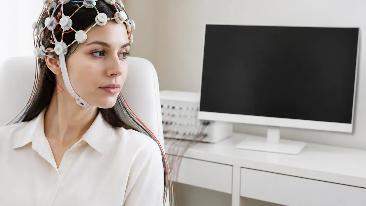
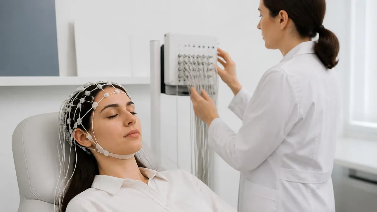
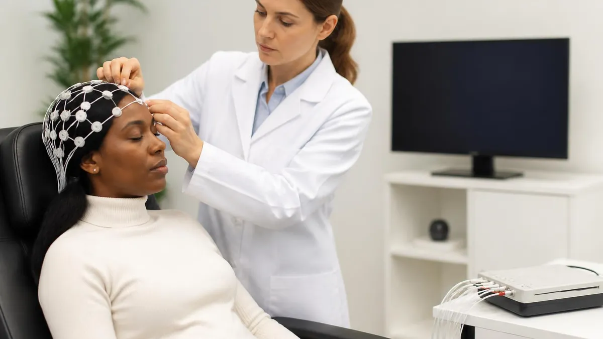
If you are wondering what is an EEG test in practical terms, think of it as an electrical map of your brain's activity captured over a defined period. During a standard routine EEG, you will sit or lie quietly while a trained EEG technologist applies electrodes to standardized locations on your scalp following the internationally recognized 10-20 electrode placement system. The recording typically lasts between 20 and 40 minutes for a routine study, though prolonged or ambulatory studies can extend for 24 to 72 hours or even longer in hospital-based monitoring units.
One of the most common questions patients ask is about EEG test side effects. The good news is that a standard EEG is entirely non-invasive and painless. The electrodes detect electrical signals — they do not transmit any electricity into your brain. The electrode gel used to ensure good contact may feel slightly cool or sticky, and some patients experience mild skin irritation at electrode sites after a prolonged study, but serious adverse effects are extremely rare.
The only notable risk during activation procedures such as hyperventilation or photic stimulation is the possibility of provoking a seizure in someone who already has a seizure disorder, which is actually the intended diagnostic purpose of those procedures.
Understanding EEG test cost is important for patients navigating healthcare in the United States. The price of an EEG varies considerably based on the type of study ordered, the facility where it is performed, and your insurance coverage. A routine outpatient EEG at a hospital or neurology clinic typically ranges from $200 to $700 before insurance adjustments.
More complex studies — such as video-EEG monitoring that may last several days — can cost several thousand dollars. Many insurance plans cover EEG when it is medically indicated, but patients should verify benefits and any prior authorization requirements with their insurer before scheduling.
This article walks you through everything you need to know about EEG monitoring: the science behind brain wave patterns, the step-by-step experience of having the test, what different results mean, how long the study takes, realistic cost expectations, and how to prepare so you get the most accurate reading possible. Whether you are a patient preparing for your first EEG or a student studying for an EEG credentialing examination, the detailed information below will give you a thorough grounding in this essential neurophysiological tool.
EEG Testing by the Numbers
How EEG Monitoring Works: Step by Step
Electrode Preparation and Placement
Baseline Recording
Activation Procedures
Drowsiness and Sleep Recording
Signal Processing and Review
If you have been wondering what to expect during an EEG medical test, the experience is generally straightforward and comfortable. When you arrive at the EEG lab, the technologist will review your medical history, confirm any preparation instructions were followed, and explain the procedure before starting. You will be asked to sit or recline in a chair or on an examination table, and the room is typically quiet and dimly lit to minimize external stimulation that could interfere with the recording.
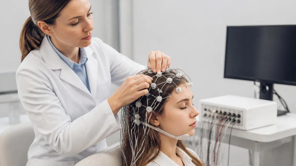
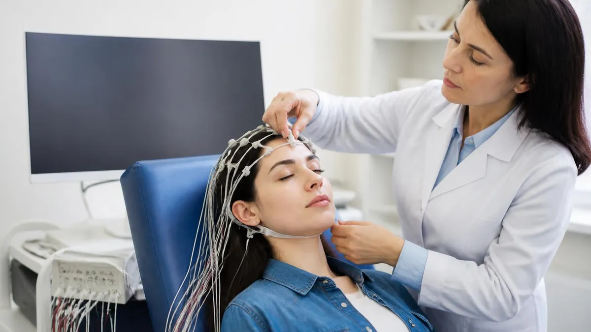
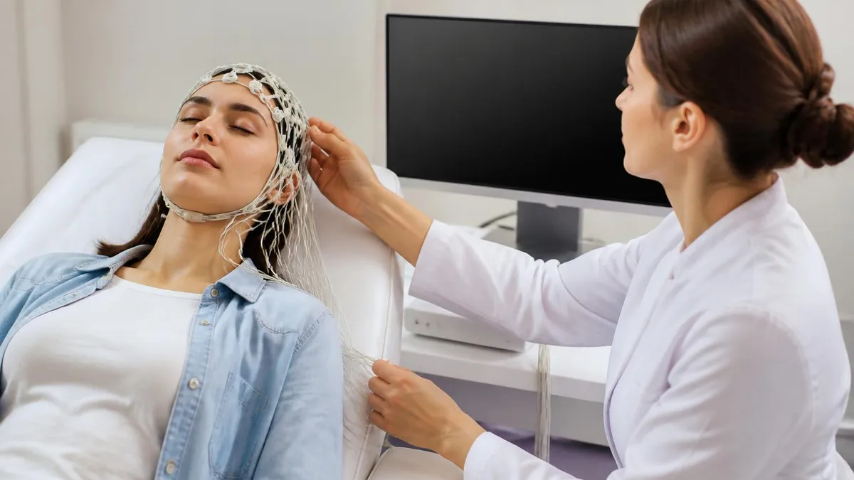
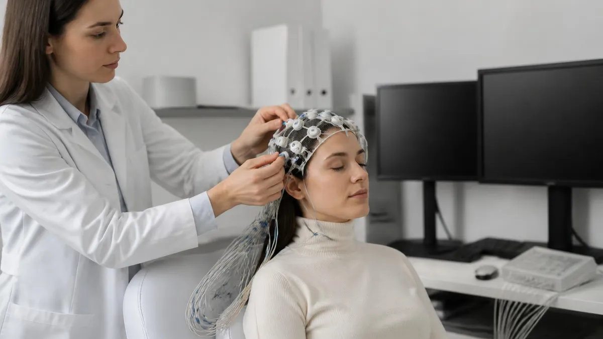
Electrode application is the most time-consuming part of the setup, typically taking 20 to 30 minutes. The technologist measures specific distances on your scalp to ensure electrodes land at the correct standardized positions. Each electrode site is gently abraded to remove oils and dead skin cells, then filled with conductive gel before the electrode is secured. Some labs use a pre-made elastic electrode cap that can reduce application time significantly, particularly for long-term monitoring or research studies.
Once recording begins, you will be asked to lie still and breathe normally. Movement, chewing, swallowing, and eye blinks all generate electrical artifacts that can obscure the underlying brain signal, so minimizing physical activity during the recording improves data quality. The technologist will communicate with you throughout the study, providing clear instructions when it is time to open or close your eyes, begin hyperventilating, or look at the strobe light during photic stimulation.
Hyperventilation typically lasts three minutes and involves taking deep, slow breaths at a rate of approximately 20 per minute. Many patients notice tingling in their hands and feet or feel slightly lightheaded — these are normal physiological responses to reduced carbon dioxide in the blood and resolve quickly once normal breathing resumes. In children and in patients with certain types of epilepsy, hyperventilation frequently provokes characteristic high-amplitude slow wave activity or generalized spike-wave discharges that are diagnostically important.
After the recording is complete, the technologist will remove the electrodes and help you clean the conductive gel from your hair and scalp. Some gel residue may remain despite rinsing, so many patients prefer to shampoo their hair at home after the appointment. The entire appointment — from arrival to departure — typically takes 60 to 90 minutes for a routine outpatient EEG, accounting for both the setup and recording time.
Patients frequently ask how long is an EEG test when they are scheduled for a prolonged or video-EEG study. In an epilepsy monitoring unit, electrodes may be left in place for several days while clinicians wait to capture a patient's habitual seizures. During this time, patients are continuously recorded on both EEG and synchronized video so that clinical behaviors can be correlated with electrographic changes. These long-term studies are essential for pre-surgical epilepsy evaluation and for distinguishing epileptic seizures from non-epileptic events.
The waiting period between completing the study and receiving results can range from a few days to two weeks, depending on the facility and the urgency of the referral. In intensive care or intraoperative settings where continuous EEG monitoring is used, a neurophysiologist may review the recording in real time or within hours and communicate critical findings directly to the treating team. For routine outpatient studies, the interpreting physician's report is typically sent to your referring neurologist or primary care provider, who will discuss the findings with you at a follow-up appointment.
EEG Practice Test Questions
Prepare for the EEG - Electroencephalography exam with our free practice test modules. Each quiz covers key topics to help you pass on your first try.
EEG Abnormal Epileptiform Patterns
EEG Exam Questions covering Abnormal Epileptiform Patterns. Master EEG Test concepts for certification prep.
EEG Activation Procedures
Free EEG Practice Test featuring Activation Procedures. Improve your EEG Exam score with mock test prep.
EEG Artifact Identification
EEG Test Prep for Artifact Identification. Practice EEG Quiz questions and boost your score.
EEG Electrode Placement and Montages
EEG Questions and Answers on Electrode Placement and Montages. Free EEG practice for exam readiness.
EEG - Electroencephalography Abnormal Epil...
EEG Mock Test covering - Electroencephalography Abnormal Epileptiform Patterns. Online EEG Test practice with instant feedback.
EEG - Electroencephalography Activation Pr...
Free EEG Quiz on - Electroencephalography Activation Procedures. EEG Exam prep questions with detailed explanations.
EEG - Electroencephalography Artifact Iden...
EEG Practice Questions for - Electroencephalography Artifact Identification. Build confidence for your EEG certification exam.
EEG - Electroencephalography Electrode Pla...
EEG Test Online for - Electroencephalography Electrode Placement and Montages. Free practice with instant results and feedback.
EEG - Electroencephalography Instrumentati...
EEG Study Material on - Electroencephalography Instrumentation and Calibration. Prepare effectively with real exam-style questions.
EEG - Electroencephalography Neuroanatomy ...
Free EEG Test covering - Electroencephalography Neuroanatomy and Physiology. Practice and track your EEG exam readiness.
EEG - Electroencephalography Non-Epileptif...
EEG Exam Questions covering - Electroencephalography Non-Epileptiform Abnormalities. Master EEG Test concepts for certification prep.
EEG - Electroencephalography Normal EEG Wa...
Free EEG Practice Test featuring - Electroencephalography Normal EEG Waveforms. Improve your EEG Exam score with mock test prep.
EEG - Electroencephalography Patient Safet...
EEG Mock Exam on - Electroencephalography Patient Safety and Ethics. EEG Study Guide questions to pass on your first try.
EEG - Electroencephalography Pediatric and...
EEG Test Prep for - Electroencephalography Pediatric and Neonatal EEG. Practice EEG Quiz questions and boost your score.
EEG ICU and Continuous EEG Monitoring
EEG Questions and Answers on ICU and Continuous EEG Monitoring. Free EEG practice for exam readiness.
EEG Instrumentation and Calibration
EEG Mock Test covering Instrumentation and Calibration. Online EEG Test practice with instant feedback.
EEG Neuroanatomy and Physiology
Free EEG Quiz on Neuroanatomy and Physiology. EEG Exam prep questions with detailed explanations.
EEG Non-Epileptiform Abnormalities
EEG Practice Questions for Non-Epileptiform Abnormalities. Build confidence for your EEG certification exam.
EEG Normal EEG Waveforms
EEG Test Online for Normal EEG Waveforms. Free practice with instant results and feedback.
EEG Patient Safety and Ethics
EEG Study Material on Patient Safety and Ethics. Prepare effectively with real exam-style questions.
Types of EEG Medical Tests Explained
A routine outpatient EEG is the most commonly ordered type of EEG test. The study lasts 20 to 40 minutes and includes baseline recording, hyperventilation, and photic stimulation. It is performed in a clinical lab with the patient awake and is best suited for detecting generalized epileptiform abnormalities, encephalopathic slow-wave changes, or focal asymmetries suggesting a structural lesion. Sleep deprivation before the study increases the probability of capturing drowsiness and light sleep, which substantially improves the diagnostic yield for temporal lobe epilepsy and other focal epilepsies.
The limitation of a routine EEG is that brain electrical activity between seizures is often completely normal — studies report that up to 50% of patients with confirmed epilepsy have a normal first routine EEG. For this reason, neurologists frequently order repeat studies, request sleep-deprived recordings, or escalate to prolonged monitoring when clinical suspicion remains high. Despite its limitations, the routine EEG remains the first-line neurophysiological test for evaluating new-onset seizures, syncope of uncertain origin, and altered mental status.
EEG Test Advantages and Limitations
- +Non-invasive and painless — no needles, radiation, or contrast agents required
- +Excellent temporal resolution — captures brain electrical changes occurring in milliseconds
- +Widely available at hospitals, neurology clinics, and outpatient diagnostic centers
- +Relatively low cost compared to MRI or CT scanning, especially for routine studies
- +Can be performed at the bedside for critically ill or immobile patients
- +Ambulatory options allow recording during natural daily activities and sleep cycles
- −Low sensitivity for single routine studies — up to 50% of epilepsy patients have a normal first EEG
- −Poor spatial resolution — cannot pinpoint lesion location with the precision of neuroimaging
- −Highly susceptible to artifacts from muscle movement, eye blinks, electrode loosening, and electrical interference
- −Requires a skilled technologist for application and a specialist for accurate interpretation
- −Long-term studies are logistically demanding and expensive for both patients and healthcare systems
- −Normal EEG does not rule out a seizure disorder or other neurological condition
How to Prepare for Your EEG Test
- ✓Wash your hair the night before but do not apply conditioner, styling products, oils, or hairspray on the morning of the test.
- ✓Ask your doctor whether to continue or hold any medications before the study — never stop seizure medications without medical guidance.
- ✓Follow sleep deprivation instructions if ordered: typically staying up until midnight and rising by 4–5 AM before a morning appointment.
- ✓Eat a full meal before your appointment to maintain stable blood sugar and prevent fainting during hyperventilation.
- ✓Avoid caffeine for at least 8 hours before the study, as it can alter alertness levels and suppress sleep during the recording.
- ✓Arrive on time and allow 60–90 minutes total for the appointment including electrode setup, recording, and cleanup.
- ✓Wear comfortable, loose-fitting clothing and avoid turtlenecks or high collars that could interfere with electrode placement.
- ✓Bring a list of all current medications including doses and timing so the technologist can note them in your study record.
- ✓Inform the technologist about any scalp conditions, recent head injuries, or surgical hardware that could affect electrode placement.
- ✓Arrange transportation home if you were sleep-deprived for the study, as extreme fatigue can affect safe driving ability.
Why Doctors Order Sleep-Deprived EEGs
Research consistently shows that sleep deprivation before an EEG increases the probability of recording drowsiness and light sleep during the study, states in which epileptiform discharges occur far more frequently than during wakefulness. A sleep-deprived EEG can increase diagnostic sensitivity for temporal lobe epilepsy by 30–50% compared to a standard rested study, making it one of the most cost-effective ways to improve yield before escalating to prolonged monitoring.
EEG test cost is one of the most searched aspects of this procedure among patients in the United States, and for good reason — healthcare costs vary enormously depending on where and how the study is performed. A routine outpatient EEG at a freestanding neurology clinic typically costs between $200 and $500 before insurance adjustments.
The same study performed in a hospital outpatient department may be billed at $500 to $1,200 or more due to the separate facility fee that hospitals charge in addition to the physician interpretation fee. Understanding this billing structure can help patients make informed decisions about where to schedule their study.
For patients with commercial insurance, EEG test cost out-of-pocket depends heavily on the specific plan's deductible, copay, and coinsurance requirements, as well as whether the ordering physician and facility are both in-network. Many plans require prior authorization before approving coverage for EEG, particularly for ambulatory or long-term monitoring studies. Patients should contact their insurance carrier before scheduling to confirm coverage, obtain authorization if required, and request an estimate of their cost-sharing obligation. Hospital financial counselors and the ordering physician's billing department can also help navigate these questions.
For patients without insurance or with high-deductible plans, several strategies can reduce EEG test price. Scheduling at a freestanding diagnostic center rather than a hospital outpatient department often yields significantly lower costs for the same study. Federally qualified health centers and community health clinics offer EEG services on a sliding-fee scale based on income. Some academic medical centers have financial assistance programs that cover or reduce the cost for patients who meet income eligibility requirements. Asking the ordering physician's office for a cash-pay discount is also a reasonable approach that many smaller independent practices accommodate.
Long-term video-EEG monitoring in an epilepsy monitoring unit represents the high end of the EEG cost spectrum. A 3–5 day hospital admission for epilepsy monitoring can generate total charges ranging from $5,000 to $30,000 or more, incorporating the costs of the inpatient stay, continuous EEG recording equipment rental or facility charges, daily professional interpretation fees, and nursing care. Insurance typically covers these studies when the medical indication is clearly documented — usually pre-surgical epilepsy evaluation or characterization of seizure type in patients failing medical management — but prior authorization is almost always required.
Ambulatory EEG occupies a middle ground in terms of cost, typically ranging from $300 to $1,000 depending on the duration of the study and the billing entity. Because ambulatory EEG is performed outpatient without the overhead of an inpatient hospitalization, it is considerably less expensive than epilepsy monitoring unit admissions while providing substantially more recording time than a routine outpatient EEG. For patients with suspected focal epilepsy or paroxysmal events that have not been captured on routine studies, ambulatory EEG offers a cost-effective intermediate step before escalating to inpatient monitoring.
Intraoperative neurophysiological monitoring — which includes continuous EEG as one component alongside other modalities such as somatosensory evoked potentials and electromyography — adds a separate billing layer for surgeries involving the brain, spinal cord, or peripheral nervous system. The professional fees for intraoperative neurophysiology services are typically billed separately from the surgeon's and anesthesiologist's fees, and vary based on the modalities used and the duration of the case. Patients scheduled for neurosurgical procedures should discuss intraoperative monitoring with their surgical team and verify insurance coverage in advance.
It is worth noting that the cost of an EEG test should be considered in the context of its diagnostic value relative to alternative investigations. A brain MRI, which is often ordered alongside an EEG for seizure evaluation, typically costs $500 to $2,000 or more depending on whether contrast is used and where it is performed. A CT scan of the brain costs $300 to $1,000. The EEG provides unique information about dynamic brain electrical activity that no structural imaging modality can replicate, making it an essential and generally cost-effective component of the neurological diagnostic workup when used appropriately.
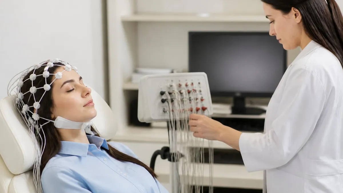
Many insurance plans require prior authorization before approving coverage for EEG studies, especially ambulatory or long-term monitoring. Scheduling without authorization can result in denial of the claim and full patient responsibility for the bill. Always verify coverage and obtain authorization before your appointment, and keep a record of the authorization number provided by your insurer.
Understanding EEG results requires familiarity with the basic categories of findings that a neurophysiologist evaluates when reviewing a recording. The most fundamental assessment is whether the background activity is normal for the patient's age and level of alertness. In a healthy awake adult, the EEG shows a well-organized posterior alpha rhythm at 8 to 13 cycles per second, faster beta activity over the frontal regions, and low-voltage mixed-frequency activity elsewhere. Deviations from this expected pattern — such as diffuse background slowing, focal delta activity, or loss of normal sleep architecture — provide important diagnostic clues.
Epileptiform abnormalities are the findings most specifically associated with seizure disorders. These include interictal epileptiform discharges (IEDs) such as spikes, sharp waves, and spike-and-slow-wave complexes that occur between seizures and indicate cortical hyperexcitability. The distribution and morphology of these discharges help localize the epileptogenic zone — for example, generalized 3-Hz spike-wave discharges suggest childhood absence epilepsy, while right temporal sharp waves point toward a focal seizure origin in the right mesial temporal region. Identifying these patterns guides both the selection of antiseizure medication and the consideration of surgical candidacy.
An ictal recording — one that captures an actual seizure — provides the most definitive electrographic information. Ictal patterns typically begin with a rhythmic focal or generalized discharge that evolves in frequency, amplitude, and spatial distribution over the course of the clinical event. Correlating the electrographic onset with the patient's symptoms on video allows clinicians to determine whether the clinical event represents a true epileptic seizure or a non-epileptic event. This distinction has profound implications for treatment, since antiseizure medications are effective only for the former and can actually delay appropriate psychological treatment in patients with psychogenic non-epileptic seizures.
Encephalopathic EEG patterns reflect diffuse dysfunction of the cerebral cortex or its subcortical connections and are commonly seen in critically ill patients. Generalized slowing — with excess theta (4–7 Hz) or delta (0.5–3 Hz) activity replacing the normal faster rhythms — indicates varying degrees of cerebral dysfunction and correlates roughly with the level of clinical impairment. More severe patterns include generalized suppression, burst-suppression, and electrocerebral inactivity, the last representing the EEG criterion for brain death. Tracking the evolution of encephalopathic patterns over serial recordings helps clinicians gauge the trajectory of a patient's neurological recovery or deterioration.
Specific EEG patterns carry eponymous names that reflect their distinct morphology and clinical significance. Triphasic waves are high-amplitude, frontally predominant complexes associated with metabolic encephalopathies such as hepatic or uremic encephalopathy. Periodic lateralized epileptiform discharges (PLEDs, now termed LPDs) are seen in the setting of acute cortical lesions such as stroke or herpes encephalitis. FIRDA (frontal intermittent rhythmic delta activity) indicates diffuse encephalopathy or deep midline structural dysfunction. Recognizing these named patterns requires substantial pattern-recognition experience and is a core competency tested on EEG credentialing examinations.
Normal variant patterns represent a category of EEG findings that can mimic pathological discharges but are actually benign phenomena seen in healthy individuals. Examples include wicket spikes, small sharp spikes of sleep (benign epileptiform transients of sleep or BETS), the mu rhythm, and lambda waves. Misidentifying these normal variants as epileptiform abnormalities leads to false-positive diagnoses and unnecessary treatment. Conversely, misclassifying true epileptiform discharges as normal variants can delay appropriate therapy. This diagnostic precision requires the kind of expert training that professional credentialing bodies assess through their examination programs.
Following a report of abnormal EEG findings, the next steps depend entirely on the clinical context. An abnormal EEG alone is never sufficient to diagnose epilepsy — the diagnosis requires a clinical history consistent with unprovoked seizures, interpreted in conjunction with the EEG results and any structural neuroimaging findings. Conversely, a normal EEG does not exclude epilepsy. Your neurologist will integrate all available information to formulate a diagnosis and treatment plan. If antiseizure medication is initiated, follow-up EEG studies may be ordered to track treatment response or to monitor for medication-related EEG changes.
For students and working professionals preparing for EEG credentialing examinations, understanding the full scope of EEG monitoring is essential. The Registered EEG Technologist (R.EEG.T.) credential awarded by the American Board of Registration of Electroencephalographic and Evoked Potential Technologists (ABRET) tests candidates on a comprehensive body of knowledge that spans neuroanatomy, electronics and instrumentation, artifact recognition, normal EEG patterns across the lifespan, activation procedures, abnormal EEG findings, and the technical aspects of long-term and ambulatory monitoring.
Preparation for the R.EEG.T. examination typically requires 6 to 12 months of focused study in addition to the clinical hours required for eligibility. Candidates who pass on their first attempt consistently report that practicing large volumes of EEG pattern recognition questions — rather than passively reading textbooks — is the most effective preparation strategy. The ability to rapidly and accurately classify waveform patterns under examination conditions requires repeated, active engagement with practice material drawn from authentic EEG recordings and high-yield clinical scenarios.
One area of the examination that many candidates underestimate is the technical and instrumentation content. A thorough understanding of amplifier gain and sensitivity settings, filter characteristics and their effects on waveform morphology, electrode impedance testing, digital sampling rates, and the differential amplifier principle is essential for both the written examination and clinical practice. EEG technologists who understand why technical problems occur — not just how to identify them on the screen — are better equipped to troubleshoot quickly and maintain recording quality in demanding clinical environments such as the operating room or ICU.
Activation procedures constitute another high-yield examination topic that rewards deep conceptual understanding. Candidates should be able to explain the physiological mechanism by which hyperventilation provokes EEG changes (cerebral vasoconstriction secondary to hypocapnia), articulate the contraindications to hyperventilation (sickle cell disease, severe cardiopulmonary disease, moyamoya disease), describe the standardized photic stimulation protocol including the range of flash frequencies tested and the significance of a photoparoxysmal response, and recognize the EEG patterns associated with drowsiness and sleep stages that make sleep a valuable activation state.
The ambulatory EEG credentialing pathway — the Certified Ambulatory EEG Technologist (CAEEG) credential — has its own specific content domain that emphasizes the unique technical and operational aspects of extended outpatient recording. Candidates pursuing this credential should develop particular expertise in electrode application techniques optimized for long-term wear, patient and caregiver education about event button use and activity diary completion, recognition of the artifact patterns that arise during ambulatory monitoring, and the data management workflows involved in reviewing and marking lengthy ambulatory studies.
Continuing education requirements keep credentialed EEG professionals current with advances in the field. ABRET requires credential holders to accumulate a defined number of continuing education credits during each renewal cycle, covering both technical and clinical content. Professional organizations such as the American Society of Electroneurodiagnostic Technologists (ASET) provide conferences, webinars, and online learning modules that fulfill these requirements. Staying engaged with the evolving literature — particularly regarding quantitative EEG, high-density recording systems, and artificial intelligence applications in EEG interpretation — positions professionals for advancement into specialized roles in epilepsy monitoring units, research settings, and neurodiagnostic laboratory management.
The career trajectory available to skilled EEG professionals reflects the growing clinical importance of neurophysiological monitoring. Entry-level technologist positions offer competitive starting salaries, with experienced and credentialed professionals in high-demand markets commanding substantially higher compensation. Lead technologist, supervisor, and laboratory director roles are available to those who combine technical expertise with leadership skills. Specialized roles in neurodiagnostic education, medical device industry training, and clinical research coordination offer additional career paths for professionals seeking to expand beyond direct patient care while leveraging their neurophysiology knowledge base.
EEG Questions and Answers
EEG Tech Salary 2026: Pay by State, Experience, and Certification
EEG Images: A Visual Guide to Brain Wave Patterns, Recordings, and Test Results
EEG for Seizures: How the Test Diagnoses Epilepsy and What to Expect
Ambulatory EEG: 24-72 Hour Home Recording for Suspected Seizures
EEG Electrode Placement: The Complete Guide to the 10-20 System, Scalp Mapping, and How an EEG Test Works
About the Author

Educational Psychologist & Academic Test Preparation Expert
Columbia University Teachers CollegeDr. Lisa Patel holds a Doctorate in Education from Columbia University Teachers College and has spent 17 years researching standardized test design and academic assessment. She has developed preparation programs for SAT, ACT, GRE, LSAT, UCAT, and numerous professional licensing exams, helping students of all backgrounds achieve their target scores.