
LVAD vs ECMO: Understanding Extracorporeal Membrane Oxygenation Support Devices
🎯 LVAD vs ECMO explained: extracorporeal membrane oxygenation procedure, circuit, costs & clinical use for neonates and adults.

When critically ill patients require mechanical circulatory or respiratory support, clinicians must choose between powerful but distinct technologies. The debate over lvad ecmo — left ventricular assist devices versus extracorporeal membrane oxygenation — is one of the most consequential decisions in modern critical care. Extracorporeal membrane oxygenation in neonates has saved thousands of lives since the 1970s, and today the technology extends to adults facing refractory cardiac or pulmonary failure. Understanding the fundamental differences between these two support systems is essential for nurses, perfusionists, respiratory therapists, and physicians preparing for certification examinations or daily clinical practice.
An LVAD, or left ventricular assist device, is a surgically implanted mechanical pump that helps the left ventricle move blood from the heart to the aorta. It is typically used as a bridge to transplant or as destination therapy for patients with end-stage heart failure who are ineligible for transplant. The device sits inside the chest, draws blood from the left ventricle apex, and delivers it into the ascending aorta, essentially bypassing a failing left ventricle. Unlike ECMO, an LVAD does not oxygenate blood — it provides purely hemodynamic support without addressing gas exchange deficits.
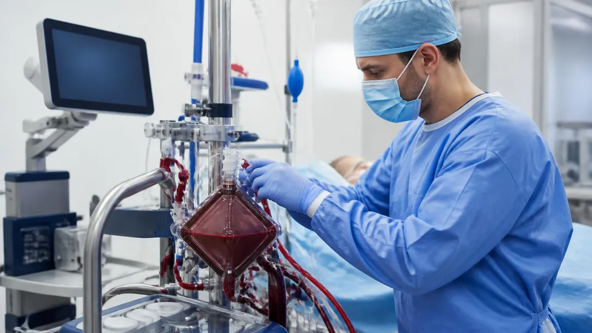
Extracorporeal membrane oxygenation, by contrast, is a temporary extracorporeal life support system capable of providing both cardiac and respiratory support simultaneously. The extracorporeal membrane oxygenation circuit consists of a pump, an oxygenator membrane, and a series of cannulas that drain deoxygenated blood from the patient, pass it through an artificial lung, and return oxygenated blood back into the circulation. This makes ECMO uniquely versatile: it can support a patient in profound cardiogenic shock, refractory ARDS, or both conditions at once, which an LVAD alone cannot accomplish. Visit lvad vs ecmo for a deeper look at cannulation strategies for veno-arterial configurations.
The clinical indications for each technology overlap but diverge in important ways. LVADs are primarily indicated for chronic, severe left ventricular dysfunction — New York Heart Association class IIIb or IV patients who remain symptomatic despite optimal guideline-directed medical therapy. ECMO, on the other hand, is reserved for acute, potentially reversible conditions: massive pulmonary embolism, post-cardiotomy cardiogenic shock, refractory ventricular fibrillation during cardiac arrest (ECPR), fulminant myocarditis, and severe acute respiratory distress syndrome. The acuity and anticipated duration of support fundamentally separates the two modalities.
Patient selection criteria reflect these differences sharply. LVAD candidates typically undergo months of evaluation by multidisciplinary heart failure teams, including cardiology, cardiac surgery, palliative care, and social work. ECMO, in contrast, is often deployed emergently within hours of deterioration, sometimes within minutes during a resuscitation. The urgency of ECMO cannulation means that comprehensive pre-procedure evaluation is often impossible, placing greater emphasis on rapid bedside assessment of reversibility and contraindications such as severe aortic regurgitation or end-organ damage too advanced to recover.
Cost and resource considerations also differ enormously. The extracorporeal membrane oxygenation machine price for a complete system ranges from approximately $100,000 to $350,000 for capital equipment, with per-day circuit and consumable costs ranging from $1,500 to $5,000 in many US centers. LVAD implantation procedures cost between $150,000 and $300,000 all-in, but the device itself then supports the patient for years without requiring daily consumable replacement. These economic realities influence institutional capacity planning, insurance coverage decisions, and the feasibility of maintaining active ECMO programs in community versus tertiary care settings.
For clinicians studying for ECMO specialist certifications such as those offered by ELSO or preparing for critical care board examinations, a thorough mastery of both technologies — including their mechanisms, indications, contraindications, complications, and management protocols — is non-negotiable. The nuances of patient selection, circuit troubleshooting, anticoagulation management, and weaning strategies appear repeatedly on high-stakes assessments, and understanding the comparative framework of LVAD versus ECMO provides an excellent conceptual anchor for organizing that knowledge effectively.
LVAD vs ECMO by the Numbers

How the Extracorporeal Membrane Oxygenation Circuit Works
Venous Drainage Cannula
Centrifugal Pump
Membrane Oxygenator
Heat Exchanger
Arterial Return Cannula
The distinction between venovenous extracorporeal membrane oxygenation and veno-arterial ECMO is the single most important conceptual divide in ECMO practice, and it maps directly onto the clinical difference between purely respiratory failure and combined cardiorespiratory failure. In venovenous ECMO, blood is drained from and returned to the venous circulation, meaning the heart still pumps all of the oxygenated blood through the pulmonary and systemic circulations. The ECMO circuit simply pre-oxygenates and decarbonates the venous blood before the heart receives it, augmenting — but not replacing — native cardiac output.
Veno-arterial ECMO, by contrast, drains blood from the venous side and returns oxygenated blood directly into the arterial circulation, effectively bypassing both the heart and lungs. This configuration provides both respiratory and hemodynamic support simultaneously, making it the appropriate choice for patients in cardiogenic shock, cardiac arrest with ECPR, or post-cardiotomy failure. However, VA-ECMO introduces unique complications absent from VV-ECMO, most notably limb ischemia from arterial cannulation, north-south syndrome (also called Harlequin syndrome) when native cardiac output recovers before ECMO flows are reduced, and left ventricular distension from retrograde aortic flow opposing a still-beating but weak heart.
The Harlequin syndrome, or differential hypoxemia, deserves particular attention for ECMO examination candidates. It occurs during peripheral VA-ECMO when the recovering native heart begins ejecting blood through the aortic valve, but that natively ejected blood is poorly oxygenated because the lungs remain diseased. Meanwhile, the ECMO return cannula in the femoral artery delivers fully oxygenated blood retrograde up the descending aorta.
The result is well-oxygenated blood perfusing the lower body and legs while the upper body — including the coronary arteries, right arm, and brain — receives deoxygenated blood from the recovering but still-failing lungs. This can be detected by pulse oximetry on the right hand showing paradoxically lower saturations than the left foot.
Left ventricular venting is another critical management consideration unique to VA-ECMO that has no direct analogue in LVAD management. When VA-ECMO returns blood retrograde into the aorta, the pressure increase can prevent the aortic valve from opening adequately, causing blood to accumulate in the left ventricle, raising left ventricular end-diastolic pressure, and distending the chamber.
Unrelieved LV distension improves pulmonary capillary wedge pressure, worsens pulmonary edema, increases myocardial oxygen consumption, and impairs myocardial recovery. Venting strategies include insertion of an Impella device (creating a hybrid ECMO-Impella configuration sometimes called ECPELLA), atrial septostomy, or surgical left ventricular vent placement via the left atrium.
Understanding how to transition a patient from VA-ECMO to an LVAD — or when to escalate from LVAD to VA-ECMO — is a growing area of clinical practice as mechanical circulatory support programs mature. A patient on an LVAD who develops acute right ventricular failure may require right-sided VA-ECMO or a right ventricular assist device (RVAD) in addition to the existing LVAD, creating a biventricular assist configuration.
Conversely, a patient initially stabilized on VA-ECMO whose myocardium shows no sign of recovery may be transitioned to a durable LVAD as a bridge to transplant, requiring careful hemodynamic assessment to ensure adequate right ventricular function before LVAD implantation.
Anticoagulation management represents one of the most challenging and complication-prone aspects of ECMO that distinguishes it sharply from LVAD management. ECMO circuits require continuous systemic anticoagulation, typically with unfractionated heparin titrated to an anti-Xa level of 0.3–0.7 IU/mL or an activated clotting time of 180–220 seconds, depending on institutional protocol.
The risk of both thrombosis — clot formation in the circuit leading to oxygenator failure or thromboembolic events — and hemorrhage — intracranial bleeding, surgical site bleeding, cannula site oozing — must be balanced continuously. LVADs also require anticoagulation, but the targets and monitoring approaches differ, and ECMO circuits generally carry a higher thromboinflammatory burden due to the larger surface area of the extracorporeal circuit.
For healthcare professionals pursuing ECMO specialist credentials, mastering the hemodynamic signatures of VV-ECMO versus VA-ECMO is foundational. On VV-ECMO, arterial blood pressure and pulse pressure remain driven entirely by native cardiac function; the clinician uses circuit flows and sweep gas settings only to optimize gas exchange. On VA-ECMO, circuit flows directly determine mean arterial pressure and systemic perfusion. Reducing VA-ECMO flow during a weaning trial causes measurable hemodynamic deterioration if the native heart cannot compensate, providing the clinical team with real-time information about myocardial recovery — a physiologic assessment that is central to both clinical management and ECMO examination content.
Extracorporeal Membrane Oxygenation Treatment: Neonates, Adults & COVID-19
Extracorporeal membrane oxygenation in neonates remains one of the most established applications of this technology, with indications including meconium aspiration syndrome, congenital diaphragmatic hernia, persistent pulmonary hypertension of the newborn, and neonatal sepsis with refractory respiratory failure. Eligibility criteria typically require a gestational age of at least 34 weeks, birth weight above 2 kg, and an oxygenation index greater than 40 or an acute deterioration index exceeding 0.4, calculated from mean airway pressure, FiO2, PaO2, and PaCO2 measurements.
Neonatal ECMO circuits are scaled down to accommodate small body weights and low blood volumes, using shorter tubing, smaller cannulas (8–14 French), and reduced priming volumes that may still represent 60–100% of the infant's estimated circulating blood volume, necessitating a circuit prime with packed red blood cells to prevent profound hemodilution upon initiation. Survival rates for neonatal ECMO range from 75–85% for meconium aspiration syndrome to 50–60% for congenital diaphragmatic hernia, according to ELSO registry data — making neonatal ECMO one of the highest-yield success areas in all of extracorporeal life support.

LVAD vs ECMO: Advantages and Limitations
- +ECMO provides simultaneous cardiac and respiratory support, unlike LVAD which addresses only left ventricular failure
- +ECMO can be initiated emergently at bedside without a formal operating room in experienced centers
- +VV-ECMO preserves native cardiac output and avoids arterial cannulation risks when lungs are the primary failing organ
- +ECMO is reversible and can be discontinued within minutes once the patient recovers, unlike surgically implanted LVADs
- +Neonatal and pediatric ECMO has decades of registry data with survival rates of 75–85% for select diagnoses
- +ECMO supports biventricular failure, right ventricular failure, and mixed shock that LVADs cannot address alone
- −ECMO requires continuous intensive bedside nursing and perfusionist presence, consuming significant staffing resources 24/7
- −The extracorporeal membrane oxygenation circuit introduces major hemorrhagic and thrombotic risks requiring expert anticoagulation management
- −ECMO is a temporary bridge with typical durations of days to weeks, not a long-term destination therapy like LVAD
- −VA-ECMO raises left ventricular afterload and can worsen LV distension in patients with residual cardiac function
- −Peripheral VA-ECMO via femoral artery risks ipsilateral limb ischemia requiring distal perfusion cannulas in up to 20% of cases
- −ECMO machine price and consumable costs are extremely high, with daily circuit costs of $1,500–$5,000 limiting broad accessibility
Pros and Cons at a Glance
| Pros | Cons |
|---|---|
| ECMO provides simultaneous cardiac and respiratory support, unlike LVAD which addresses only left ventricular failure | The extracorporeal membrane oxygenation circuit introduces major hemorrhagic and thrombotic risks requiring expert anticoagulation management |
| ECMO can be initiated emergently at bedside without a formal operating room in experienced centers | ECMO is a temporary bridge with typical durations of days to weeks, not a long-term destination therapy like LVAD |
| VV-ECMO preserves native cardiac output and avoids arterial cannulation risks when lungs are the primary failing organ | VA-ECMO raises left ventricular afterload and can worsen LV distension in patients with residual cardiac function |
ECMO Clinical Decision Checklist: Key Assessment Points
- ✓Confirm the primary indication — isolated respiratory failure favors VV-ECMO; cardiogenic shock or arrest favors VA-ECMO.
- ✓Assess reversibility of the underlying condition before initiating ECMO to establish a realistic endpoint for support.
- ✓Verify absence of absolute contraindications: uncontrolled bleeding, severe aortic regurgitation, and irreversible end-organ failure.
- ✓Confirm gestational age ≥34 weeks and birth weight ≥2 kg before initiating extracorporeal membrane oxygenation in neonates.
- ✓Select cannula size and configuration based on patient weight, flow requirements, and venous anatomy using echo guidance.
- ✓Prime the circuit and establish target anticoagulation (anti-Xa 0.3–0.7 IU/mL) before initiation to reduce early thrombotic events.
- ✓Place a distal perfusion cannula in the ipsilateral superficial femoral artery immediately after femoral arterial cannulation for VA-ECMO.
- ✓Obtain baseline chest X-ray, echocardiogram, and head ultrasound (neonates) within the first 24 hours of ECMO support.
- ✓Monitor and document ECMO flows, sweep gas settings, temperatures, and pressures at minimum every hour throughout the run.
- ✓Establish clear weaning criteria and conduct daily spontaneous breathing or cardiac function trials to avoid unnecessarily prolonged support.
The ECPELLA Configuration: Combining ECMO and Impella
When VA-ECMO alone causes progressive left ventricular distension — evidenced by rising pulmonary artery pressures, a non-opening aortic valve on echocardiography, and worsening pulmonary edema — adding a transvalvular microaxial flow pump (Impella) to vent the left ventricle can dramatically improve myocardial recovery rates. Studies from high-volume cardiogenic shock centers report 30-day survival improvements of 10–15 percentage points with ECPELLA versus VA-ECMO alone in refractory cardiogenic shock, establishing this combination as the preferred configuration at many US cardiac centers.
The extracorporeal membrane oxygenation procedure itself begins with a careful patient positioning and sterile preparation of the cannulation sites. In most adult VA-ECMO cases using a peripheral approach, the right femoral vein and right or left femoral artery are the preferred cannulation sites, though axillary artery cannulation is increasingly favored for ambulatory ECMO or when prolonged support is anticipated because it avoids femoral vessel complications and permits early patient mobilization. Ultrasound guidance for vessel access and fluoroscopic confirmation of guidewire and cannula positioning are standard of care at experienced ECMO centers.
Once cannulas are placed and secured, the circuit is connected, de-aired thoroughly using a systematic protocol to prevent air embolism, and flow is gradually increased from an initial 1–2 L/min to target flow over 5–10 minutes. Target ECMO blood flow is typically 60–80 mL/kg/min in neonates and 50–60 mL/kg/min in adults, calibrated to achieve SvO2 greater than 70% on VV-ECMO or adequate mean arterial pressure on VA-ECMO.
Simultaneously, mechanical ventilator settings are reduced to lung-protective or lung-rest parameters — typically FiO2 of 0.3, PEEP of 10 cmH2O, driving pressure below 15 cmH2O — to allow the injured lungs to recover without ongoing ventilator-induced injury.
Daily ECMO management involves a multidisciplinary team that may include bedside nurses with specialized ECMO training, perfusionists monitoring circuit function, respiratory therapists managing the ventilator interface, intensivists or ECMO specialists overseeing overall patient care, and bedside echocardiographers performing serial assessments of cardiac recovery. Point-of-care testing for activated clotting time, anti-Xa levels, plasma-free hemoglobin (a marker of hemolysis), fibrinogen, and platelet counts guides anticoagulation adjustments and helps identify early circuit failure such as oxygenator thrombosis, which manifests as increasing transmembrane pressure gradient and worsening gas exchange efficiency.
Oxygenator failure is the most common circuit complication requiring emergent component replacement during an ECMO run. Clot formation within the oxygenator fiber bundle progressively impairs both oxygen transfer and carbon dioxide removal, while simultaneously increasing the risk of thromboembolic events.
The decision to change an oxygenator is guided by transmembrane pressure gradient trends, post-oxygenator blood gas values, and visual inspection of the oxygenator chamber for visible clot. Circuit change-outs require a coordinated team response, with a new pre-primed backup circuit available at the bedside in all active ECMO programs, and are performed in under five minutes by experienced teams to minimize interruption of support.
Renal replacement therapy is required in 40–60% of adult ECMO patients due to acute kidney injury from the underlying illness, the systemic inflammatory response triggered by blood contact with the extracorporeal circuit, and hemolysis-associated renal tubular injury from plasma-free hemoglobin. Continuous renal replacement therapy (CRRT) can be run in-line with the ECMO circuit using a filter placed between the pump outlet and the oxygenator inlet, avoiding the need for separate vascular access and simplifying fluid management. However, in-line CRRT introduces additional anticoagulation complexity and a potential pressure drop that must be accounted for in circuit pressure monitoring.
Neurological complications represent among the most devastating outcomes of ECMO support, occurring in 10–20% of neonatal cases and up to 25–30% of adult VA-ECMO cases in published series. Intracranial hemorrhage, ischemic stroke, seizures, and diffuse hypoxic-ischemic injury can each result from the coagulopathy of ECMO, systemic embolization of microthrombi generated in the circuit, or hemodynamic instability during initiation or weaning. For neonates, serial cranial ultrasounds every 24–48 hours during the ECMO run are standard practice to detect intraventricular or parenchymal hemorrhage that may necessitate discontinuation of ECMO even before the primary indication has resolved.
Weaning from ECMO is approached differently depending on the configuration. VV-ECMO weaning involves progressively reducing sweep gas flow — often called a CPAP or cap trial — to zero while maintaining full pump flow, then observing whether the patient can maintain acceptable SpO2 and PaCO2 on native ventilatory support alone.
VA-ECMO weaning involves progressive reduction of pump flow in 0.5 L/min decrements while monitoring hemodynamic and echocardiographic parameters for evidence of adequate native cardiac output. Serial echocardiography showing improved ejection fraction, lateral mitral annular systolic velocity, and aortic velocity-time integral during flow reduction trials provides objective evidence of myocardial recovery guiding the timing of decannulation.

Significant aortic regurgitation is an absolute contraindication to peripheral VA-ECMO because retrograde aortic flow from the femoral return cannula will force blood backward through the incompetent aortic valve, causing rapid and severe left ventricular distension that can precipitate irreversible myocardial injury. Any patient with more than mild aortic regurgitation who requires mechanical circulatory support must be evaluated for central cannulation, LV venting strategies, or alternative devices such as intra-aortic balloon pump or Impella, with echocardiographic assessment mandatory before VA-ECMO initiation whenever clinical circumstances permit.
The financial reality of ECMO programs has significant implications for healthcare systems, insurance payers, and the patients who need this technology. The extracorporeal membrane oxygenation machine price varies by manufacturer and configuration — a complete adult ECMO system including the pump console, oxygenator, tubing pack, and monitoring equipment typically costs between $100,000 and $350,000 in capital expenditure.
This figure does not include the ongoing costs of disposable circuit components, which run $5,000–$15,000 per circuit setup, or the personnel costs of 24-hour bedside ECMO specialist coverage, which some estimates place at an additional $2,000–$4,000 per patient per day in labor costs alone at high-volume US centers.
Insurance coverage for ECMO in the United States is generally available for medically appropriate indications, with Medicare and Medicaid covering ECMO under DRG payment structures that reimburse at higher rates for cases involving ECMO. However, the reimbursement rarely covers actual costs at most hospitals, particularly for prolonged runs, meaning ECMO programs often operate at a financial loss per case. This economic reality concentrates ECMO capability in large academic and quaternary medical centers, creating geographic access disparities that advocacy groups and professional societies such as ELSO have highlighted as a patient safety and equity concern for rural and underserved communities.
For students and clinicians preparing for the ECMO specialist certification examination administered by ELSO's certifying body, understanding the full scope of LVAD versus ECMO comparisons — including indications, contraindications, circuit components, hemodynamic physiology, complication recognition, and management algorithms — is essential. The certification examination covers approximately 170 questions across domains including patient selection, initiation, ongoing management, troubleshooting, and transport of ECMO patients. A thorough understanding of how ECMO differs from and complements LVAD therapy provides a strong framework for mastering these domains systematically rather than memorizing isolated facts.
The extracorporeal membrane oxygenation diagram most commonly encountered in teaching materials depicts the blood flow path from venous drainage cannula, through the pump, through the oxygenator with sweep gas interface, through the heat exchanger, and back to the patient via the return cannula.
For VV-ECMO, both cannulas access the venous circulation, typically the right internal jugular vein and the right femoral vein, or via a single dual-lumen bicaval cannula (such as the Avalon Elite) placed through the right internal jugular vein. Familiarity with these diagrams and the ability to trace physiologic variables through each component of the circuit is a core competency tested on ECMO specialist examinations and directly applicable to bedside troubleshooting.
Regional ECMO retrieval and transport programs have become increasingly important as evidence accumulates that outcomes are better at high-volume centers. Mobile ECMO teams — typically composed of a cardiothoracic surgeon or interventional cardiologist, an ECMO perfusionist, and an intensivist — travel to referring hospitals, cannulate patients who meet criteria, and transport them on ECMO back to the ECMO center. Transport on ECMO requires specialized battery-powered pumps, portable gas supplies, and careful circuit management to handle the vibrations and positioning changes of ground or air transport without triggering circuit alarms or hemodynamic instability.
The trajectory of ECMO technology points toward miniaturization, greater automation, and reduced anticoagulation requirements. Next-generation oxygenators with heparin-bonded or phosphorylcholine-coated surfaces aim to reduce thromboinflammatory activation and permit lower systemic anticoagulation targets, potentially expanding ECMO eligibility to patients with pre-existing coagulopathies or recent intracranial surgery who would currently be excluded. Automated closed-loop ECMO management systems — integrating continuous blood gas monitoring, pump flow adjustment, and sweep gas titration — are in clinical trials at several European and US centers and may eventually reduce the cognitive burden on bedside ECMO teams while improving consistency of gas exchange management.
For comprehensive exam preparation covering the comparison of lvad vs ecmo support modalities, practitioners should focus on mastering the hemodynamic signatures of each ECMO configuration, the physiologic rationale for venting strategies in VA-ECMO, the specific criteria for extracorporeal membrane oxygenation in neonates, and the systematic approach to troubleshooting circuit complications. These topics consistently appear across ECMO specialist, critical care, and advanced cardiovascular nursing examinations, and a conceptual rather than purely factual approach — understanding the underlying physiology deeply enough to reason through novel clinical scenarios — is the most reliable strategy for high-stakes examination success.
Practical preparation for ECMO specialist certification or critical care board examinations covering ECMO content requires a structured, multi-modal study approach that goes well beyond passive reading of reference texts. The most effective exam candidates combine conceptual mastery of underlying physiology with extensive practice question exposure, simulation training when available, and regular self-assessment using spaced repetition techniques. Beginning with a systematic review of ELSO guidelines — particularly the ELSO General Guidelines and disease-specific guidelines for respiratory, cardiac, and neonatal ECMO — provides the authoritative evidence base from which examination questions are drawn.
Flashcard-based spaced repetition is particularly effective for memorizing the specific threshold values that appear on examinations: oxygenation index cutoffs for ECMO candidacy, anti-Xa target ranges for anticoagulation, flow rate goals by patient weight and configuration, oxygenator pressure drop thresholds, and neonatal eligibility criteria by gestational age and birth weight. These numerical thresholds are high-yield examination content because they represent the clinical decision points at which management changes — knowing that an oxygenation index of 40 triggers ECMO consideration in neonates, for example, is more testable than a vague understanding that ECMO is used for severe respiratory failure.
Case-based learning is the second pillar of effective ECMO examination preparation. Working through clinical scenarios — a neonate with meconium aspiration syndrome, an adult with post-cardiotomy cardiogenic shock, a COVID-19 patient on VV-ECMO who develops worsening hemodynamics suggesting myocardial involvement — builds the pattern recognition skills needed to answer higher-order examination questions that present complex clinical vignettes rather than simple knowledge recall. Many ECMO certification programs and critical care fellowship curricula now incorporate high-fidelity simulation with actual ECMO circuits to build hands-on troubleshooting competencies alongside theoretical knowledge.
Team dynamics and communication represent an underappreciated examination domain that reflects the intensely collaborative nature of real ECMO care. Questions about closed-loop communication during emergencies, roles and responsibilities of each team member during initiation or circuit emergencies, and structured handoff frameworks appear in both ECMO specialist and critical care nursing examinations. The Joint Commission's emphasis on TeamSTEPPS frameworks and ISBAR-structured communication resonates in ECMO contexts where breakdowns in communication during high-acuity circuit events can result in catastrophic patient harm within seconds.
Pharmacology is another high-yield domain requiring dedicated preparation, particularly the pharmacokinetics of drugs administered during ECMO. The extracorporeal circuit significantly alters drug distribution and metabolism for many commonly used medications. Lipophilic drugs with large volumes of distribution — including sedatives like midazolam and fentanyl — sequester extensively in PVC tubing and oxygenator membranes, requiring substantially higher loading doses and infusion rates than standard dosing guides recommend.
Hydrophilic drugs are less affected by circuit sequestration but may be subject to volume of distribution changes from hemodilution during circuit priming, particularly in neonatal ECMO where priming volumes approach or exceed total blood volume.
Infection prevention during prolonged ECMO runs requires vigilant attention to catheter-associated bloodstream infection prevention bundles, as the large-bore ECMO cannulas represent high-risk indwelling devices in an immunocompromised patient population. Central line-associated bloodstream infections during ECMO carry mortality rates far exceeding those in non-ECMO patients and can precipitate circuit thrombosis by inducing systemic inflammatory coagulopathy. Daily assessment of cannula sites, strict aseptic technique for all circuit access, antimicrobial dressings, and routine surveillance cultures as clinically indicated are standard components of high-quality ECMO nursing care that appear across professional certification competency frameworks.
Finally, end-of-life care and ethical decision-making represent topics that experienced ECMO clinicians navigate regularly but that examination candidates sometimes overlook. The decision to discontinue ECMO in a patient who is not recovering — recognizing futility while supporting the patient and family through the dying process — requires a structured ethical framework, clear documentation of goals of care conversations, and institutional policies governing ECMO withdrawal.
Examination questions in this domain test candidates' knowledge of surrogate decision-making, the ethical permissibility of ECMO discontinuation when consistent with patient goals and values, and the role of palliative care teams in supporting families through these profound clinical transitions.
ECMO Questions and Answers

VA ECMO Cannulation: Complete Guide to Veno-Arterial Extracorporeal Membrane Oxygenation

ECMO vs Life Support: Understanding Extracorporeal Membrane Oxygenation and Conventional Life-Sustaining Therapies

Neonatal ECMO: Extracorporeal Membrane Oxygenation in Newborns — Complete Guide

ECMO Types: Complete Guide to Extracorporeal Membrane Oxygenation Configurations
ECMO Patient Guide: What to Expect, Care, Recovery & Outcomes
About the Author

Educational Psychologist & Academic Test Preparation Expert
Columbia University Teachers CollegeDr. Lisa Patel holds a Doctorate in Education from Columbia University Teachers College and has spent 17 years researching standardized test design and academic assessment. She has developed preparation programs for SAT, ACT, GRE, LSAT, UCAT, and numerous professional licensing exams, helping students of all backgrounds achieve their target scores.
Join the Discussion
Connect with other students preparing for this exam. Share tips, ask questions, and get advice from people who have been there.
View discussion (9 replies)