
Does MRI Show Nerve Damage? What the Scan Reveals and What It Misses
Does MRI show nerve problems? Learn what MRI detects, which sequences work best, and when other tests are needed for nerve damage diagnosis. 💯
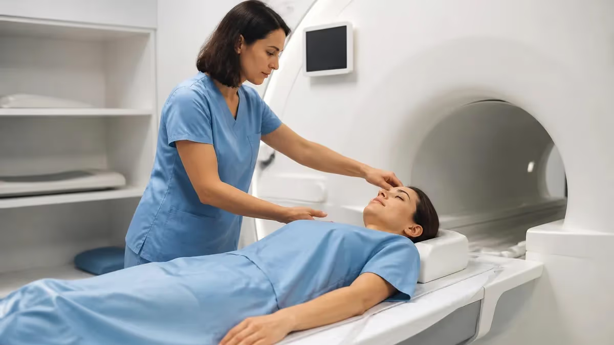
Does MRI show nerve problems clearly enough to guide treatment? The short answer is yes — but with important caveats every patient and clinician should understand. Magnetic resonance imaging is currently one of the most powerful non-invasive tools available for visualizing the nervous system, and modern high-field scanners at 1.5T and 3T can detect structural changes, compression, inflammation, and even early demyelination in peripheral and central nerves. For many patients experiencing numbness, weakness, or radiating pain, an MRI is the first imaging study ordered — and often the most informative.
The reason MRI excels at nerve evaluation comes down to its superior soft-tissue contrast. Unlike X-rays or CT scans, which excel at imaging bone, MRI uses radiofrequency pulses and magnetic field gradients to generate contrast between water-rich tissues. Nerve tissue, myelin sheaths, and surrounding fat all have different T1 and T2 relaxation times, which means a skilled radiologist can distinguish healthy nerve from inflamed, compressed, or damaged nerve tissue using the right pulse sequences and imaging planes.
That said, MRI is not a perfect nerve imaging tool. Small peripheral nerves — those with diameters under 3 mm — can be difficult to resolve even on high-resolution scanners. Functional nerve damage that causes pain or weakness without structural change may appear completely normal on MRI, which leads some patients to frustration when their scan comes back unremarkable despite debilitating symptoms. Understanding what MRI can and cannot show is essential for setting realistic expectations and choosing the right diagnostic pathway.
The spine is where MRI proves most decisive for nerve-related diagnoses. Herniated discs compressing nerve roots, spinal stenosis narrowing the neural canal, and foraminal narrowing impinging on exiting nerves are all clearly visible on standard sagittal and axial sequences. Radiologists look for nerve root effacement, loss of epidural fat signal, and T2 hyperintensity within the cord or roots as signs of significant compression or injury. These findings directly correlate with the patient's dermatomal symptoms and guide surgical or interventional planning.
Beyond the spine, a specialized technique called magnetic resonance neurography (MRN) has transformed the imaging of peripheral nerves. MRN uses fat-suppressed T2 sequences and diffusion-tensor imaging to trace individual nerves through the extremities and pelvis, detecting enlargement, increased signal, and fascicular disruption that standard MRI misses. Conditions such as piriformis syndrome, thoracic outlet syndrome, brachial neuritis, and sciatic nerve entrapment can now be imaged with remarkable specificity using 3T MRN protocols.
For those preparing for radiology or MRI technologist certification exams, understanding nerve imaging protocols is a high-yield topic. Questions about pulse sequence selection, imaging planes for specific nerve territories, and the difference between myelopathy and radiculopathy on MRI appear frequently on registry exams. Reviewing how T1, T2, STIR, and DWI sequences each contribute to nerve assessment will strengthen both exam performance and clinical practice. If you want to test your knowledge right now, you can review resources at does mri show nerve damage to deepen your understanding of spinal nerve root imaging.
This article covers the full spectrum of MRI nerve imaging: what sequences detect damage, how central and peripheral findings differ, the role of contrast agents, limitations of the technique, and practical guidance for patients and technologists alike. Whether you are a patient seeking answers about your own scan or a student preparing for board exams, this comprehensive guide provides the evidence-based detail you need to understand MRI's role in nerve damage diagnosis.
MRI and Nerve Damage by the Numbers
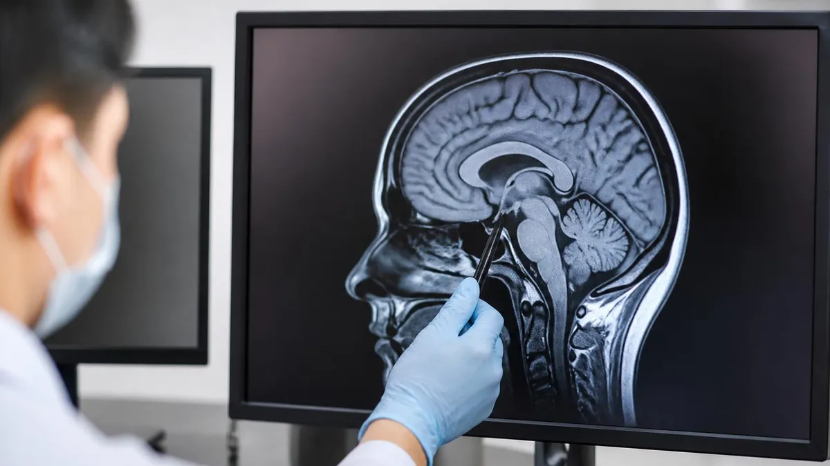
How MRI Detects Nerve Damage Step by Step
Patient Positioning and Coil Selection
Localizer and Scout Images
T1-Weighted Sequence Acquisition
T2 and STIR Sequence Acquisition
Diffusion and Advanced Sequences
Radiologist Interpretation and Report
Selecting the correct MRI sequences is the single most important technical decision in nerve imaging, and the choice depends heavily on which nerve territory is being evaluated and what pathology is suspected. For spinal nerve roots, the standard protocol includes sagittal T1, sagittal T2, and axial T2 sequences through the levels of interest.
These three sequences together give the radiologist anatomical context, disc morphology, and nerve root compression status. The sagittal T2 is particularly valuable because it displays the entire spinal cord as a bright structure, making cord edema, contusion, or myelomalacia immediately visible as a focal bright signal within the cord parenchyma.
Short Tau Inversion Recovery (STIR) sequences deserve special attention in any nerve imaging protocol. STIR suppresses fat signal, which in normal anatomy surrounds and obscures peripheral nerves. When fat signal is nulled, an injured nerve with increased water content stands out brightly against a dark background. This makes STIR the sequence of choice for screening for brachial plexus injury, lumbosacral plexus pathology, and entrapment neuropathies in the extremities. Many radiologists consider STIR the most sensitive single sequence for detecting active nerve inflammation or injury anywhere in the body.
Contrast-enhanced T1 sequences using gadolinium-based agents add another diagnostic layer. Normal nerves do not enhance significantly because the blood-nerve barrier restricts gadolinium entry. When that barrier breaks down — as it does in Guillain-Barré syndrome, chronic inflammatory demyelinating polyneuropathy (CIDP), neuritis, or nerve tumors — enhancement becomes visible and diagnostically specific. Radiologists specifically look for enhancement of nerve roots, the cauda equina, or peripheral nerve fascicles as evidence of active inflammatory or neoplastic disease that requires urgent management.
For peripheral nerve entrapment syndromes, the 3D isotropic sequences have revolutionized diagnosis. Sequences like 3D SPACE, CUBE, or VISTA acquire thin slices that can be reformatted in any plane, allowing the radiologist to follow a nerve from its proximal origin to the point of entrapment. In carpal tunnel syndrome, for example, 3D sequences clearly show median nerve flattening and increased T2 signal at the fibrous flexor retinaculum — findings that correlate directly with nerve conduction study severity and help surgeons plan the release procedure.
Diffusion-tensor imaging represents the frontier of nerve MRI. By measuring water diffusion along multiple directions, DTI generates fiber tractography maps that visualize individual nerve fascicles as colored streamlines. Fractional anisotropy (FA) values quantify the directional coherence of diffusion — normal peripheral nerves have high FA values because water moves preferentially along the axon. After axonal injury, FA drops measurably. Studies have shown DTI can distinguish axonotmesis from neuropraxia in traumatic nerve injuries, information that is crucial for predicting recovery and timing surgical intervention.
The choice of magnetic field strength significantly affects nerve imaging quality. While 1.5T scanners are adequate for spinal nerve root evaluation, 3T scanners offer twice the signal-to-noise ratio, enabling higher spatial resolution, thinner slices, and shorter acquisition times. The improved resolution at 3T is essential for imaging small peripheral nerves like the common peroneal nerve at the fibular head, the ulnar nerve at the cubital tunnel, and the digital nerves in the hand. Ultra-high-field 7T MRI, currently used primarily in research, can resolve individual nerve fascicles and has demonstrated remarkable detail in studies of the brachial plexus and lumbosacral plexus.
MRI technologists and radiologists preparing for certification exams should be comfortable explaining the rationale for sequence selection in nerve imaging. Understanding why STIR outperforms conventional fat-sat T2 in the presence of magnetic field inhomogeneity, or why diffusion imaging must be acquired before contrast administration, demonstrates the kind of protocol knowledge that distinguishes competent practitioners. These principles also appear regularly in registry exam questions, where candidates must select the most appropriate sequence for a given clinical scenario involving nerve pathology.
MRI Practice Test Questions
Prepare for the MRI - Magnetic Resonance Imaging exam with our free practice test modules. Each quiz covers key topics to help you pass on your first try.
MRI Knowledge
MRI Exam Questions covering Knowledge. Master MRI Test concepts for certification prep.
MRI Physics
Free MRI Practice Test featuring Physics. Improve your MRI Exam score with mock test prep.
MRI Anatomy and Pathology
MRI Test Prep for MRI Anatomy and Pathology. Practice MRI Quiz questions and boost your score.
MRI Anatomy and Positioning
MRI Questions and Answers on MRI Anatomy and Positioning. Free MRI practice for exam readiness.
MRI Contrast Agents
Free MRI Quiz on MRI Contrast Agents. MRI Exam prep questions with detailed explanations.
MRI Patient Care and Positioning
MRI Practice Questions for MRI Patient Care and Positioning. Build confidence for your MRI certification exam.
Central vs. Peripheral Nerve Damage on MRI
MRI is the gold standard for evaluating spinal cord injury, myelopathy, and nerve root compression. On T2-weighted sequences, the spinal cord appears bright, and any focal area of increased signal within the cord indicates edema, contusion, demyelination, or ischemia. Cervical spondylotic myelopathy typically shows a pencil-like T2 hyperintensity at the level of maximum canal stenosis, while multiple sclerosis plaques appear as oval periventricular lesions oriented perpendicular to the cord axis. Nerve root compression from disc herniation shows root effacement and loss of the surrounding epidural fat on axial T2 images.
Radiologists grade nerve root compression on a standardized scale from 0 to 3, where grade 0 is no compression, grade 1 shows mild deformation with preserved CSF signal, grade 2 shows moderate deformation with obliterated CSF signal, and grade 3 shows severe deformation with cord displacement. This grading system, validated across multiple studies, correlates strongly with clinical severity and guides neurosurgical decision-making. STIR sequences add sensitivity for detecting foraminal stenosis and detecting subtle edema within compressed roots that T2 alone can miss in areas of susceptibility artifact.
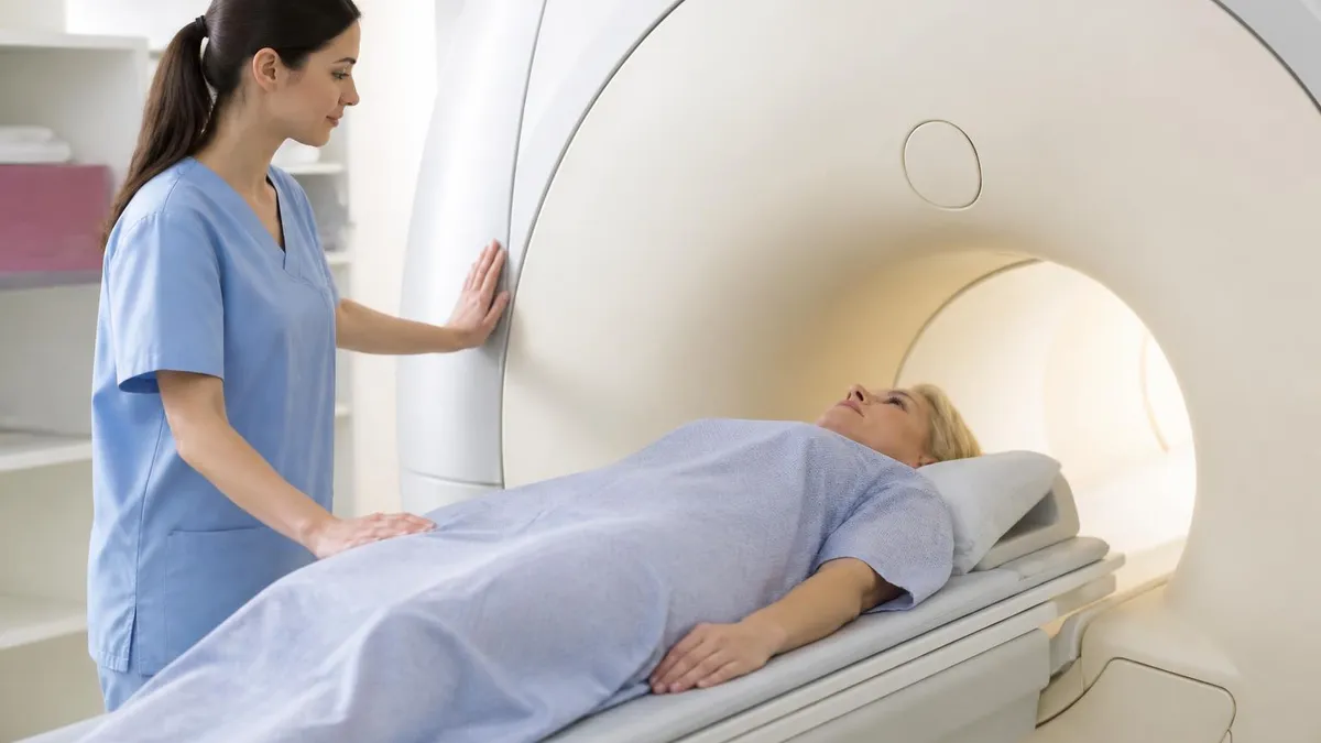
MRI for Nerve Damage: Strengths and Weaknesses
- +No ionizing radiation — safe for repeated imaging during treatment follow-up
- +Superior soft-tissue contrast clearly visualizes nerve compression, edema, and signal changes
- +Can image the entire spinal cord, nerve roots, plexuses, and peripheral nerves in one session
- +Gadolinium contrast distinguishes active inflammation from structural compression
- +Diffusion-tensor imaging provides quantitative nerve integrity measurements
- +3D isotropic sequences allow multiplanar reconstruction to follow nerve anatomy precisely
- −Cannot directly measure nerve conduction velocity or electrophysiological function
- −Small nerves under 3 mm diameter can fall below spatial resolution limits even at 3T
- −Functional nerve damage without structural changes produces a normal-appearing MRI
- −Metal implants, pacemakers, and claustrophobia limit patient eligibility for the scan
- −Cost is significantly higher than ultrasound for peripheral nerve evaluation
- −Motion artifact and susceptibility artifact near bone can obscure nerve detail at key sites
What to Check Before Your Nerve MRI Scan
- ✓Confirm you have removed all metal jewelry, piercings, and hairpins before entering the MRI room.
- ✓Inform the technologist of any metal implants, surgical hardware, or electronic devices in your body.
- ✓Disclose all previous surgeries, especially spinal fusions, joint replacements, or aneurysm clip placements.
- ✓Tell the technologist if you are pregnant or may be pregnant before contrast administration is considered.
- ✓Notify staff of kidney disease or reduced GFR if gadolinium contrast has been ordered for your scan.
- ✓Review your referring physician's clinical notes to ensure the correct nerve territory is being imaged.
- ✓Ask the ordering provider whether gadolinium contrast is needed and confirm it is included in the protocol.
- ✓Wear comfortable, loose clothing with no metal fasteners to simplify the pre-scan preparation process.
- ✓Plan for the scan to last 30 to 90 minutes depending on the region being imaged and protocol complexity.
- ✓Arrange transportation if sedation or anxiolytic medication has been prescribed to help manage claustrophobia.
A Normal MRI Does Not Rule Out Nerve Damage
One of the most important facts for patients and clinicians to understand is that a structurally normal MRI does not exclude clinically significant nerve dysfunction. Small fiber neuropathy, early axonal degeneration, and functional conduction block can all cause severe symptoms while producing no visible MRI abnormality. In these cases, nerve conduction studies and electromyography (EMG) are essential complementary tests that assess the electrical function of nerves that appear normal on imaging.
Understanding the limitations of MRI for nerve damage diagnosis is as important as understanding its strengths. One of the most clinically significant limitations is MRI's inability to image very small peripheral nerves with the resolution necessary to detect subtle pathological changes. The digital nerves of the hand, the small branches of the lateral femoral cutaneous nerve, and the fine terminal branches of the pudendal nerve are all below the practical spatial resolution limit of even high-field clinical scanners.
Conditions affecting these structures — such as Morton's neuroma, meralgia paresthetica, and pudendal neuralgia — require clinical diagnosis supplemented by nerve blocks, ultrasound-guided assessment, or specialized high-resolution MRI protocols developed primarily in academic research centers.
Functional nerve damage without structural change represents another critical limitation. Small fiber neuropathy, which affects only the unmyelinated C-fibers and thinly myelinated A-delta fibers responsible for pain and temperature sensation, produces no detectable MRI abnormality. Patients with small fiber neuropathy often undergo extensive imaging workups that return normal results, leading to delayed diagnosis and significant patient frustration. The diagnosis of small fiber neuropathy requires skin punch biopsy to count intraepidermal nerve fiber density, a test that is entirely independent of MRI. This is an important concept for both clinicians ordering MRI and patients interpreting their results.
Diabetic peripheral neuropathy, one of the most prevalent nerve disorders in the United States affecting an estimated 50% of people with long-standing diabetes, also presents a diagnostic challenge for MRI. The diffuse, length-dependent axonal degeneration typical of diabetic polyneuropathy shows subtle and nonspecific changes on standard MRI.
High-resolution MRN can demonstrate increased T2 signal and enlarged caliber in the sural and common peroneal nerves in advanced cases, but early diabetic neuropathy is largely invisible on imaging. EMG and nerve conduction studies remain the diagnostic standard for quantifying the severity of diabetic neuropathy, with MRI reserved for ruling out compressive or structural causes of worsening symptoms.
Artifacts pose a significant challenge to nerve imaging quality at anatomically complex sites. The cervicothoracic junction, brachial plexus origin zone, and lumbosacral plexus are all areas where susceptibility artifact from adjacent bone, pulsation artifact from nearby vascular structures, and motion artifact from breathing or swallowing can degrade image quality. Experienced MRI technologists apply specific artifact mitigation strategies — including cardiac gating, respiratory triggering, spatial saturation bands, and optimized echo times — to preserve nerve detail at these challenging locations. Understanding these techniques is essential for technologists performing neurography protocols and frequently appears in registry exam questions.
The cost and availability of advanced nerve MRI protocols is another practical limitation. Standard spinal MRI is widely available at most radiology facilities and is covered by insurance for indicated diagnoses. However, dedicated MR neurography protocols requiring 3T scanners, specialized surface coils, and radiologists with subspecialty neurography training are available at far fewer centers. Patients in rural or underserved areas may need to travel significant distances for these advanced studies. The cost differential between standard MRI and full-body MR neurography can be substantial, and insurance coverage for the advanced protocol is not always guaranteed without prior authorization.
Interpretation variability among radiologists is a limitation that patients and referring physicians rarely consider. The detection of subtle nerve signal changes, focal fascicular enlargement, and early enhancement on nerve-dedicated sequences requires significant subspecialty experience. Studies comparing radiologist performance in peripheral nerve imaging have shown substantial inter-reader variability for findings like mild nerve T2 hyperintensity and equivocal enhancement. Seeking interpretation from a musculoskeletal or neuroradiologist with specific experience in neurography protocols — rather than a general radiologist reading a high volume of routine studies — can meaningfully improve diagnostic accuracy and report quality.
Despite these limitations, MRI remains indispensable in the nerve damage diagnostic workup. Its ability to simultaneously assess multiple anatomical structures — bone, disc, ligament, and nerve — in a single examination provides a comprehensive overview that no other modality matches.
When a patient presents with radiculopathy, myelopathy, or plexopathy, MRI can identify the structural cause, quantify severity, and guide the treatment decision in a way that pure functional testing cannot. The key is recognizing when MRI is the right tool, when it needs to be supplemented by electrophysiology or ultrasound, and when a negative MRI result means the diagnostic process needs to continue rather than conclude.
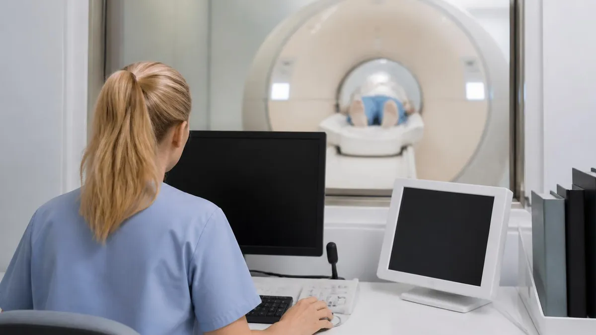
Standard spinal MRI may miss peripheral nerve entrapment, plexus injuries, and inflammatory neuropathies. If your symptoms persist despite a normal lumbar or cervical MRI report, ask your referring physician specifically about dedicated MR neurography at a 3T center, nerve conduction studies with EMG, or ultrasound-guided nerve assessment. A normal routine MRI does not exclude these conditions, and a targeted advanced protocol may reveal the diagnosis that changes your treatment plan.
Comparing MRI to other nerve diagnostic tests helps clinicians and patients choose the right combination of studies for each clinical presentation. Nerve conduction studies (NCS) and electromyography (EMG) are the most widely used complementary tests, and together they assess the electrical function of peripheral nerves and muscles.
NCS measures conduction velocity, amplitude, and latency of motor and sensory nerve potentials, providing quantitative data that distinguishes axonal loss from demyelination — a distinction with major implications for prognosis and treatment. EMG samples the electrical activity of individual muscles at rest and during contraction, detecting denervation potentials that indicate nerve injury even before structural changes appear on MRI.
The relationship between MRI and electrodiagnostic testing is complementary rather than competitive. MRI identifies structural causes of nerve damage — compression, tumor, vascular malformation, demyelinating plaques — while NCS/EMG quantifies functional severity and localizes the level of injury along the nerve. In clinical practice, the combination of MRI and electrodiagnostic testing provides a more complete picture than either test alone. For example, a patient with cervical radiculopathy might have an MRI showing mild C6-7 foraminal stenosis and an EMG confirming C7 root dysfunction — together these findings support surgical intervention far more convincingly than either study in isolation.
Ultrasound has emerged as a valuable and underutilized tool for peripheral nerve imaging, particularly at superficial sites accessible to high-frequency probes. Musculoskeletal ultrasound can image the median nerve at the wrist, the ulnar nerve at the elbow, the peroneal nerve at the fibular head, and the radial nerve in the spiral groove with excellent spatial resolution and real-time dynamic assessment.
The ability to perform nerve compression and movement testing under ultrasound guidance — watching the nerve sublux over the medial epicondyle during elbow flexion, for example — provides functional information that static MRI cannot capture. Ultrasound is also significantly cheaper and faster than MRI, making it attractive for initial assessment of suspected peripheral nerve entrapment.
However, ultrasound has significant limitations compared to MRI for deeper structures. The brachial plexus origin, lumbosacral plexus, sciatic nerve in the deep gluteal space, and spinal nerve roots are all inaccessible or poorly visualized with standard ultrasound probes. For these regions, MRI is unambiguously superior. Additionally, ultrasound is highly operator-dependent — image quality varies substantially with the skill and experience of the sonographer, and subtle nerve signal changes visible on MRI are beyond the resolving power of ultrasound. For complex cases involving deep plexus or root-level pathology, MRI remains the examination of choice.
CT myelography, once the dominant technique for spinal nerve root imaging, has been largely replaced by MRI but retains specific indications. In patients with contraindications to MRI — including certain cardiac devices, cochlear implants, and orbital metallic foreign bodies — CT myelography provides excellent depiction of nerve root compression, arachnoiditis, and dural ectasia.
The procedure involves lumbar puncture with intrathecal contrast injection followed by CT acquisition, which carries procedural risks including post-lumbar puncture headache, nerve root irritation, and rare infection. For patients who can safely undergo MRI, the non-invasive nature of MRI makes it clearly preferable, but CT myelography remains an important fallback when MRI is contraindicated.
Positron emission tomography (PET) scanning is increasingly used in oncological contexts to evaluate nerve tumors and perineural tumor spread. FDG-PET can identify metabolically active nerve sheath tumors, distinguish benign from malignant peripheral nerve sheath tumors in neurofibromatosis patients, and detect cranial nerve involvement by head and neck cancers that might be morphologically subtle on MRI.
PET-MRI fusion imaging, now available at major academic centers, combines metabolic and structural information in a single examination, offering the most comprehensive assessment of neoplastic nerve involvement available. This hybrid modality is likely to play an expanding role in nerve tumor diagnosis and treatment response assessment over the coming decade.
For MRI technologists and radiologists studying for board certification, comparative knowledge of these diagnostic modalities — including indications, contraindications, sensitivity, specificity, and the clinical scenarios where each excels — is a high-yield exam topic. The ability to recommend the appropriate next diagnostic step after an inconclusive MRI, or to explain why EMG is needed despite a structurally normal nerve on imaging, demonstrates the integrative clinical thinking that certification exams consistently test and that separates competent practitioners from those who simply execute protocols mechanically.
Practical guidance for patients undergoing an MRI for suspected nerve damage starts with understanding what information your referring physician is seeking. Before your scan, ask your doctor which nerve territory is being evaluated, whether contrast will be used, and whether you are being referred to a standard MRI facility or a specialized neurography center.
This information helps you prepare appropriately and ensures the radiology facility has the correct protocol queued before your appointment. Arriving at a general imaging center for a brachial plexus MRN when only a standard cervical spine MRI protocol is available will produce a far less informative examination than one performed at a center equipped for dedicated neurography.
Preparation for the MRI itself involves several practical steps. Complete the MRI safety screening questionnaire thoroughly and honestly, disclosing all implants, surgical history, and occupational metal exposures. Many patients are unaware that metallic fragments from machining, welding, or military service can present serious hazards in the MRI environment, and orbital X-rays may be required before scanning these individuals.
Bring a list of all medications, particularly any anticoagulants or antiplatelet agents if a lumbar puncture or nerve block procedure is anticipated alongside the imaging. If you have claustrophobia, contact the facility in advance to discuss anxiolytic medication options — an incomplete scan due to patient motion is far less useful than a well-executed study under mild sedation.
After your MRI, understanding your radiology report is the next challenge. Reports describing nerve damage typically use terminology like T2 hyperintensity, nerve root effacement, foraminal stenosis, and enhancement. T2 hyperintensity in a nerve indicates increased water content, which can mean edema, inflammation, or injury. Effacement means compression has flattened the nerve root. Enhancement after gadolinium indicates blood-nerve barrier disruption, suggesting active inflammation or tumor. If your report contains these findings without adequate clinical explanation from your referring physician, request a formal consultation with a neurosurgeon, neurologist, or pain medicine specialist who regularly reviews MRI findings for nerve conditions.
For patients whose MRI appears normal despite persistent symptoms, understanding the diagnostic gap is crucial. As discussed throughout this article, MRI cannot detect functional nerve damage, small fiber neuropathy, or mild conduction slowing. If your symptoms include burning pain, allodynia, temperature insensitivity, or autonomic dysfunction alongside normal MRI findings, raise the possibility of small fiber neuropathy specifically with your neurologist.
The diagnosis requires a skin punch biopsy and is entirely missed by MRI-only workups. Seeking a second opinion at a neuromuscular disease center or academic medical center with a dedicated peripheral nerve program is reasonable and often productive for patients with unexplained neuropathic symptoms.
MRI technologists in clinical practice should advocate for protocol optimization whenever a nerve-specific examination is ordered. This means checking that the correct coil is selected, confirming field strength availability for the requested protocol, ensuring adequate slice coverage for the entire nerve territory, and performing quality-control checks on STIR fat suppression before releasing the patient from the scanner.
Incomplete fat suppression is the most common technical failure in peripheral nerve MRI and can render an otherwise well-positioned examination non-diagnostic. Taking an extra two minutes to assess fat suppression quality on the first STIR sequence and adjusting the shimming if needed routinely prevents the need for patient recall.
Students and technologists preparing for the ARRT MRI registry or ARMRIT certification should focus their nerve imaging study on a few key areas: the pathophysiology of demyelination versus axonal injury and how each appears differently on MRI, the indications for gadolinium contrast in nerve imaging and contraindications related to renal function, the technical principles behind STIR fat suppression and why it outperforms chemical fat saturation in areas of field inhomogeneity, and the clinical presentations that warrant MR neurography versus standard spinal MRI.
These concepts appear in multiple domains of the registry exam, including patient care, imaging procedures, and image production, and a solid conceptual foundation in nerve imaging will reinforce performance across all sections.
The field of nerve MRI is evolving rapidly, with artificial intelligence-assisted image analysis, quantitative MRI biomarkers, and ultra-high-field neurography all advancing the diagnostic capabilities available in clinical practice. AI algorithms trained on large neurography datasets can now automatically segment peripheral nerves, measure cross-sectional area and signal intensity along their entire length, and flag abnormal segments for radiologist review — reducing interpretation time and potentially improving consistency.
As these tools move from research into clinical implementation, MRI technologists and radiologists will need to understand their capabilities and limitations to use them responsibly and to explain their outputs meaningfully to referring clinicians and patients.
MRI Questions and Answers

MRI of Cervical Spine: Complete Guide to Imaging, Anatomy, Pathology, and Patient Preparation

Knee MRI Images: A Complete Guide to Reading, Understanding, and Interpreting Knee Scans
Ace Your Exam with MRI Practice Test 2026 Prep

MRI Tech School 2026 — Programs, Requirements, and What to Expect
MRI Procedure: What to Expect Before, During and After Your Scan
About the Author

Medical Laboratory Scientist & Clinical Certification Expert
Johns Hopkins UniversityDr. Sandra Kim holds a PhD in Clinical Laboratory Science from Johns Hopkins University and is certified as a Medical Technologist (MT) and Medical Laboratory Scientist (MLS) through ASCP. With 16 years of clinical laboratory experience spanning hematology, microbiology, and molecular diagnostics, she prepares candidates for ASCP board exams, MLT, MLS, and specialist certification tests.
Join the Discussion
Connect with other students preparing for this exam. Share tips, ask questions, and get advice from people who have been there.
View discussion (8 replies)