MRI Neurography: A Complete Guide to Peripheral Nerve Imaging 2026 July
💯 MRI neurography explained — sequences, indications, anatomy, and protocols for high-resolution peripheral nerve imaging in clinical practice.
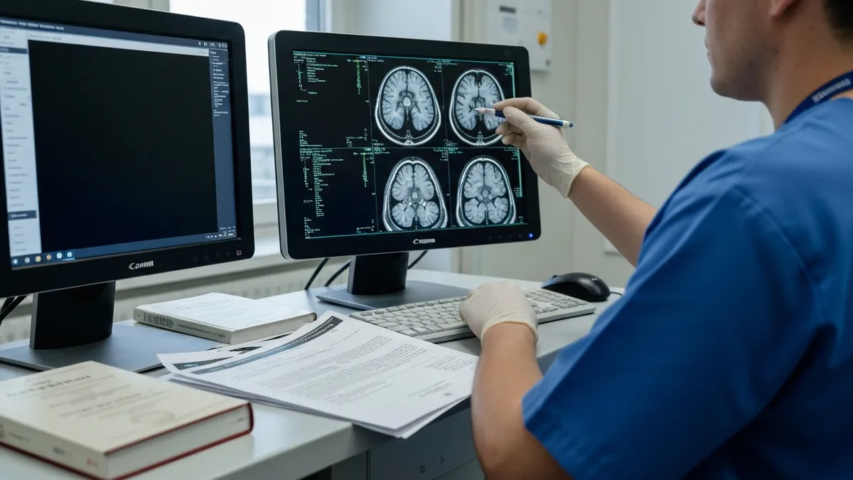
MRI neurography is a specialized magnetic resonance imaging technique designed to visualize peripheral nerves with unprecedented clarity. Unlike conventional MRI sequences that focus on muscles, bones, or vascular structures, mri neurography uses tailored pulse sequences, fat suppression, and high field strengths to isolate the nerve signal from surrounding tissue. The result is a detailed image that lets radiologists assess nerve caliber, fascicular architecture, signal intensity, and continuity in ways that were impossible just two decades ago.
The clinical demand for peripheral nerve imaging has grown rapidly as referrers recognize the limitations of electrodiagnostic studies alone. EMG and nerve conduction tests are excellent for confirming functional deficits, but they cannot localize a lesion with millimeter precision, identify the underlying cause, or characterize space-occupying lesions like schwannomas. MRI neurography bridges that gap by providing anatomic detail that complements physiologic data, helping clinicians plan surgical decompression, biopsy, or targeted injection therapy.
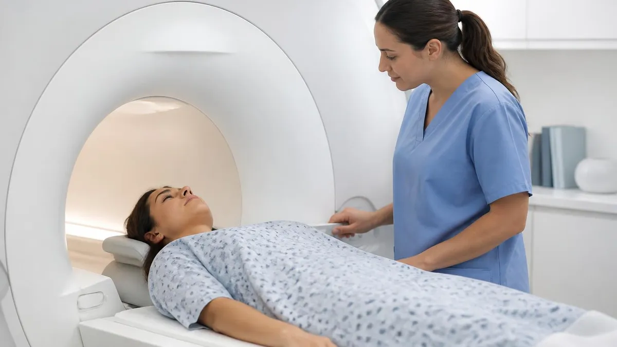
Performing a successful neurography study requires meticulous attention to technique. Coil selection, slice orientation, voxel size, and contrast strategy all influence whether subtle nerve abnormalities will be visible. Most modern protocols rely on 3T scanners because the higher signal-to-noise ratio allows submillimeter in-plane resolution. Three-dimensional sequences with isotropic voxels enable multiplanar reformatting, so the radiologist can trace a nerve along its entire course without re-scanning the patient — a critical workflow advantage in busy outpatient settings.
The technique evolved out of foundational research in the 1990s, when investigators first showed that diffusion-weighted and short-tau inversion recovery sequences could selectively brighten peripheral nerves. Since then, vendor-specific implementations such as 3D STIR SPACE, DESS, and reverse FOCUS DWI have refined image quality further. A solid grounding in the history of MRI helps trainees appreciate why these advances took so long and why neurography remains an active research area.
Indications for mri neurography are broad and continue to expand. Brachial plexopathy after trauma or radiation, lumbosacral plexus involvement in pelvic malignancy, suspected piriformis syndrome, and entrapment neuropathies of the median, ulnar, or peroneal nerves all benefit from dedicated imaging. Increasingly, neurography is also requested for diabetic neuropathy research, immune-mediated polyneuropathies like CIDP, and post-surgical evaluation of nerve grafts. Each indication has its own protocol nuances, and the technologist must understand them to deliver diagnostic images.
This guide walks through the physics, hardware, protocol design, anatomy, and clinical applications of peripheral nerve MRI. Whether you are a radiology resident preparing for board exams, an MRI technologist building protocols for a new service line, or a referring clinician trying to understand what your report means, the sections below will demystify how modern neurography works. Expect concrete numbers, sequence parameters, and clinical pearls drawn from real-world practice rather than vague generalities.
By the end of this article, you will understand why certain sequences highlight nerves while suppressing background tissue, how to recognize the difference between a normal hyperintense nerve and a pathologic one, and what pitfalls — magic angle artifact, vascular pulsation, partial volume averaging — can mimic disease. Neurography is technique-dependent, so a confident interpretation always begins with a confident acquisition.
MRI Neurography by the Numbers
Core Sequences in MRI Neurography
A heavily T2-weighted, fat-suppressed 3D sequence with variable flip-angle refocusing. Produces isotropic voxels for multiplanar reformat and selectively highlights edematous or pathologic nerves against suppressed fat and muscle background.
Dual-echo steady-state sequences provide excellent fascicular detail in cranial and brachial plexus imaging. They combine T2-like contrast with high spatial resolution, making them ideal for fine structural assessment of nerve bundles.
Diffusion-weighted neurography uses high b-values and vascular suppression to produce maximum-intensity projections that look like a digital subtraction angiogram of the nerve, useful for brachial and lumbosacral plexus tractography.
Standard T1 sequences provide anatomic context, demonstrate perineural fat planes, and help identify space-occupying lesions like schwannomas or neurofibromas. They are essential reference sequences for nerve localization.
Gadolinium-enhanced fat-suppressed T1 imaging is reserved for tumor evaluation, inflammatory neuropathies, and post-surgical assessment. Normal peripheral nerves do not enhance, so any uptake is significant.
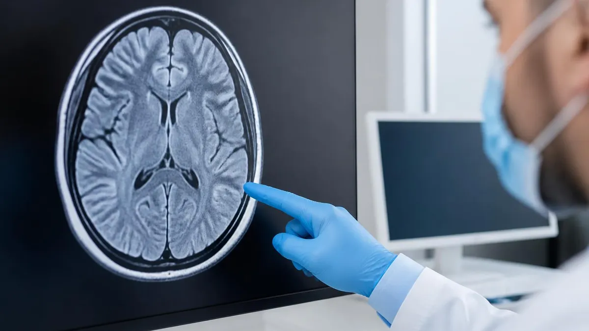
Peripheral nerve anatomy on MRI is more nuanced than it first appears. A normal nerve appears as a tubular structure with internal fascicular pattern — small round hypointensities representing nerve fascicles surrounded by hyperintense endoneurial and perineurial connective tissue on T2-weighted sequences. On axial images, this gives a characteristic honeycomb appearance that radiologists use to confirm they are tracking a nerve rather than a small vessel or tendon. Loss of fascicular pattern is one of the earliest signs of significant pathology.
Indications for neurography fall into broad categories: trauma, entrapment, inflammation, neoplasm, and post-surgical assessment. Traumatic injury includes stretch lesions, transections, and neuroma formation. Entrapment syndromes account for the largest referral volume — carpal tunnel, cubital tunnel, tarsal tunnel, and thoracic outlet syndrome being the most common. Inflammatory and immune-mediated neuropathies like Parsonage-Turner syndrome or CIDP show diffuse nerve hyperintensity and sometimes enhancement. Each indication shapes the protocol you select.
The brachial plexus is one of the most technically demanding regions because it courses through complex anatomy from cervical roots to the axilla. A complete brachial plexus protocol requires coronal oblique imaging aligned with the C5-T1 roots, axial coverage from the foramina to the cords, and often coronal 3D STIR or DWI reformats to demonstrate continuity. Patients with suspected radiation plexopathy or post-traumatic injury benefit most from this comprehensive approach, and report turnaround often depends on the radiologist's familiarity with the territory.
The lumbosacral plexus presents similar challenges with the added complication of bowel motion, vascular pulsation, and the deep retroperitoneal location. Saturation bands, breath-hold or respiratory-triggered acquisitions, and pelvic phased-array coils help mitigate these issues. Lumbosacral neurography is increasingly requested for piriformis syndrome, endometriosis-related nerve involvement, and obstetric injuries to the lumbosacral trunk, expanding the technique well beyond its original niche.
Distal extremity nerves — median, ulnar, radial, common peroneal, tibial, sural — are typically imaged with surface coils that wrap snugly around the limb. Resolution requirements are higher because these nerves are smaller, often 2 to 4 millimeters in diameter at points of entrapment. Voxel sizes of 0.3 to 0.5 millimeters in-plane are typical, with slice thicknesses of 2 to 3 millimeters for 2D acquisitions or isotropic 0.6 millimeter voxels for 3D approaches. The trade-off is always between resolution, coverage, and acquisition time.
mri of cervical spine matters more than many technologists realize. Patients should be positioned to minimize motion, with cushioning to relieve pressure on the limb of interest. Pain medication may be needed for patients with severe neuropathic symptoms who cannot lie still for 40 minutes. Understanding when contrast is needed is also important — many neurography studies are performed without contrast, and you can review MRI with and without contrast to understand the clinical reasoning behind that decision.
Communication with the referring physician is the final piece of indication management. A vague request like "rule out neuropathy" is much less useful than "evaluate for median nerve entrapment at carpal tunnel, patient with positive Tinel sign and abnormal NCV." The more specific the clinical question, the more targeted the protocol, and the more diagnostic the resulting study. Many large centers have a dedicated neurography intake form to extract this information up front.
MRI Practice Test Questions
Prepare for the MRI - Magnetic Resonance Imaging exam with our free practice test modules. Each quiz covers key topics to help you pass on your first try.
MRI Knowledge
MRI Exam Questions covering Knowledge. Master MRI Test concepts for certification prep.
MRI Physics
Free MRI Practice Test featuring Physics. Improve your MRI Exam score with mock test prep.
MRI Anatomy and Pathology
MRI Test Prep for MRI Anatomy and Pathology. Practice MRI Quiz questions and boost your score.
MRI Anatomy and Positioning
MRI Questions and Answers on MRI Anatomy and Positioning. Free MRI practice for exam readiness.
MRI Contrast Agents
Free MRI Quiz on MRI Contrast Agents. MRI Exam prep questions with detailed explanations.
MRI Patient Care and Positioning
MRI Practice Questions for MRI Patient Care and Positioning. Build confidence for your MRI certification exam.
Technical Approach for MRI Neurography by Region
Brachial plexus imaging is performed with a dedicated neurovascular or large surface coil array spanning the lower neck, supraclavicular fossa, and axilla. Coronal oblique 3D STIR SPACE is the workhorse sequence, aligned with the C5-T1 roots to demonstrate the nerves from foramina to cords. Axial T2 fat-suppressed and T1 sequences provide cross-sectional anatomy. Diffusion-weighted neurography with vascular suppression produces tractographic reformats that referring surgeons find intuitive.
Common indications include traumatic injury after motor vehicle accidents, radiation-induced plexopathy in breast or lung cancer survivors, Parsonage-Turner syndrome, and thoracic outlet syndrome with neurogenic compression. Imaging in dynamic positions — arm abducted versus adducted — is sometimes added to characterize positional compression. Reports should describe individual trunk, division, and cord involvement using consistent terminology so surgeons can plan exploration.
MRI Neurography vs Conventional Workup
- +Provides direct anatomic visualization of peripheral nerves with submillimeter detail
- +Localizes lesions precisely along the nerve course, guiding surgical planning
- +Identifies space-occupying lesions like schwannomas and neurofibromas that EMG cannot detect
- +Allows non-invasive follow-up after nerve repair or decompression surgery
- +Demonstrates inflammatory and immune-mediated neuropathies through nerve hyperintensity patterns
- +Multiplanar 3D reformats enable surgeons to plan approach without exploratory dissection
- −Long acquisition times of 35 to 50 minutes can be difficult for patients in pain
- −Requires 3T scanner and dedicated coils not available at all centers
- −Operator-dependent — protocol design and coil placement strongly affect image quality
- −Magic angle, vascular pulsation, and partial volume artifacts can mimic pathology
- −Higher cost than EMG and nerve conduction studies, with variable insurance coverage
- −Cannot directly measure nerve conduction velocity or motor unit function the way EMG can
MRI Neurography Pre-Scan Checklist
- ✓Confirm clinical question and target nerve with the referring physician before scheduling
- ✓Screen patient for MRI safety contraindications including implants and pacemakers
- ✓Select the appropriate coil — surface coil for distal extremities, phased array for plexus
- ✓Position the patient symmetrically and pad pressure points to minimize motion
- ✓Mark anatomic landmarks externally to assist with slice prescription
- ✓Verify scanner field strength is 3T whenever possible for optimal SNR
- ✓Load the validated neurography protocol with 3D STIR and high-resolution T2 sequences
- ✓Decide on contrast administration based on suspected pathology — usually not needed for entrapment
- ✓Educate the patient on breath-holding or stillness requirements during long acquisitions
- ✓Plan multiplanar reformats while the patient is still on the table in case re-scan is needed
Suppress the fat, see the nerve
Peripheral nerves are surrounded by epineurial fat that, on conventional T2 imaging, masks the subtle hyperintensity of pathology. Robust fat suppression — STIR, Dixon, or spectral — is the single most important technical decision in mri neurography. Without it, even severe nerve edema can be invisible against bright background fat.
Interpreting a neurography study begins with anatomy. Before commenting on signal abnormalities, the radiologist must locate every relevant nerve, confirm its course, and assess its caliber. Asymmetry between sides is the most reliable internal control, so bilateral imaging is preferred when feasible. A nerve that is two to three times larger than its contralateral counterpart, or that demonstrates a sudden caliber change at a fibrous arcade, is usually pathologic regardless of signal intensity. Caliber assessment is therefore the first interpretive step.
Signal intensity comes next. A normal nerve is mildly hyperintense to muscle on T2-weighted images, reflecting endoneurial water content. Marked hyperintensity approaching that of cerebrospinal fluid suggests significant edema from compression, inflammation, or denervation. The pattern matters: focal hyperintensity at a known anatomic chokepoint — the carpal tunnel, the cubital tunnel, the spinoglenoid notch — supports entrapment. Diffuse, segmental, or skip-pattern hyperintensity raises suspicion for immune-mediated polyneuropathy like CIDP or vasculitis.
Fascicular pattern preservation is a more subtle but important sign. In a healthy nerve, individual fascicles are resolvable as small dark dots on high-resolution T2-weighted axial images. Loss of this pattern — fascicular effacement — indicates severe injury, often grade 3 or higher Sunderland classification, and predicts incomplete spontaneous recovery. When combined with absent contraction on EMG, fascicular effacement helps surgeons decide whether to explore and graft a nerve rather than wait.
Denervation changes in downstream muscles provide indirect but powerful evidence of nerve injury. Acute denervation shows muscle edema on T2 fat-suppressed sequences within two to four weeks of injury. Subacute changes include further edema and early fatty replacement. Chronic denervation manifests as fatty atrophy on T1-weighted images, with the muscle shrinking and replaced by bright signal fat. Identifying the specific muscles involved helps confirm which nerve and which branch are affected, particularly useful when neurography itself is equivocal.
Space-occupying lesions are usually straightforward but require careful description. Schwannomas appear as well-circumscribed, fusiform masses intimately associated with the nerve, often with a target sign on T2 and avid enhancement. Neurofibromas have similar features but are less well-defined and may be multiple in neurofibromatosis. Perineuriomas are rarer and tend to cause segmental fusiform thickening with preserved fascicular pattern. Each entity has implications for biopsy planning, given the risk of nerve injury during sampling.
Comparison with prior imaging is essential when available. Post-surgical patients are particularly challenging because expected post-operative changes — fibrosis, granulation tissue, suture artifact — overlap with recurrent pathology. A baseline study within three months of surgery, when feasible, provides a reference point for future comparison. Many surgeons now request a routine post-operative neurography at six months to document anatomic restoration even when the patient is asymptomatic.
The structured report is the final interpretive product. A useful neurography report includes the nerve evaluated, its caliber relative to the contralateral side, signal intensity, fascicular pattern, course, surrounding tissue, downstream muscle status, and any space-occupying lesion. It then offers a probability-weighted differential and an actionable next step — surgical referral, EMG correlation, follow-up imaging, or biopsy. Reports that simply describe findings without integration are far less valuable to clinicians.
When peripheral nerves run at approximately 55 degrees to the main magnetic field, they can appear artifactually hyperintense on short TE sequences, mimicking pathology. This is most common in the brachial plexus and around the elbow. Always verify suspicious findings on long-TE T2 sequences before reporting nerve injury.
Artifacts and pitfalls in mri neurography deserve dedicated discussion because they cause more interpretive errors than any other category. Magic angle artifact, mentioned above, is only the most famous example. It occurs when a structure containing ordered collagen — like nerve fascicles, tendons, or ligaments — orients at approximately 55 degrees to the static magnetic field. The result is artifactual signal increase on sequences with short echo times, particularly proton density and gradient echo. Long TE sequences are essentially immune to the effect, so always confirm questionable hyperintensity on a T2 sequence with TE greater than 60 milliseconds.
Partial volume averaging is another common pitfall. When voxel size is large relative to nerve diameter, the nerve signal is averaged with surrounding tissue, blurring fascicular detail and potentially masking subtle disease. This is most problematic in distal extremity imaging where target nerves may be only two to three millimeters thick. The solution is to push for submillimeter in-plane resolution and to acquire thin slices, accepting longer scan times as the cost of diagnostic quality. 3D acquisitions with isotropic voxels mitigate the issue by allowing reformat in any plane.
Vascular pulsation can create ghosting artifacts that overlay nerves and either mimic pathology or obscure real findings. Brachial and lumbosacral plexus imaging are most affected because of proximity to the subclavian and iliac vessels. Saturation bands, vascular suppression pulses, and cardiac gating can help, though gating adds significant scan time. Diffusion-weighted neurography is particularly susceptible to vascular contamination, and dedicated vascular-suppression preparation pulses are usually built into modern sequences.
Chemical shift and fat suppression failure plague neurography more than most MRI applications because the technique relies so heavily on fat saturation. STIR is robust but has lower SNR than spectral fat saturation; spectral saturation is more efficient but vulnerable to B0 inhomogeneity at field edges or near metallic implants. Dixon-based fat-water separation has become popular because it provides reliable fat suppression even in difficult anatomy. Knowing the strengths and weaknesses of each approach lets the technologist choose appropriately for each region.
Susceptibility artifact from surgical hardware is a frequent challenge in post-operative neurography. Suture anchors, screws, and plates can create signal voids that obscure adjacent nerves. Multi-acquisition with variable resonance image combination (MAVRIC), slice encoding for metal artifact correction (SEMAC), and view angle tilting (VAT) techniques have improved imaging near metal substantially. When ordering imaging on a post-operative patient, confirming hardware composition with the surgeon helps the technologist select the optimal protocol — see also the broader role of MRI equipment like coils and gradients in artifact management.
Patient motion remains the single most common cause of non-diagnostic studies. Long scan times, painful positioning, and the inability of some patients to remain still all contribute. Strategies include shorter accelerated sequences using parallel imaging or compressed sensing, prone or oblique positioning for the brachial plexus, light sedation in selected cases, and clear pre-scan communication with the patient about expectations. Reviewing image quality periodically during the exam allows re-acquisition of motion-degraded sequences before the patient leaves.
Reporting language should reflect uncertainty when warranted. Rather than calling a possibly artifactual finding definitive nerve injury, use phrases like "focal signal abnormality of uncertain significance, consider correlation with EMG" or "finding could represent partial volume averaging; recommend follow-up if symptoms persist." Overcalling subtle findings damages clinical trust and leads to unnecessary intervention. Undercalling real disease delays diagnosis. The radiologist's job is to communicate confidence calibrated to the evidence on the image.
Building a successful neurography service requires more than just acquiring the right equipment. Protocol development should be collaborative — radiologists, technologists, and referring clinicians work together to define which sequences are essential and which can be omitted when scan time is limited. A tiered protocol approach helps: a core mandatory sequence list plus an optional extension based on clinical scenario. This keeps studies focused, reduces patient burden, and ensures every acquisition contributes to the diagnostic question.
Technologist training is critical and often underestimated. Even an excellent scanner with optimal sequences produces poor images if the technologist does not understand nerve anatomy, slice prescription strategy, or how to recognize a motion-degraded acquisition. Continuing education modules, ride-alongs with experienced neuro MR technologists, and case review sessions all help build the expertise needed. Some centers designate one or two technologists as neurography specialists to maintain consistent quality across a large team.
Quality assurance should be ongoing. Track turnaround time, percentage of non-diagnostic studies, and inter-reader agreement on key findings. Phantom studies confirm that resolution and fat suppression remain within specification across the lifespan of the scanner. Regular case conferences where radiologists discuss difficult studies with surgeons and neurologists build collective expertise and identify protocol weaknesses before they cause clinical problems. Treating neurography as a quality-driven program, rather than a one-off service line, pays long-term dividends.
Patient communication starts before the scan and continues after the report is finalized. Patients often arrive anxious because they have been in pain for months and feel unheard by previous providers. A brief pre-scan explanation of what neurography is, why it takes longer than a standard MRI, and what the radiologist will look for goes a long way toward cooperation during the exam. Many centers have developed printed handouts and short video explainers that referring offices can share at the time of order — a small investment that improves the overall experience.
Cost and access remain genuine barriers. Neurography requires 3T capability, specialized coils, and trained interpretation, none of which are uniformly available. Independent outpatient centers have begun to fill the gap in metropolitan areas, often with shorter scheduling lead times than hospital-based programs. Patients in rural areas may need to travel two to three hours for a dedicated neurography exam, which influences referral patterns and follow-up adherence. Explore options like MRI imaging centers that provide outpatient access to advanced MR techniques.
Looking forward, several technological advances are reshaping neurography. Deep-learning reconstruction is reducing scan times by 30 to 50 percent without quality loss, making longer protocols more tolerable. Quantitative imaging — T2 relaxometry, diffusion tensor metrics, magnetization transfer ratio — is moving from research into limited clinical use, offering objective measures rather than subjective signal interpretation. Compressed sensing and parallel imaging acceleration techniques continue to evolve, and 7T systems, while still investigational for peripheral nerves, hint at future resolution gains.
The bottom line for clinicians, technologists, and trainees is that mri neurography is now a mature clinical tool, not an experimental technique. Its successful use requires investment in equipment, training, and protocol design, but the diagnostic and surgical-planning benefits justify the effort. Whether you are interpreting your first brachial plexus study or designing a new tertiary referral service, the principles outlined in this guide — careful coil selection, robust fat suppression, multiplanar acquisition, structured reporting, and ongoing quality assurance — will serve as a reliable foundation.
MRI Questions and Answers

The History of MRI: From Discovery to Modern Medicine

MRI With and Without Contrast: How It Works, What to Expect

MRI Medical Abbreviation: What MRI Stands For and Why It Matters
MRI Imaging Centers: Complete Guide to Independent Outpatient MRI Facilities
MRI Equipment: A Complete Guide to Scanners, Coils, and Components
About the Author

Medical Laboratory Scientist & Clinical Certification Expert
Johns Hopkins UniversityDr. Sandra Kim holds a PhD in Clinical Laboratory Science from Johns Hopkins University and is certified as a Medical Technologist (MT) and Medical Laboratory Scientist (MLS) through ASCP. With 16 years of clinical laboratory experience spanning hematology, microbiology, and molecular diagnostics, she prepares candidates for ASCP board exams, MLT, MLS, and specialist certification tests.
Join the Discussion
Connect with other students preparing for this exam. Share tips, ask questions, and get advice from people who have been there.
View discussion (6 replies)