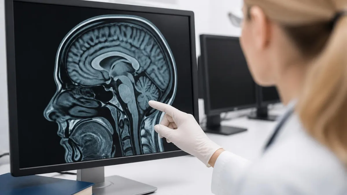
Will MRI Show Nerve Damage? What an MRI Can and Cannot Detect 2026
Will MRI show nerve damage? Learn what an MRI can detect, 📗 its limits, when EMG is needed, and how nerves appear on scans in this complete guide.

Will MRI show nerve damage is one of the most common questions patients ask before they slide into the scanner, and the honest answer is: sometimes yes, sometimes no, and it depends heavily on which nerve, what kind of damage, and how severe it is. Magnetic resonance imaging excels at showing the structures that surround nerves and the larger nerve roots themselves, but it is not a perfect window into every microscopic injury. Understanding this distinction helps you set realistic expectations before your appointment. Practice with the free MRI - Magnetic Resonance Imaging practice test on PracticeTestGeeks (11 questions with answer explanations).
An MRI is fundamentally an anatomical imaging test. It produces detailed cross-sectional pictures of soft tissue using a powerful magnet and radio waves, with no ionizing radiation involved. Because nerves are soft tissue, larger nerves and nerve roots do appear on the images. What MRI shows best, however, is compression, swelling, tumors, and inflammation affecting nerves rather than the electrical conduction failure that defines many nerve injuries. That difference is the key to interpreting your results.
For example, if a herniated disc in your lower back is pressing on a nerve root, an MRI will usually show the bulging disc and the pinched nerve clearly. If you have a tumor wrapped around a nerve, that mass will likely be visible. But if your nerve fibers are damaged on a cellular level from diabetes, chemotherapy, or a metabolic condition, the nerve may look entirely normal on MRI even while it functions poorly. The structure is intact; the wiring inside is not.
This is why doctors often pair MRI with other tests. Electromyography (EMG) and nerve conduction studies measure how well nerves actually carry electrical signals, catching functional problems that imaging misses entirely. Think of MRI as the photograph of the road and EMG as the traffic report. One shows you the physical layout; the other tells you whether anything is actually moving through. Together they give a far more complete picture of nerve health than either alone.
Whether you are a patient trying to make sense of a referral or a radiology student preparing for boards, knowing the strengths and blind spots of MRI matters. If you are studying the modality formally, working through practice scenarios can sharpen your judgment about when imaging answers the clinical question and when it does not. You can review realistic cases and will mri show nerve damage style questions to test how well you understand the relationship between anatomy and function.
Throughout this guide we will walk through exactly what MRI detects, the types of nerve damage it sees well, the kinds it tends to miss, how specialized sequences like MR neurography push the limits, and what complementary tests fill the gaps. By the end you will know what to expect from your scan, what questions to ask your physician, and why a normal MRI does not always mean your nerves are healthy.
Free MRI - Magnetic Resonance Imaging Study Tools
Interactive MRI - Magnetic Resonance Imaging resources on PracticeTestGeeks, free to start:
- Free MRI Knowledge Questions and Answers — 11 questions with instant scoring and answer explanations
- MRI - Magnetic Resonance Imaging flashcards — review key terms and concepts card by card
- MRI - Magnetic Resonance Imaging study guide — topic-by-topic overview of what the exam covers
- MRI - Magnetic Resonance Imaging cheat sheet — one-page printable summary for last-minute review
MRI: upcoming dates and official changes
- Aug 17, 2026: ARRT launched a separate entry-level Imaging Assistant, Magnetic Resonance (I.A.) credential. Applications made Aug. 17–Oct. 14, 2026 paid a reduced $75 fee for testing Oct. 15–Nov. 30, 2026. Applicants after Oct. 14, 2026 pay $225 and can test on or after Jan. 1, 2027. The I.A. exam also requires a scaled section score of 7.5 or higher in MRI Screening and Safety.[3]
- The ARRT MRI postprimary exam has 220 questions: 200 scored and 20 unscored pilot items.[1]
- The ARRT MRI postprimary exam allows 210 minutes of test time within a 230-minute (3 hour 50 minute) appointment.[1]
- ARRT exams, including MRI, are scored on a 1–99 scale, and candidates need a total scaled score of 75 to pass.[2]
- The ARRT MRI postprimary application fee is $225, or $450 for candidates using an NMTCB or ARDMS credential as the supporting category.[3]
All verified MRI facts and sources
Numbers in brackets link to the official sources listed under Sources & last verified. Dates that have passed are removed automatically.
MRI and Nerve Damage by the Numbers

What an MRI Can Actually Detect About Nerves
MRI clearly reveals when a herniated disc, bone spur, or narrowed canal presses on a nerve root or peripheral nerve, showing the exact level and severity of the pinch.
Schwannomas, neurofibromas, and other growths along nerves show up as discrete masses. MRI helps locate them, measure size, and assess involvement of surrounding tissue.
Inflamed, swollen, or edematous nerves often appear brighter on certain sequences. This signal change can hint at neuritis, recent trauma, or active irritation of the nerve.
Complete or partial nerve transections, severe stretch injuries, and traumatic neuromas can be visualized, especially with dedicated MR neurography focused on the affected limb.
When a nerve has been damaged for weeks, the muscles it supplies may show denervation edema or fatty atrophy on MRI, giving indirect evidence of nerve injury.
To understand whether an MRI will show nerve damage in your specific case, it helps to know how nerves themselves appear on the images. Large nerves and nerve roots are soft-tissue structures with a characteristic signal intensity that radiologists learn to recognize. On standard T1-weighted images, healthy nerves appear as smooth, uniform cords of intermediate signal, often nestled in bright fat that outlines their borders. On fluid-sensitive T2 sequences, a healthy nerve is relatively dark compared to surrounding fluid.
When a nerve is injured, irritated, or compressed, its appearance can change in predictable ways. An inflamed or acutely damaged nerve frequently becomes brighter, or hyperintense, on T2-weighted and fat-suppressed images because of edema and increased water content. The nerve may also enlarge, lose its normal smooth contour, or show abnormal enhancement after contrast is given. These signal and shape changes are the breadcrumbs radiologists follow to infer that something is wrong, even before the precise cause is identified.
Nerve roots in the spine are particularly well seen because they are bathed in cerebrospinal fluid, which provides excellent natural contrast. A pinched root from a herniated disc often appears displaced, flattened, or swollen at the point of compression. This is why MRI is the workhorse test for radiculopathy and sciatica. The combination of clear roots and visible discs lets clinicians pinpoint exactly which level is generating a patient's leg or arm symptoms with impressive accuracy.
Peripheral nerves in the arms and legs are smaller and harder to image, but modern scanners and dedicated coils have improved this dramatically. MR neurography, a specialized technique optimized for peripheral nerves, can trace nerves like the sciatic, median, or brachial plexus along their course and flag focal abnormalities. This is increasingly used when a peripheral nerve problem is suspected but the cause is unclear from physical examination alone.
It is worth emphasizing again that a normal-looking nerve on MRI does not guarantee a healthy nerve. Many forms of damage occur at the level of individual axons and myelin sheaths, far below the resolution of even the best clinical scanner. The nerve can be electrically failing while its overall structure remains intact and unremarkable on imaging. Radiologists and referring physicians keep this firmly in mind when interpreting an unremarkable study against a patient's clear symptoms.
Students and technologists learning to read these studies benefit enormously from repetition and pattern recognition. Recognizing normal nerve signal, spotting subtle T2 hyperintensity, and distinguishing a swollen root from a normal one all take practice. Working through annotated cases and timed questions builds the instinct to notice when a nerve simply does not look right, which is often the first clue that prompts a more targeted functional workup or referral to a specialist.
For a fuller set of questions on this topic, see our open bore mri resource.
For a fuller set of questions on this topic, see our mri machine pictures resource.
MRI vs EMG: Will MRI Show Nerve Damage Alone?
MRI is an anatomical test. It shows the physical state of nerves and everything around them, including discs, bones, tumors, and inflammation. It excels at finding the cause of nerve compression, such as a herniated disc or a mass pressing on a nerve root, and it can reveal swelling or signal change within larger nerves.
What MRI does not measure is electrical function. A nerve can look perfectly normal on the scan while conducting signals poorly. That is why a clean MRI in a symptomatic patient often leads doctors to order additional functional testing rather than stopping the workup right there.

Pros and Cons of Using MRI to Evaluate Nerve Damage
- +Excellent for showing nerve compression from discs, bone spurs, and stenosis
- +Detects tumors, cysts, and masses pressing on or arising from nerves
- +Uses no ionizing radiation, making it safe to repeat over time
- +Shows surrounding muscles, which may reveal indirect signs of denervation
- +MR neurography can trace peripheral nerves along their entire course
- +Provides the structural roadmap surgeons need before any operation
- −Cannot measure electrical conduction or nerve function directly
- −Often normal in diffuse neuropathies from diabetes or chemotherapy
- −Microscopic axon and myelin damage falls below image resolution
- −More expensive and slower than nerve conduction studies
- −Metal implants and motion can degrade images of small nerves
- −Findings can be incidental and not the true source of symptoms
Pros and Cons at a Glance
| Pros | Cons |
|---|---|
| Excellent for showing nerve compression from discs, bone spurs, and stenosis | Cannot measure electrical conduction or nerve function directly |
| Detects tumors, cysts, and masses pressing on or arising from nerves | Often normal in diffuse neuropathies from diabetes or chemotherapy |
| Uses no ionizing radiation, making it safe to repeat over time | Microscopic axon and myelin damage falls below image resolution |
| Shows surrounding muscles, which may reveal indirect signs of denervation | More expensive and slower than nerve conduction studies |
| MR neurography can trace peripheral nerves along their entire course | Metal implants and motion can degrade images of small nerves |
| Provides the structural roadmap surgeons need before any operation | Findings can be incidental and not the true source of symptoms |
Checklist: Preparing for an MRI to Evaluate Nerve Damage
- ✓Tell the technologist about any metal implants, pacemakers, or surgical clips.
- ✓List all symptoms and exactly where you feel numbness, tingling, or weakness.
- ✓Bring prior imaging and EMG results so the radiologist can compare.
- ✓Ask whether contrast dye will be used and disclose any allergies.
- ✓Mention pregnancy or possible pregnancy before the scan begins.
- ✓Remove all jewelry, watches, hairpins, and metal-containing clothing.
- ✓Inform staff if you have claustrophobia so sedation can be arranged.
- ✓Confirm the body region being scanned matches your symptoms.
- ✓Ask your doctor whether an EMG should be ordered alongside the MRI.
- ✓Stay still during the scan, as motion blurs fine nerve detail.
A normal MRI does not always mean healthy nerves
MRI shows structure, not function. If your scan is clean but symptoms persist, that is a strong reason to pursue nerve conduction studies or EMG rather than assuming nothing is wrong. Many real nerve injuries are invisible to imaging because they live at the microscopic level of axons and myelin.
Now that we have covered the strengths, it is important to be candid about the limits and blind spots of MRI when it comes to nerve damage. The single biggest limitation is that MRI cannot see electrical dysfunction. Diffuse peripheral neuropathies, the kind caused by diabetes, alcohol use, kidney disease, vitamin deficiencies, or chemotherapy, typically produce a completely normal MRI even when a patient has significant numbness, burning, and weakness. The damage is real, but it is microscopic and functional rather than structural.
Resolution is the second major constraint. Even a powerful 3 Tesla scanner cannot resolve individual axons or myelin sheaths, which are measured in micrometers. A nerve can have substantial fiber loss and demyelination while its overall cord remains smooth and normal in caliber on the images. This is why early or mild nerve injuries frequently slip past imaging entirely. The scanner sees the cable, not the thousands of tiny wires inside it that actually carry the signals.
Timing also matters more than many patients realize. In the first days after an acute nerve injury, the nerve may look normal because the changes that MRI detects, such as muscle denervation edema, take time to develop. Scanning too early can produce a falsely reassuring result. Conversely, scanning very late may show only chronic fatty muscle atrophy without revealing the original cause, which by then may have resolved or become subtle and hard to identify.
Artifacts present a practical hurdle as well. Metal hardware from prior surgery, dental work, or implants can create signal voids and distortion that obscure nearby nerves. Patient motion, breathing, and even normal pulsation of blood vessels can blur the fine detail needed to evaluate small peripheral nerves. Technologists use specialized sequences to reduce these effects, but in some cases the relevant nerve simply cannot be assessed confidently, and an alternative test becomes necessary.
Incidental findings are another double-edged sword. MRI is so sensitive to anatomy that it often reveals bulging discs, small cysts, or signal changes that are not actually causing the patient's symptoms. A disc bulge seen on the scan may be an innocent bystander while the true problem lies elsewhere. This is why imaging should always be interpreted alongside the clinical picture, the physical exam, and functional testing rather than in isolation.
Finally, not every nerve is equally accessible. Tiny sensory nerves, autonomic fibers, and nerves deep within complex anatomy may be beyond reliable visualization even with neurography. For these, clinicians lean on functional studies, careful examination, and sometimes diagnostic nerve blocks. Recognizing where MRI ends and other tools begin is the mark of a thoughtful workup, and it protects patients from both false reassurance and unnecessary procedures based on misleading images.

If you have clear symptoms of nerve damage but your MRI comes back normal, do not assume the problem is imaginary. Ask your physician about nerve conduction studies, EMG, or referral to a neurologist. Functional nerve injuries are common and routinely invisible on imaging alone.
So when should you actually get an MRI to evaluate possible nerve damage, and when is a different test the smarter first move? The decision usually hinges on whether your symptoms point to a structural problem that imaging can capture. If you have sharp, radiating pain down one arm or leg, sudden weakness in a specific muscle group, or symptoms that follow a single nerve root's distribution, MRI is often the ideal first study because it can reveal a disc, tumor, or compression causing the trouble.
On the other hand, if your symptoms are diffuse, symmetric, and gradual, such as burning feet, glove-and-stocking numbness, or tingling in both hands at night, the underlying issue is more likely a functional neuropathy. In those situations, nerve conduction studies and EMG are usually more informative than imaging, and many physicians will start there. An MRI may still follow if those tests suggest a focal lesion, but it would not typically be the opening move in the workup.
Red-flag symptoms always change the calculus. Loss of bladder or bowel control, rapidly progressing weakness, saddle numbness, or symptoms following major trauma warrant urgent imaging regardless of the usual algorithm. These can signal serious compression of the spinal cord or cauda equina, where time matters enormously and an MRI can be the difference between full recovery and permanent deficit. In these cases the scan is not optional, and it should not wait.
Cost, availability, and prior testing also shape the timing. MRI is more expensive and less immediately available than an EMG in many settings, so insurers and clinicians often want a clear clinical rationale before ordering it. If you have already had imaging that was unrevealing, repeating it rarely helps unless something has changed. A focused conversation with your doctor about what each test can and cannot answer prevents wasted appointments and unnecessary expense.
For learners and technologists, this clinical reasoning is exactly what separates rote scanning from genuine understanding. Knowing which protocol to run, when contrast adds value, and how to recognize whether the study can even answer the clinical question is a skill that develops through study and practice. Reviewing realistic scenarios builds the judgment to advocate for the right test at the right time rather than defaulting to imaging for every complaint.
Ultimately, the question of whether MRI will show nerve damage is best answered not in isolation but as part of a thoughtful diagnostic strategy. MRI is a remarkably powerful tool for what it does well, and pairing it intelligently with functional testing gives patients the clearest possible answer. If you are preparing for credentialing or simply want to understand the modality more deeply, structured practice and review will reward you with the confidence to interpret results accurately.
To get the most out of an MRI ordered for possible nerve damage, a few practical steps make a meaningful difference in the quality and usefulness of your results. First, be precise with your symptom history. Tell the ordering physician exactly where the numbness, tingling, or weakness is, when it started, and what makes it better or worse. This guides the technologist to image the correct region with the right protocol, which is far more important than many patients realize for capturing a subtle nerve lesion.
Second, ask whether a dedicated sequence is appropriate. If a peripheral nerve problem is suspected, MR neurography focused on that nerve will reveal far more than a generic scan of the whole limb. Mentioning your specific concern, such as suspected sciatic or ulnar nerve injury, lets the team tailor the study. The same applies to spine imaging, where naming the suspected level helps focus attention on the nerve roots most likely to be involved.
Third, gather and bring your prior records. Earlier MRIs, X-rays, and especially any EMG or nerve conduction reports give the radiologist crucial context. Comparison over time often reveals whether a finding is new, stable, or worsening, which dramatically changes its significance. A swollen nerve that is unchanged for years means something very different from one that has newly enlarged, and the radiologist can only know that with the prior studies in hand.
Fourth, prepare for the scan itself. Arrive early, remove all metal, wear comfortable clothing without metal fasteners, and tell the staff about any implants or claustrophobia. Staying completely still during the acquisition is one of the simplest yet most powerful things you can do, because motion blurs exactly the fine detail needed to evaluate small nerves. If you struggle with lying still, ask in advance about mild sedation or an open-bore scanner.
Fifth, set realistic expectations about results. A radiologist needs time to interpret a nerve study carefully, and the report will describe structure rather than function. If the scan is normal but you remain symptomatic, see that not as a dead end but as a signpost toward functional testing. Discuss the findings with the physician who knows your full clinical picture, and ask specifically how the imaging fits with your exam and any other tests.
Finally, for students and aspiring technologists, treat every real or practice case as a chance to connect anatomy with clinical meaning. The best way to internalize when MRI answers the nerve-damage question is through repetition: reviewing normal nerves, recognizing abnormal signal, and pairing imaging logic with functional reasoning. Consistent, focused practice turns scattered facts into reliable instinct, which is exactly what employers, examiners, and ultimately patients depend on.
Sample MRI - Magnetic Resonance Imaging Practice Questions
Try these questions from our free MRI - Magnetic Resonance Imaging practice tests. The correct answer and an explanation follow each question.
These materials are employed as T2 contrast agents because they show positive susceptibility, are stronger than paramagnetic materials but weaker than ferromagnetic materials.
- A. Paramagnetic
- B. Diamagnetic
- C. Ferromagnetic
- D. Superpapmagnetic
Answer: D. Superpapmagnetic
Superparamagnetic materials possess magnetic properties stronger than paramagnetic substances but, unlike ferromagnetic materials, they do not retain magnetism once the external field is removed. They are often used as T2 contrast agents in MRI because their large magnetic moments create significant local field inhomogeneities, leading to rapid T2 decay and signal loss, which enhances contrast in certain tissues.
Following an RF burst, protons will
- A. As per T2*, recover along B0, and thereafter dephase in accordance with T1.
- B. Dephase in accordance with T2*, then recover in accordance with T1 along B0.
- C. As per T2*, dephase in accordance with T1 and then recover along B0.
- D. As instructed by T1, recover along B0, and then dephase as instructed by T2*.
Answer: B. Dephase in accordance with T2*, then recover in accordance with T1 along B0.
Immediately following an RF burst, the excited protons begin to lose their phase coherence, causing the transverse magnetization to decay. This dephasing is governed by T2* relaxation, which includes both intrinsic molecular interactions and static magnetic field inhomogeneities. Simultaneously, the longitudinal magnetization, which was tipped away from B0, begins to recover along the B0 field, a process governed by T1 relaxation.
In a spin-echo sequence, what is the role of the 180° refocusing pulse?
- A. It excites protons from equilibrium into the transverse plane
- B. It reverses phase dispersion caused by static field inhomogeneities to form an echo
- C. It suppresses fat signal by inverting its magnetization
- D. It encodes spatial information along the frequency direction
Answer: B. It reverses phase dispersion caused by static field inhomogeneities to form an echo
The 180° pulse flips the transverse magnetization so that faster and slower precessing spins rephase, forming a spin echo and eliminating fixed inhomogeneity effects.
In k-space, what does the center of k-space primarily determine?
- A. Image contrast
- B. Image resolution
- C. Slice position
- D. Echo time
Answer: A. Image contrast
The center of k-space contains low spatial frequencies that carry the bulk of image contrast and overall signal intensity.
Take the full MRI - Magnetic Resonance Imaging practice test
MRI Questions and Answers
Sources & last verified
- assets-us-01.kc-usercontent.com — number of questions, time limit (official source). “MRI Scored Items 200 Pilot Items 20 Total Items 220 Test Time 210 min” Verified Oct 16, 2026.
- The American Registry of Radiologic Technologists — passing score (official source). “Our exam scoring scale ranges from 1 to 99, and you’ll need a total score of 75 to pass the exam.” Verified Oct 18, 2026 · source checked Oct 18, 2026.
- The American Registry of Radiologic Technologists — exam fee, other (official source). “Postprimary (M, CT, MR, BD, CI, VI, VS, BS, R.R.A.): $225 Postprimary (CT, MR, BD, or VS—using NMTCB as supporting category): $450 Postprimary (MR, VS,…” Verified Oct 18, 2026 · source checked Oct 18, 2026.
How we verify: editorial & fact-checking policy. Report an error: contact us.
MRI Practice Test
MRI of Cervical Spine: Complete Guide to Imaging, Anatomy, Pathology, and Patient Preparation
Knee MRI Images: A Complete Guide to Reading, Understanding, and Interpreting Knee Scans
White Spots on Brain MRI: Causes, What They Mean, and When to Worry