
Dental Implants and MRI: What Patients and Techs Need to Know About Safety, Artifacts, and Imaging
Dental implants and MRI explained: are titanium implants MRI safe, why artifacts appear, scan prep, and what techs and patients should know. 📗
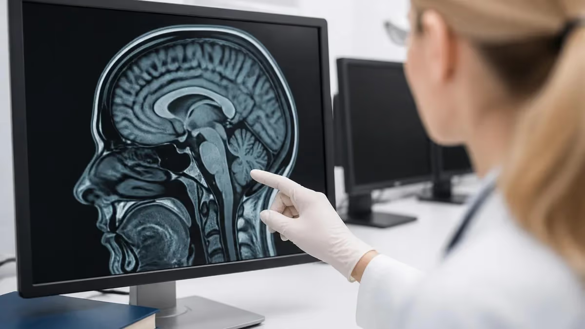
The relationship between dental implants and mri imaging is one of the most common questions patients ask before stepping into the scanner, and it is a topic every MRI technologist must understand thoroughly. The short answer reassures most people: the overwhelming majority of dental implants placed in the United States are made from titanium or titanium alloys, which are non-ferromagnetic and considered MRI conditional or MRI safe. That means the magnet will not yank an implant out of your jaw, and you can almost always be scanned without removing anything at all. Practice with the free MRI - Magnetic Resonance Imaging practice test on PracticeTestGeeks (200+ questions with answer explanations).
Still, the picture is more nuanced than a simple yes or no. Dental implants, crowns, bridges, orthodontic hardware, and metal fillings can all interact with the powerful magnetic field and radiofrequency pulses inside an MRI machine. While serious safety incidents are extremely rare, these dental materials frequently create image artifacts, which are distortions on the scan that can obscure nearby anatomy. For a brain, sinus, or head-and-neck study, that distortion matters a great deal, so understanding the materials in your mouth is genuinely important.
For radiologic technologists and aspiring MRI techs, dental hardware is a daily screening consideration. Every patient intake form asks about implants, plates, screws, and prosthetics for good reason. Knowing which dental devices are ferromagnetic, which merely cause artifacts, and which require no special handling separates a confident technologist from a hesitant one. If you are preparing for certification, working through realistic mri and dental implants scenarios will sharpen your screening judgment considerably over time.
This guide walks through the full landscape: implant materials and their magnetic behavior, why artifacts form and how they appear on different sequences, the safety screening process, what patients should tell their care team, and the practical techniques radiologists and technologists use to salvage diagnostic images when metal gets in the way. We will keep the language plain enough for patients while including the technical depth that students and working techs genuinely need.
It helps to start by separating two distinct concerns that people often blur together. The first is safety, meaning whether the device can move, heat dangerously, or malfunction in the magnetic field. The second is image quality, meaning whether the device degrades the diagnostic value of the pictures. A modern titanium implant scores very well on safety but can still score poorly on image quality near the scan region. Keeping these two ideas separate prevents a lot of needless anxiety and cancelled appointments.
Throughout this article you will also find references to free practice quizzes covering MRI safety, physics, and registry-style questions. Whether you are a nervous patient trying to understand your upcoming scan or a student building toward the ARRT or AMRIT credential, the goal here is the same: replace uncertainty with clear, accurate, and clinically grounded information about how dental implants and MRI scanners genuinely interact in everyday practice across American imaging centers, year after year.
MRI: upcoming dates and official changes
- Aug 17, 2026: ARRT launched a separate entry-level Imaging Assistant, Magnetic Resonance (I.A.) credential. Applications made Aug. 17–Oct. 14, 2026 paid a reduced $75 fee for testing Oct. 15–Nov. 30, 2026. Applicants after Oct. 14, 2026 pay $225 and can test on or after Jan. 1, 2027. The I.A. exam also requires a scaled section score of 7.5 or higher in MRI Screening and Safety.[4]
- The ARRT MRI postprimary exam has 220 questions: 200 scored and 20 unscored pilot items.[1]
- The ARRT MRI primary pathway exam has 230 questions: 200 scored and 30 unscored pilot items.[2]
- The ARRT MRI postprimary exam allows 210 minutes of test time within a 230-minute (3 hour 50 minute) appointment.[1]
- The ARRT MRI primary pathway exam allows 230 minutes of test time within a 250-minute (4 hour 10 minute) appointment.[2]
- ARRT exams, including MRI, are scored on a 1–99 scale, and candidates need a total scaled score of 75 to pass.[3]
- The ARRT MRI postprimary application fee is $225, or $450 for candidates using an NMTCB or ARDMS credential as the supporting category.[4]
- The ARRT MRI primary pathway application fee is $225.[4]
- ARRT MRI technologists holding an R.T. credential must complete Continuing Qualifications Requirements (CQR) every 10 years.[5]
- ARRT MRI credential holders renew every year for $65 and must complete 24 CE credits every two-year biennium.[5]
Numbers in brackets link to the official sources listed under Sources & last verified. Dates that have passed are removed automatically.
MRI Exam at a Glance
| Item | Detail | Source · Verified |
|---|---|---|
| Number of questions (Postprimary pathway) | 220 questions (200 scored + 20 pilot)[1] | assets-us-01.kc-usercontent.com · Oct 16, 2026 |
| Number of questions (Primary pathway) | 230 questions (200 scored + 30 pilot)[2] | assets-us-01.kc-usercontent.com · Oct 16, 2026 |
| Time limit (Postprimary pathway) | 210 minutes test time (230-minute appointment)[1] | assets-us-01.kc-usercontent.com · Oct 16, 2026 |
| Time limit (Primary pathway) | 230 minutes test time (250-minute appointment)[2] | assets-us-01.kc-usercontent.com · Oct 16, 2026 |
| Passing score | Scaled score of 75 (scale 1–99)[3] | The American Registry of Radiologic Technologists · Oct 16, 2026 |
| Exam fee (Postprimary pathway) | $225 application fee ($450 with an NMTCB or ARDMS supporting credential)[4] | The American Registry of Radiologic Technologists · Oct 16, 2026 |
| Exam fee (Primary pathway) | $225 application fee[4] | The American Registry of Radiologic Technologists · Oct 16, 2026 |
| Validity | Continuing Qualifications Requirements (CQR) every 10 years[5] | assets-us-01.kc-usercontent.com · Oct 16, 2026 |
| Renewal | Annual renewal, $65/year; 24 CE credits every 2 years[5] | assets-us-01.kc-usercontent.com · Oct 16, 2026 |
| Delivery | Computer-based at Pearson VUE test centers[2] | assets-us-01.kc-usercontent.com · Oct 16, 2026 |
| Eligibility (Postprimary pathway) | Supporting credential (ARRT, NMTCB or ARDMS) + structured education + clinical experience[1] | assets-us-01.kc-usercontent.com · Oct 16, 2026 |
| Eligibility (Primary pathway) | ARRT-verified MRI program + associate degree or higher + ethics[2] | assets-us-01.kc-usercontent.com · Oct 16, 2026 |
| Score release | Preliminary score at the test center; official results in about 10 business days[3] | The American Registry of Radiologic Technologists · Oct 16, 2026 |
| Testing window | One-year exam window per application[2] | assets-us-01.kc-usercontent.com · Oct 16, 2026 |
Free MRI - Magnetic Resonance Imaging Study Tools
Interactive MRI - Magnetic Resonance Imaging resources on PracticeTestGeeks, free to start:
- MRI - Magnetic Resonance Imaging practice test — 200+ questions with instant scoring and answer explanations
- MRI - Magnetic Resonance Imaging flashcards — review key terms and concepts card by card
- MRI - Magnetic Resonance Imaging study guide — topic-by-topic overview of what the exam covers
- MRI - Magnetic Resonance Imaging cheat sheet — one-page printable summary for last-minute review
Dental Implants and MRI by the Numbers
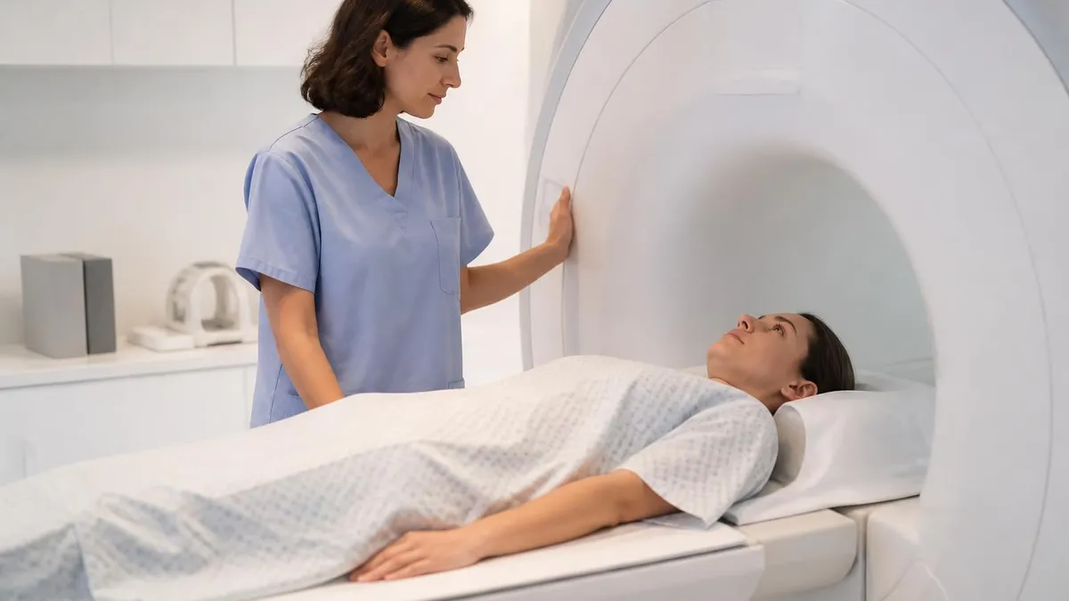
Implant Materials and Their Magnetic Behavior
The gold standard for dental implants and abutments. Titanium and its alloys are non-ferromagnetic and biocompatible, so they will not move in the magnet. They are MRI conditional and produce only mild local artifacts on most clinical sequences.
A ceramic, metal-free alternative gaining popularity for esthetics. Zirconia is non-magnetic and produces minimal MRI artifact, making it excellent for head and neck imaging. It poses essentially no safety concern in the scanner environment whatsoever.
Gold, porcelain-fused-to-metal, and base-metal alloy restorations vary widely. Most are non-ferromagnetic, but some contain nickel or cobalt-chromium that intensifies artifacts. Safety risk is low, but image distortion can be significant near the restoration itself.
Braces, archwires, and retainers may use stainless steel that is weakly ferromagnetic. These rarely pose a movement hazard but cause large artifacts and can heat slightly. Removable retainers should be taken out before scanning whenever possible.
Silver amalgam contains no iron and is not ferromagnetic, so it is MRI safe. It can still create small susceptibility artifacts near the tooth but rarely affects diagnostic imaging of regions away from the immediate dental area.
When patients ask whether their dental work makes an MRI dangerous, the honest answer is that genuine safety hazards from dental implants are exceedingly rare. The primary safety concerns in any MRI exam are ferromagnetic attraction, where a magnetic object is pulled toward the bore, and radiofrequency heating, where conductive material absorbs energy and warms up. Titanium and zirconia implants, which make up the vast majority of modern placements, are essentially immune to the first concern and produce only negligible heating under standard clinical scanning conditions today.
That said, not every metal object in the mouth behaves identically. Some older or imported restorations, certain orthodontic stainless steel components, and a small number of magnetic denture retention systems can be weakly ferromagnetic. Magnetic dentures are the one category that genuinely deserves attention, because they use small permanent magnets to hold the prosthesis in place. These should generally be removed before scanning, both because they can experience torque and because they can become demagnetized, ruining the appliance entirely.
Radiofrequency heating is the second technical concern worth understanding. Long, looped, or elongated conductive structures can act like antennas and concentrate RF energy. Most dental implants are small and compact, so they do not form efficient antennas, and measured temperature increases are clinically insignificant. Orthodontic archwires are longer and theoretically more capable of heating, but real-world studies consistently show only minor temperature changes that fall well within accepted safety thresholds for routine imaging procedures everywhere.
The screening process is the safety net that catches the rare problematic device. Before any scan, technologists review a detailed questionnaire and, when uncertain, consult device documentation or the radiologist. The MRI conditional label that accompanies most implants specifies the exact field strength, gradient, and RF limits under which the device was tested. A diligent tech treats unknown or undocumented hardware conservatively until its safety can be confirmed through manufacturer records or supplementary imaging studies.
For patients, the reassuring takeaway is that you almost never need to do anything special. You will not feel your implant during the scan, it will not move, and it will not heat to any noticeable degree. The exceptions, magnetic dentures and certain removable orthodontic appliances, are easy to handle simply by taking them out beforehand. Permanent titanium implants stay exactly where your oral surgeon placed them, completely undisturbed by the powerful magnetic field around you.
Technologists studying for certification should commit the conditional labeling system to memory, because exam questions frequently test the distinction between MRI safe, MRI conditional, and MRI unsafe. Understanding that a titanium implant is conditional rather than unconditionally safe, and knowing what those conditions actually specify, demonstrates the kind of precise knowledge that registry examinations reward and that real patients quietly depend on every single day inside the imaging suite.
Understanding MRI Artifacts From Dental Hardware
MRI artifacts from dental work arise mainly from magnetic susceptibility differences between metal and surrounding tissue. Metal distorts the local magnetic field, causing the scanner to mismap signal location. The result is signal voids, bright pile-up regions, and geometric warping immediately around the implant, crown, or filling that can obscure the very anatomy a radiologist needs to evaluate carefully.
The severity depends on the material, its mass, and the imaging sequence chosen. Dense base-metal alloys distort far more than titanium or zirconia. Gradient-echo sequences are much more vulnerable to susceptibility artifact than spin-echo sequences, which is exactly why sequence selection becomes a deliberate clinical strategy when dental hardware sits near the target region of interest for a given study.
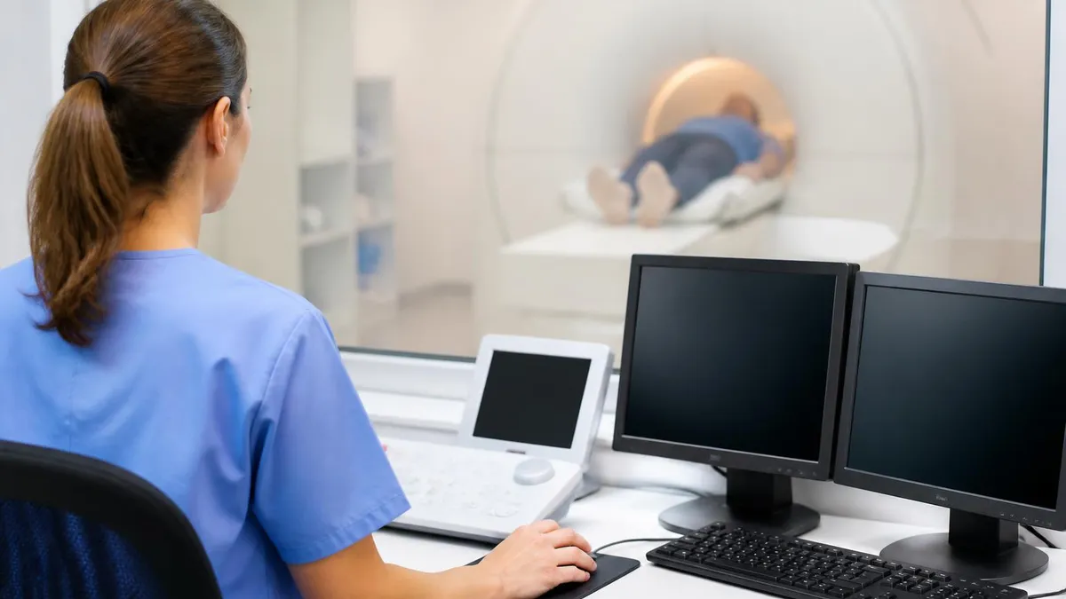
Getting an MRI With Dental Implants: Benefits and Limitations
- +Titanium and zirconia implants are non-ferromagnetic and will not move in the magnet
- +No need to remove permanently fixed implants before scanning
- +Documented dislodgement of standard dental implants in MRI is essentially nonexistent
- +Heating from compact dental implants is clinically negligible
- +Most body and limb scans are completely unaffected by dental hardware
- +Metal-artifact-reduction sequences can recover diagnostic images near implants
- −Susceptibility artifacts can obscure nearby brain, sinus, and head-and-neck anatomy
- −Higher field strength scanners produce larger artifacts from the same metal
- −Base-metal alloy crowns distort images more than titanium or zirconia
- −Magnetic denture attachments may need removal and can become demagnetized
- −Metal-reduction protocols add scan time and sometimes reduce image resolution
- −Undocumented or unknown implant materials require conservative extra screening
Pros and Cons at a Glance
| Pros | Cons |
|---|---|
| Titanium and zirconia implants are non-ferromagnetic and will not move in the magnet | Susceptibility artifacts can obscure nearby brain, sinus, and head-and-neck anatomy |
| No need to remove permanently fixed implants before scanning | Higher field strength scanners produce larger artifacts from the same metal |
| Documented dislodgement of standard dental implants in MRI is essentially nonexistent | Base-metal alloy crowns distort images more than titanium or zirconia |
| Heating from compact dental implants is clinically negligible | Magnetic denture attachments may need removal and can become demagnetized |
| Most body and limb scans are completely unaffected by dental hardware | Metal-reduction protocols add scan time and sometimes reduce image resolution |
| Metal-artifact-reduction sequences can recover diagnostic images near implants | Undocumented or unknown implant materials require conservative extra screening |
Dental Implants and MRI Patient Screening Checklist
- ✓Tell your technologist about every implant, crown, bridge, and denture you have
- ✓Mention any orthodontic braces, wires, or retainers, even removable ones
- ✓Disclose magnetic denture attachments, which usually must be removed
- ✓Bring any implant identification card or device documentation you received
- ✓Note the approximate date and location where implants were placed
- ✓Remove all removable dental appliances before entering the scan room
- ✓Report any prior MRI where you felt warmth or discomfort near your mouth
- ✓Tell the team if your scan targets the head, sinuses, jaw, or neck
- ✓Ask whether metal-artifact-reduction sequences will be used
- ✓Confirm the field strength of the scanner if you have extensive metalwork
- ✓Alert staff to any non-dental implanted devices such as pacemakers
- ✓Ask the radiologist about expected image limitations near your restorations
Fixed titanium implants stay; magnetic dentures come out
The single most useful distinction to remember is that permanently osseointegrated titanium implants never need removal and pose no movement risk. The genuine exception is a denture or partial held in place by small magnets. Those magnetic attachments should be removed before your scan to protect both your safety and the appliance itself.
When dental hardware threatens to ruin an otherwise necessary scan, radiologists and technologists have a deep toolbox of techniques to reduce metal artifact. The first and simplest lever is sequence selection. Spin-echo and fast spin-echo sequences are inherently far more resistant to susceptibility artifact than gradient-echo sequences, because the refocusing pulse recovers signal that the metal would otherwise scramble. Choosing the right base sequence often makes the difference between a non-diagnostic study and a perfectly readable one for the radiologist.
Beyond basic sequence choice, manufacturers offer dedicated metal-artifact-reduction techniques, sometimes branded with names like SEMAC, MAVRIC, or VAT. These specialized acquisitions use extra encoding steps and view-angle tilting to correct the spatial distortion that metal introduces. They cost additional scan time and can slightly reduce resolution or signal-to-noise, but for a patient whose target anatomy sits near dense restorations, the diagnostic payoff is enormous and well worth the few extra minutes involved.
Technologists also adjust acquisition parameters thoughtfully throughout the exam. Increasing receiver bandwidth shrinks the spatial spread of susceptibility artifact, tightening the dark blooming region around metal. Using thinner slices, smaller voxels, and a higher matrix can sharpen the boundary of an artifact so it intrudes less on adjacent anatomy. Each adjustment trades something away, so the technologist balances bandwidth, time, and resolution against the specific clinical question being asked by the referring doctor.
Field strength selection is another strategic decision. Because susceptibility artifact scales with field strength, imaging a heavily restored head-and-neck region at 1.5 Tesla rather than 3 Tesla can meaningfully reduce distortion. Many imaging centers that operate both magnets will deliberately route a patient with extensive dental metal to the lower-field scanner when the clinical target lies close to the mouth and artifact would otherwise dominate the entire picture being acquired.
Orientation and frequency-encoding direction offer subtler control over the result. Susceptibility artifact spreads preferentially along the frequency-encoding axis, so swapping the phase and frequency directions can push the worst distortion away from the critical anatomy. An experienced technologist will angle slices and reposition encoding directions specifically to shift artifact into a region where it does the least possible harm to the diagnostic interpretation of the study.
Finally, the radiologist interprets within the limits of what the physics allows. A skilled reader recognizes the characteristic geometry of metal artifact and avoids mistaking it for disease, correlates with other sequences and prior imaging, and clearly documents any region rendered non-diagnostic. When artifact truly defeats the MRI, the team may recommend an alternative such as CT, which tolerates dental metal differently, ensuring the patient still receives an accurate and complete diagnostic evaluation despite the hardware.
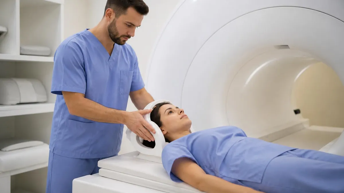
Some dentures and overdentures are retained by small permanent magnets that can experience torque in the MRI field and may become permanently demagnetized. Always tell your technologist and remove these appliances before scanning. Failing to disclose them risks both image degradation and damage to an expensive prosthesis.
Good communication between patients and the imaging team prevents the vast majority of dental-related MRI problems before they ever happen. The most valuable thing a patient can do is answer the screening questionnaire fully and honestly, then volunteer details that forms sometimes miss. If you have had multiple crowns, a full-arch implant bridge, lingering orthodontic wires, or a magnetically retained denture, say so plainly. None of these will likely stop your scan, but each piece of information helps the technologist plan the optimal protocol before you lie down.
Patients often worry needlessly because they confuse a dental implant with the kind of electronic implant that genuinely demands caution, such as a pacemaker or neurostimulator. A dental implant is an inert post of titanium or ceramic anchored in bone. It has no battery, no circuitry, and no moving parts whatsoever. Explaining this distinction to anxious patients is part of compassionate care, and it reflects the same conceptual clarity that mri and dental implants practice questions reinforce for students.
If your scan targets your head, brain, sinuses, jaw, or neck, it is worth asking the team specifically how your dental work might affect the images. A thoughtful technologist will explain whether metal-reduction sequences are planned and what limitations to expect. This conversation sets realistic expectations and reduces the chance of a confusing follow-up call where the radiologist requests a repeat study or an alternative imaging method because artifact obscured the area of clinical interest.
Bring documentation when you have it available. After implant surgery, many oral surgeons provide an implant card listing the manufacturer and model. For MRI conditional devices, that information lets the technologist verify exact scanning conditions in seconds rather than treating the hardware as an unknown quantity. While dental implants rarely require this level of detail, the habit of carrying device cards serves you well for any implanted hardware throughout your life.
Patients should also feel free to report sensations during the scan itself. Although clinically significant heating from dental implants is essentially unheard of, the squeeze ball or intercom exists precisely so you can speak up. If you ever feel unusual warmth, tingling, or discomfort near your mouth, tell the technologist immediately. They can pause the sequence, reassess, and continue safely. Your comfort and your honest feedback are integral parts of a safe imaging procedure from start to finish.
For technologists and students, the patient-facing side of this topic is just as testable as the physics. Registry examinations and clinical competencies expect you to communicate risk accurately, screen systematically, and handle the rare exception confidently. Practicing realistic scenarios, where a patient mentions a magnetic denture or an unknown crown, builds the calm, methodical habits that define excellent MRI practice and protect both image quality and patient trust in every single exam you perform.
If you are a patient preparing for an upcoming scan, a few practical habits make the experience smooth. Arrive a little early so you have time to complete screening without rushing, and review your dental history beforehand so you can answer questions confidently. Remove any removable appliances, retainers, or magnetic dentures and store them safely with your other belongings. Wear comfortable clothing free of metal, and mention every implant or restoration even if you assume it is completely irrelevant to the team.
For students and new technologists, build a mental decision tree for dental hardware. First, classify the material: titanium and zirconia are reassuring, base metals and stainless steel orthodontics warrant attention, and magnetic attachments demand removal. Second, ask where the scan target sits relative to the mouth. Third, decide whether artifact-reduction strategies are warranted for this particular study. Rehearsing this sequence until it becomes automatic turns a potentially stressful screening into a routine, confident interaction that protects the patient and the study.
When studying for certification, focus your energy on the concepts that examiners love to test. Understand the difference between MRI safe, conditional, and unsafe, and be ready to apply those labels to common dental devices. Know why gradient-echo sequences worsen susceptibility artifact and why spin-echo sequences resist it. Be able to explain how field strength, bandwidth, and encoding direction each influence artifact size. These cause-and-effect relationships appear repeatedly on registry questions and translate directly into competent clinical practice.
Practice tests are one of the most effective preparation tools because they expose gaps in your reasoning, not just your memorization. Working through realistic safety scenarios trains you to spot the rare hazard among many harmless devices, which is exactly the skill the credential certifies. Combine question banks with hands-on clinical observation, and you will internalize how the textbook physics actually manifests on real images from real patients with real dental work in their mouths.
Patients and clinicians both benefit from setting realistic expectations about image quality. Even with every artifact-reduction trick applied, some studies near heavy dental metal will retain residual distortion. That is not a failure of the technologist or the equipment; it is simply the physics of magnetic susceptibility at work. A clearly documented, partially limited study that still answers the clinical question is a success. When artifact genuinely defeats the goal, recommending an alternative modality is good medicine, not a shortcoming.
Ultimately, the story of dental implants and MRI is one of reassurance grounded in real science. The hardware in your mouth is almost certainly safe in the scanner, the rare exceptions are easy to manage, and the main challenge is image quality rather than danger. With honest screening, thoughtful protocol design, and clear communication, patients get the diagnostic answers they need and technologists deliver excellent care. Keep practicing, keep asking questions, and keep treating each screening as the meaningful safety checkpoint it truly is.
For a fuller set of questions on this topic, see our open mri near me resource.
Sample MRI - Magnetic Resonance Imaging Practice Questions
Try these questions from our free MRI - Magnetic Resonance Imaging practice tests. The correct answer and an explanation follow each question.
What is the term for the MRI safety classification that indicates a device has been tested and found safe under specified conditions?
- A. MR Safe
- B. MR Conditional
- C. MR Unsafe
- D. MR Compatible
Answer: B. MR Conditional
MR Conditional means the device poses no known hazards when used within the specific conditions (field strength, SAR, gradient limits) stated by the manufacturer.
What is the main advantage of parallel imaging techniques (e.g., GRAPPA, SENSE) in MRI?
- A. Reduced acquisition time by undersampling k-space using multiple coil elements
- B. Improved SNR by adding signals from multiple coils
- C. Elimination of susceptibility artifacts
- D. Increased spatial resolution without penalty
Answer: A. Reduced acquisition time by undersampling k-space using multiple coil elements
Parallel imaging uses the spatial sensitivity profiles of phased-array coils to reconstruct missing k-space lines, allowing faster acquisitions at the cost of some SNR.
What is the primary advantage of parallel imaging techniques (e.g., GRAPPA, SENSE) in MRI?
- A. They increase SNR at the same scan time
- B. They reduce scan time or allow higher spatial resolution by using multiple coil elements to under-sample k-space
- C. They eliminate the need for gradient coils
- D. They improve T1 contrast
Answer: B. They reduce scan time or allow higher spatial resolution by using multiple coil elements to under-sample k-space
Parallel imaging exploits spatial sensitivity differences among phased-array coil elements to reconstruct fully sampled images from under-sampled k-space, reducing scan time by the acceleration factor (R).
A patient reports having a cardiac pacemaker. What is the standard MRI safety classification for most older pacemakers?
- A. MR Safe
- B. MR Conditional
- C. MR Unsafe
- D. MR Compatible
Answer: C. MR Unsafe
Most older pacemakers are classified MR Unsafe because the magnetic field can interfere with device function or cause heating.
Take the full MRI - Magnetic Resonance Imaging practice test
MRI Questions and Answers
Sources & last verified
- assets-us-01.kc-usercontent.com — number of questions, time limit, eligibility (official source). “MRI Scored Items 200 Pilot Items 20 Total Items 220 Test Time 210 min” Verified Oct 16, 2026.
- assets-us-01.kc-usercontent.com — number of questions, time limit, delivery, eligibility (official source). “MRI Scored Items 200 Pilot Items 30 Total Items 230 Test Time 230 min” Verified Oct 16, 2026.
- The American Registry of Radiologic Technologists — passing score, score release (official source). “Our exam scoring scale ranges from 1 to 99, and you’ll need a total score of 75 to pass the exam.” Verified Oct 16, 2026.
- The American Registry of Radiologic Technologists — exam fee, other (official source). “Postprimary (M, CT, MR, BD, CI, VI, VS, BS, R.R.A.): $225 Postprimary (CT, MR, BD, or VS—using NMTCB as supporting category): $450 Postprimary (MR, VS,…” Verified Oct 16, 2026.
- assets-us-01.kc-usercontent.com — validity, renewal (official source). “Completing your Continuing Qualifications Requirements (CQR) every 10 years (if you have an R.T. or R.R.A. credential)” Verified Oct 16, 2026 · source checked Oct 16, 2026.
How we verify: editorial & fact-checking policy. Report an error: contact us.
MRI Practice Test

MRI of Cervical Spine: Complete Guide to Imaging, Anatomy, Pathology, and Patient Preparation

Knee MRI Images: A Complete Guide to Reading, Understanding, and Interpreting Knee Scans
White Spots on Brain MRI: Causes, What They Mean, and When to Worry

MRI Tech School 2026 — Programs, Requirements, and What to Expect
About the Author

Medical Laboratory Scientist & Clinical Certification Expert
Johns Hopkins UniversityDr. Sandra Kim holds a PhD in Clinical Laboratory Science from Johns Hopkins University and is certified as a Medical Technologist (MT) and Medical Laboratory Scientist (MLS) through ASCP. With 16 years of clinical laboratory experience spanning hematology, microbiology, and molecular diagnostics, she prepares candidates for ASCP board exams, MLT, MLS, and specialist certification tests.