
CT Scan or MRI: Which Imaging Test Do You Actually Need? 2026 September
CT scan or MRI — learn the key differences, when doctors order each, costs, risks, and how to prepare. 🎯 Complete 2026 September guide.
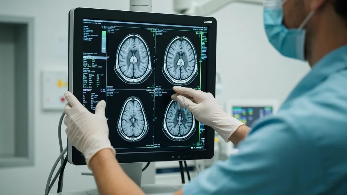
When your doctor orders imaging, one of the most common questions patients ask is: should I get a ct scan or mri? Both are powerful diagnostic tools, yet they work on completely different physical principles, excel in different clinical scenarios, and carry distinct risk profiles. Understanding those differences helps you ask better questions, prepare more effectively, and feel less anxious about whichever test your care team recommends. This guide breaks down everything you need to know in plain language backed by current clinical practice.
CT scans — short for computed tomography — use X-rays rotated around the body to generate cross-sectional images reconstructed by computer. A full chest CT delivers roughly 7 millisieverts of radiation, equivalent to about two years of background radiation exposure. Scans take only seconds to minutes, which makes CT the go-to choice in emergency departments where speed is critical and patients may not be able to lie still for extended periods. The images are excellent for bone fractures, internal bleeding, lung disease, and kidney stones.
MRI — magnetic resonance imaging — relies on powerful magnetic fields and radiofrequency pulses to excite hydrogen protons in body tissue. When those protons realign after the pulse, they emit signals that software translates into extraordinarily detailed images of soft tissue. No ionizing radiation is involved whatsoever, making MRI safer for repeated use and preferable when evaluating the brain, spinal cord, joints, tendons, ligaments, and pelvic organs. The tradeoff is time: a standard brain MRI takes 30 to 60 minutes, and some comprehensive spine studies run even longer.
The choice between the two modalities is almost never left to the patient; your physician selects based on the clinical question at hand. A neurologist suspecting a stroke will order an MRI with diffusion-weighted sequences because early infarcts are invisible on CT. An emergency physician evaluating a trauma patient with possible internal hemorrhage will order a CT because it takes three minutes instead of forty-five. Radiologists who read both types of images routinely describe MRI as providing superior soft-tissue contrast while CT offers superior speed and bone detail.
Cost is another significant consideration that influences decision-making for many Americans. The average uninsured cost for a brain CT is approximately $1,200 to $3,000, while a brain MRI typically runs $1,500 to $5,000 or more depending on the facility, geographic region, and whether contrast is used. Insurance coverage varies widely by plan and diagnosis code, so always verify benefits before scheduling. Some outpatient imaging centers offer self-pay rates that are dramatically lower than hospital-based pricing — sometimes 60 to 80 percent less for identical equipment and protocols.
Contrast agents amplify the usefulness of both modalities. CT contrast is iodine-based and injected intravenously; it highlights blood vessels and areas of active inflammation or tumor growth. MRI contrast uses gadolinium, a rare-earth metal chelate that crosses a disrupted blood-brain barrier to reveal lesions, tumors, and inflammatory foci invisible on non-contrast sequences. Patients with impaired kidney function face elevated risk with both contrast types, so creatinine levels are routinely checked before administration. Allergic reactions to iodine contrast occur in roughly one to two percent of patients and can range from mild hives to rare anaphylaxis.
For MRI technologists and radiology students preparing for board examinations, mastering the clinical indications for each modality is foundational knowledge. Understanding physics, safety protocols, and anatomy as they relate to both CT and MRI prepares you not only for credentialing exams but for the daily decisions you will make on the floor. You can explore advanced concepts such as ct scan or mri diffusion-weighted imaging sequences, which demonstrate how water molecule motion differs between healthy and infarcted tissue in ways that CT simply cannot replicate.
CT Scan vs. MRI by the Numbers
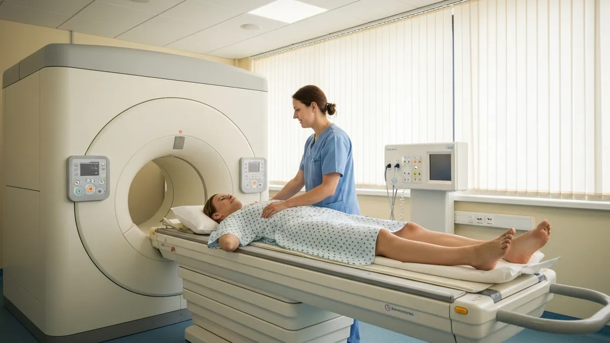
Key Differences Between CT and MRI
CT uses rotating X-ray beams and computer reconstruction to create cross-sectional images. MRI uses a strong magnetic field and radiofrequency pulses to excite hydrogen protons, producing images based on tissue water content and molecular environment.
CT delivers ionizing radiation — a chest CT equals roughly 2 years of background exposure. MRI produces zero ionizing radiation, making it the preferred modality for pregnant patients, children, and anyone requiring frequent repeat imaging.
CT scans complete in 3 to 10 minutes, critical in trauma and emergency care. Standard MRI exams run 30 to 90 minutes depending on body region and sequences ordered, requiring patients to remain still throughout the entire acquisition.
CT excels at bone fractures, pulmonary embolism, internal bleeding, kidney stones, and rapid trauma surveys. MRI is superior for brain lesions, spinal cord pathology, soft-tissue tumors, joint injuries, and pelvic organ evaluation.
CT is contraindicated in pregnancy when feasible, and contrast is restricted in renal impairment. MRI is contraindicated with most pacemakers, cochlear implants, certain aneurysm clips, and ferromagnetic foreign bodies in critical locations.
Physicians choose MRI over CT in a wide range of clinical situations where soft-tissue contrast, multiplanar capability, or radiation avoidance takes priority. The single most important deciding factor is the tissue type being evaluated. Soft tissues — the brain, spinal cord, intervertebral discs, cartilage, tendons, muscles, and pelvic organs — contain varying water concentrations that MRI translates into dramatically different signal intensities. A herniated lumbar disc compressing a nerve root is exquisitely visible on MRI but nearly invisible on plain CT without intrathecal contrast.
Neurological conditions represent perhaps the strongest clinical domain for MRI. When a patient presents with new-onset seizures, progressive cognitive decline, suspected multiple sclerosis, or a possible brain tumor, MRI is the first-line and often only imaging study needed. T1-weighted sequences define anatomy; T2-weighted sequences reveal edema and demyelination; FLAIR sequences suppress cerebrospinal fluid signal to expose periventricular plaques. Post-contrast T1 images highlight blood-brain barrier breakdown. No single CT protocol replicates this multi-parametric tissue characterization capability.
Musculoskeletal applications provide another domain where MRI clearly dominates. Orthopedic surgeons routinely order knee MRI before any intervention for suspected meniscal tears, ACL ruptures, or articular cartilage damage. The ACL is virtually invisible on CT, yet MRI can grade its integrity, assess concomitant bone bruising, and evaluate the posterolateral corner structures in a single 45-minute study. Similarly, shoulder MRI with intra-articular contrast (MR arthrography) outperforms CT arthrography for labral tear characterization in young athletes.
Pediatric imaging tilts heavily toward MRI precisely because children are more sensitive to ionizing radiation and face a lifetime of potential cumulative exposure. The developing brain and thyroid gland carry the highest radiation sensitivity. Many pediatric hospitals now follow Image Gently campaign guidelines that minimize CT use and substitute MRI wherever clinically feasible. The longer scan time in children sometimes requires sedation or general anesthesia, which adds complexity and cost, but the radiation-free benefit is considered worth it for many indications.
Cardiac MRI has emerged as the gold standard for assessing myocardial viability, cardiomyopathy phenotyping, and congenital heart disease anatomy. Late gadolinium enhancement sequences reveal scar tissue within the heart muscle with millimeter-level precision, guiding decisions about implantable defibrillators and ablation procedures. CT coronary angiography competes in the evaluation of coronary artery stenosis, but functional assessment of the myocardium remains firmly in MRI's domain. Electrophysiologists now rely on cardiac MRI to map fibrosis before complex ablations.
Oncological staging increasingly leverages MRI's superior soft-tissue resolution. Rectal cancer staging with high-resolution pelvic MRI determines whether a tumor has penetrated the mesorectal fascia, directly influencing the decision to administer neoadjuvant chemoradiation before surgery. Prostate MRI using multi-parametric protocols — combining T2-weighted, diffusion-weighted, and dynamic contrast-enhanced sequences — has transformed prostate cancer detection and active surveillance management. Breast MRI supplements mammography in high-risk women and evaluates the extent of newly diagnosed breast cancer before surgery.
Understanding these clinical distinctions matters deeply for MRI technologists sitting for the ARRT MRI examination. The registry tests not only MRI physics and safety but also clinical reasoning about appropriate protocols and contrast use. Recognizing when a patient's clinical history calls for specific sequences — such as diffusion-weighted imaging for acute stroke evaluation — demonstrates the advanced judgment that separates a competent technologist from an exceptional one. Preparation resources like board review quizzes help you internalize these distinctions across the full breadth of MRI applications.
How CT Scans and MRI Work: Physics and Process
A CT scanner houses an X-ray tube and detector array mounted on a rotating gantry. As the patient moves through the bore on a motorized table, the tube rotates continuously, firing X-ray beams through the body at thousands of angles. Detectors measure how much radiation passes through versus is absorbed by different tissue densities. Bone, air, soft tissue, and fat each have characteristic Hounsfield unit values — bone near +1,000, air near -1,000, and soft tissue between -100 and +100 — allowing precise tissue differentiation.
Modern multidetector CT (MDCT) scanners with 64 to 320 detector rows can acquire sub-millimeter slices across the entire chest in under five seconds. Reconstruction algorithms then stack these cross-sectional images into axial, coronal, and sagittal planes, plus three-dimensional volume renderings. Iterative reconstruction techniques introduced over the last decade have cut radiation doses by 30 to 50 percent compared to older filtered back-projection methods while maintaining diagnostic image quality — a major advance for patient safety.
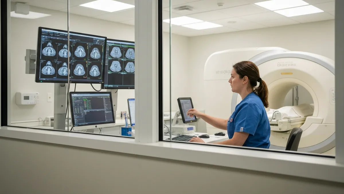
CT Scan vs. MRI: Advantages and Limitations
- +MRI provides superior soft-tissue contrast for brain, spinal cord, and joint imaging
- +MRI uses zero ionizing radiation — ideal for children, pregnant women, and repeat studies
- +CT scans complete in minutes, critical for trauma and emergency life-threatening conditions
- +CT is widely available at virtually every hospital including small community facilities
- +MRI can acquire images in any plane without repositioning the patient
- +CT is excellent for visualizing bone detail, calcifications, and lung parenchyma
- −MRI scans take 30 to 90 minutes, requiring extended patient cooperation and stillness
- −MRI is contraindicated in patients with most pacemakers and many metal implants
- −CT delivers ionizing radiation with cumulative lifetime exposure concerns
- −MRI has limited availability in rural areas and low-resource settings worldwide
- −CT soft-tissue contrast is inferior to MRI for brain lesions and spinal pathology
- −MRI is significantly more expensive than CT, with higher out-of-pocket costs for patients
Patient Preparation Checklist for CT Scan or MRI
- ✓Notify your ordering physician if you are pregnant or think you might be pregnant before scheduling either exam.
- ✓Inform the scheduling staff about any kidney disease, diabetes, or prior contrast reactions before a contrast study.
- ✓Remove all metal jewelry, piercings, and hairpins before entering the MRI suite — leave them at home if possible.
- ✓Bring a complete list of all implants, surgical hardware, and pacemaker model numbers to your MRI appointment.
- ✓Follow fasting instructions — typically nothing by mouth for 4 hours before contrast CT or sedated MRI.
- ✓Wear comfortable, loose-fitting clothing with no metal zippers, snaps, or underwire bras to expedite room preparation.
- ✓Arrive 15 minutes early to complete safety screening paperwork and allow time for IV line placement if contrast is ordered.
- ✓Tell the technologist about any claustrophobia before the exam so they can discuss open-bore options or anxiolytic protocols.
- ✓Arrange a driver if you will be taking oral sedation medication for anxiety management during MRI.
- ✓Ask your radiologist or technologist about expected wait times for results and how your physician will receive the report.
MRI and Pacemakers: The Rules Have Changed
As of 2026, many modern pacemakers and implantable cardioverter-defibrillators are labeled MR-conditional, meaning they can safely undergo 1.5T MRI under specific scanning conditions and with device interrogation before and after. However, legacy devices implanted before 2011 are typically not MR-conditional. Always provide the exact device manufacturer and model number at screening — never assume compatibility based on age alone.
Cost is one of the most anxiety-inducing aspects of medical imaging for American patients, and the price gap between CT and MRI is substantial. A hospital-based CT scan of the abdomen and pelvis with contrast can run $3,000 to $8,000 on the chargemaster list price, while the same facility might bill $5,000 to $12,000 for a comparable MRI. However, these list prices bear little relationship to what patients actually pay. Negotiated insurance rates typically reduce these figures by 40 to 70 percent, and Medicare reimbursement rates are even lower.
For uninsured or underinsured patients, the price variation across facility types is staggering. Freestanding outpatient imaging centers — which are not affiliated with hospital systems — often charge $300 to $900 for a brain CT and $400 to $1,500 for a brain MRI. These facilities use identical or comparable equipment, employ board-certified radiologists, and achieve the same diagnostic accuracy. The difference is purely a function of overhead structure and negotiating leverage. Patients should always call ahead, ask for the self-pay rate, and compare at least two or three facilities before scheduling.
Health insurance coverage depends heavily on the diagnosis code submitted with the imaging order. A CT scan ordered for a vague symptom like headache may trigger prior authorization requirements or even denial if the insurer considers it not medically necessary. An MRI ordered for the same symptom frequently encounters higher scrutiny because of its greater cost. Patients who receive a denial should request a peer-to-peer review between their ordering physician and the insurance medical director — this step reverses denials in a significant percentage of cases.
Medicare Part B covers diagnostic imaging at 80 percent of the approved amount after the deductible is met, leaving the patient responsible for 20 percent coinsurance with no cap. For a $1,000 Medicare-approved MRI, that means $200 out of pocket — manageable for most. But complex multi-sequence studies at academic medical centers can generate multiple billing components: facility fee, radiologist professional fee, and contrast administration fee separately. Patients should request an itemized estimate before the study to avoid surprise bills.
The No Surprises Act, which took effect in January 2022, provides some protection against unexpected out-of-network charges at in-network facilities. If you are referred to an in-network hospital for imaging but the radiologist reading your scan is out of network, the No Surprises Act generally limits your cost-sharing to in-network rates and prohibits balance billing. However, the law applies to emergency situations and certain non-emergency services — understanding which category your imaging falls into requires careful review of your specific plan documents.
Medical imaging financing has expanded significantly in recent years. Many imaging centers now partner with medical lending companies that offer 12 to 24 months of interest-free financing for out-of-pocket imaging costs. Nonprofit hospitals are federally required to have financial assistance programs for patients below 200 to 400 percent of the federal poverty level. Community health centers that receive federal funding offer sliding-scale fees based on income and can refer patients to contracted imaging facilities at reduced rates. No patient with a legitimate clinical need should forgo necessary imaging purely for financial reasons without first exhausting these options.
For MRI technologists and students, understanding the reimbursement landscape is increasingly important beyond purely clinical training. Radiologic technologists who understand coding, prior authorization workflows, and cost considerations can serve as better patient advocates and contribute more effectively to the administrative functions of imaging departments. Board examinations do not test billing knowledge directly, but career advancement into supervisory and management roles increasingly demands this broader operational awareness.
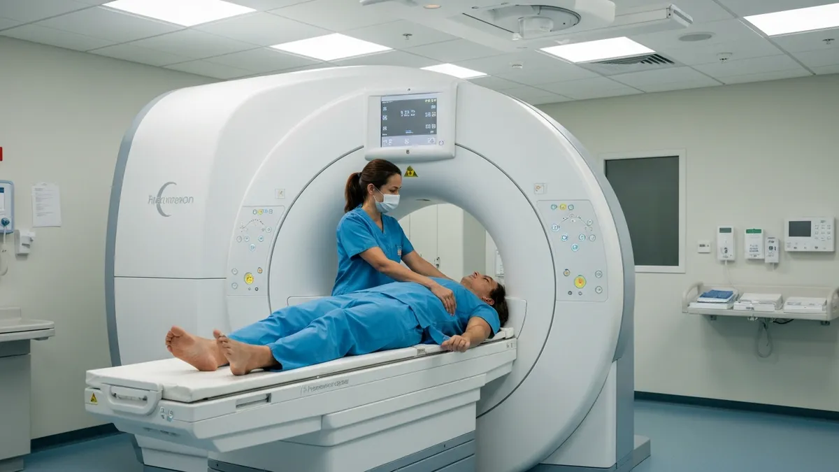
Research published since 2014 has confirmed that gadolinium-based contrast agents can deposit in brain tissue, bone, and skin following repeated MRI administrations, even in patients with normal kidney function. The FDA added a class warning to all gadolinium agents in 2017. While no neurological effects from this retention have been definitively proven in patients with normal kidneys, radiologists now apply the ALARA principle to contrast use — ordering gadolinium only when it provides clear diagnostic value beyond non-contrast sequences.
For radiology and MRI technologist students, the comparison between CT and MRI forms a cornerstone of both academic training and board examination preparation. The ARRT MRI examination blueprint allocates significant question weight to patient care and education, MRI safety, image production, and procedures — all of which require a thorough understanding of how MRI differs from CT in practice. Students who can confidently articulate why a physician would choose one modality over another demonstrate the clinical reasoning that separates knowledge recall from genuine understanding.
MRI physics questions on board examinations frequently test the interaction between magnetic field strength and signal-to-noise ratio. Higher field strength — moving from 1.5T to 3T — roughly doubles the available signal, enabling thinner slices, faster acquisitions, or improved temporal resolution for dynamic studies. However, 3T systems also amplify susceptibility artifacts, radiofrequency field inhomogeneity, and specific absorption rate (SAR) — the measure of radiofrequency energy deposited in tissue per unit mass. SAR limits set by the FDA and IEC protect patients from excessive tissue heating, particularly relevant in patients with implants that may concentrate the radiofrequency field.
Pulse sequence selection is another high-yield examination topic that directly relates to the CT versus MRI comparison. Spin-echo sequences with long repetition times and long echo times produce T2-weighted images — the workhorses of pathology detection since most diseases increase tissue water content. Short repetition and echo time combinations yield T1-weighted images ideal for anatomy and post-contrast enhancement. Inversion recovery sequences like STIR suppress fat signal to reveal bone marrow edema, while FLAIR suppresses free water to expose periventricular white matter lesions. No equivalent pulse-sequence flexibility exists in CT, where contrast is determined almost entirely by tissue electron density.
Safety screening is a domain where MRI technologists bear direct clinical responsibility that has no parallel in CT operation. Before any patient enters Zone III or Zone IV of the MRI environment, the technologist must confirm the absence of ferromagnetic foreign bodies, review implant documentation, assess claustrophobia, and evaluate renal function for contrast eligibility. The ACR Manual on MR Safety, updated periodically, provides the authoritative framework for these decisions. Technologists who have not internalized this framework risk patient injury — MRI-related deaths and serious injuries, though rare, have occurred from projectile events when ferromagnetic objects entered the bore.
Contrast administration protocols differ between CT and MRI in ways that affect both image quality and patient safety. CT contrast bolus timing is critical — a scan timed too early in the arterial phase versus venous phase yields completely different diagnostic information.
Power injectors deliver iodinated contrast at 3 to 5 mL/second; scan triggering is automated using bolus-tracking software that monitors contrast arrival in the aorta or target vessel. MRI contrast, by contrast, is injected at 1 to 2 mL/second and relies more on tissue permeability characteristics than precise vascular timing, though dynamic contrast-enhanced sequences for liver and breast MRI do require careful temporal resolution.
Artifact recognition distinguishes competent technologists from exceptional ones. CT artifacts include beam hardening streaks adjacent to dense metal (dental implants, hip prostheses), motion blur from cardiac pulsation or patient movement, and ring artifacts from detector calibration errors.
MRI artifacts are more diverse: magnetic susceptibility artifacts bloom around metal implants; chemical shift artifacts outline fat-water interfaces; wrap-around (aliasing) occurs when anatomy outside the field of view folds into the image; and Gibbs ringing appears as parallel lines adjacent to sharp signal interfaces like the spinal cord-CSF boundary. Each artifact has a known physical cause and a specific mitigation strategy that may be tested on board examinations.
Practical preparation for the MRI registry examination benefits enormously from active practice with image-based questions that present clinical scenarios requiring protocol selection and safety decision-making. The highest-performing candidates combine didactic review of physics and anatomy with sustained question-bank practice that builds pattern recognition. Many successful candidates report that working through 1,500 to 2,000 practice questions over a 12-week preparation period is a reliable benchmark for registry readiness, provided those questions are accompanied by detailed explanations that teach the underlying principle rather than just the correct answer.
Practical tips for getting the most out of your CT or MRI experience begin well before the day of the scan. If your physician orders imaging, ask specifically what clinical question the study is designed to answer. Understanding the purpose — detecting a tumor, evaluating a herniated disc, ruling out a pulmonary embolism — helps you understand why specific preparations matter and motivates compliance with potentially inconvenient instructions like fasting or stopping certain medications. Informed patients who understand the purpose of their imaging tend to cooperate better, produce fewer motion artifacts, and have shorter exam times.
On the day of a CT scan, dress simply. Avoid clothing with metal buttons, zippers, or clasps that require removal and slow preparation. If abdominal or pelvic CT is ordered with oral contrast, begin drinking the contrast preparation solution exactly as instructed — typically starting one to two hours before the scan — since underdistended bowel loops mimic pathological masses and force repeat imaging. Patients scheduled for CT coronary angiography may receive beta-blocker medication to slow the heart rate below 65 beats per minute, since cardiac motion is the enemy of sharp coronary artery visualization.
For MRI preparation, the single most impactful action you can take is arriving with complete implant documentation. A card in your wallet identifying the manufacturer and model of a pacemaker, cochlear implant, neurostimulator, or orthopedic implant eliminates uncertainty at screening and prevents scheduling delays. The MRI technologist cannot scan you without confirming implant compatibility — an undocumented implant means rescheduling while documentation is tracked down. Contact your surgeon's office, device manufacturer, or hospital records department weeks in advance if you know you have implants.
Claustrophobia management deserves attention and planning rather than last-minute improvisation. If you have experienced anxiety in enclosed spaces before, discuss this with your ordering physician before the MRI appointment. Mild oral benzodiazepines like lorazepam or diazepam, taken 30 to 60 minutes before the scan, reduce anxiety effectively for most patients. Arrange transportation since these medications impair driving. Alternatively, ask whether your imaging facility has a wide-bore (70 cm) scanner — these have become standard at many centers and significantly reduce the sense of confinement compared to older 60 cm bore systems.
Breathing techniques help reduce motion artifacts in body MRI. Technologists will coach patients through specific breath-hold instructions for liver, pancreas, and kidney sequences — typically five to 20 second breath-holds at end-expiration. Practicing at home before the exam is not excessive preparation; patients who can reliably hold their breath for 20 seconds produce sharper abdominal images than those who cannot. For patients with limited breath-hold capacity due to lung disease or anxiety, free-breathing sequences with respiratory triggering are available and should be requested at scheduling.
After imaging, understanding the result communication process reduces anxiety during what can be a stressful waiting period. Most radiology reports are finalized within 24 to 48 hours for outpatient studies, though urgent or emergent findings are communicated to the ordering physician immediately by phone.
Many health systems now deliver imaging reports directly to patients through electronic health record patient portals — sometimes before the ordering physician has reviewed them. Reading your own radiology report without clinical context can be frightening; radiologists routinely note incidental findings and recommend follow-up for lesions that are almost certainly benign. Always discuss your report with your physician before drawing conclusions.
For MRI technologist students using practice resources, integrating clinical scenario questions into your study routine dramatically improves retention compared to reading alone. When you encounter a question about which pulse sequence to use for a given pathology, pause and visualize why — what tissue property does that sequence emphasize, why does that property differ in the pathological state, and what would the image look like? Building these mental models through active recall rather than passive review is the mechanism behind effective examination preparation. Quality practice resources that include detailed rationale for every answer choice accelerate this learning process substantially.
MRI Questions and Answers
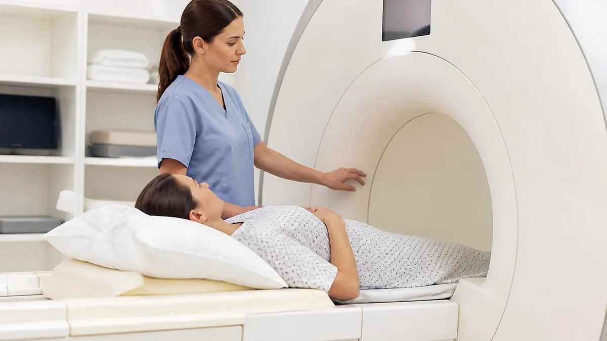
DWI MRI: Diffusion-Weighted Imaging Explained (2026 Guide)
MRI Lumbar Spine Without Contrast: Complete Guide to the Scan, What It Shows, and How to Prepare
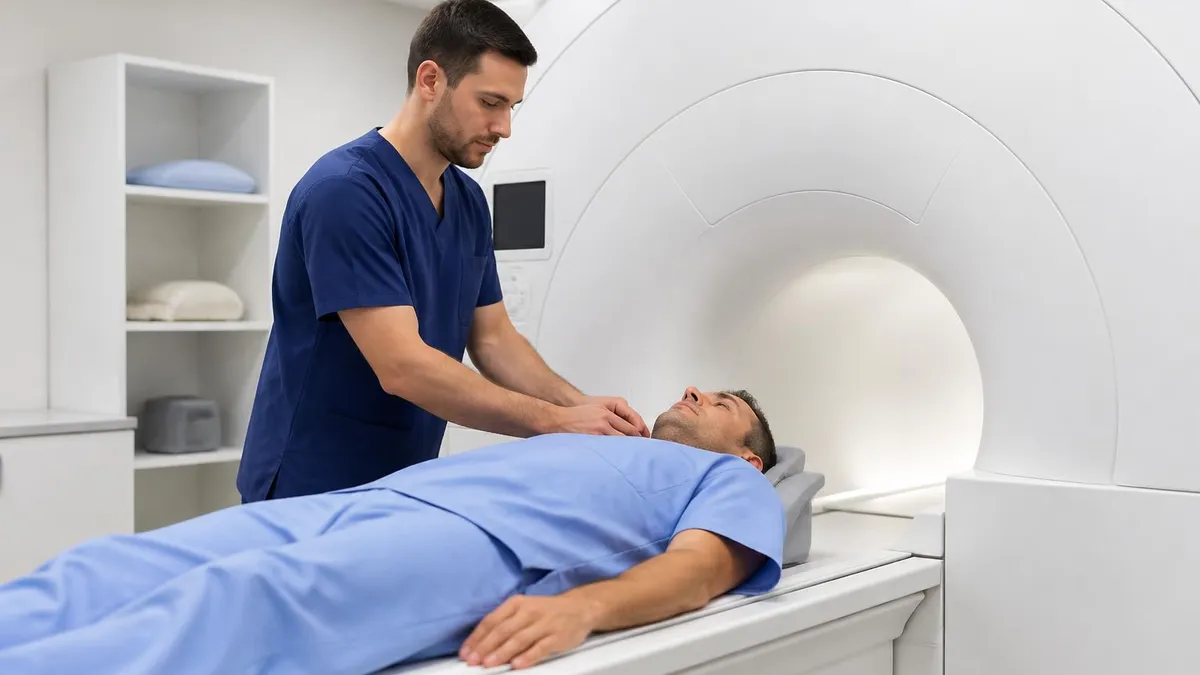
MRI History: From Nuclear Resonance to Modern Scanners

MRI of Cervical Spine: Complete Guide to Imaging, Anatomy, Pathology, and Patient Preparation
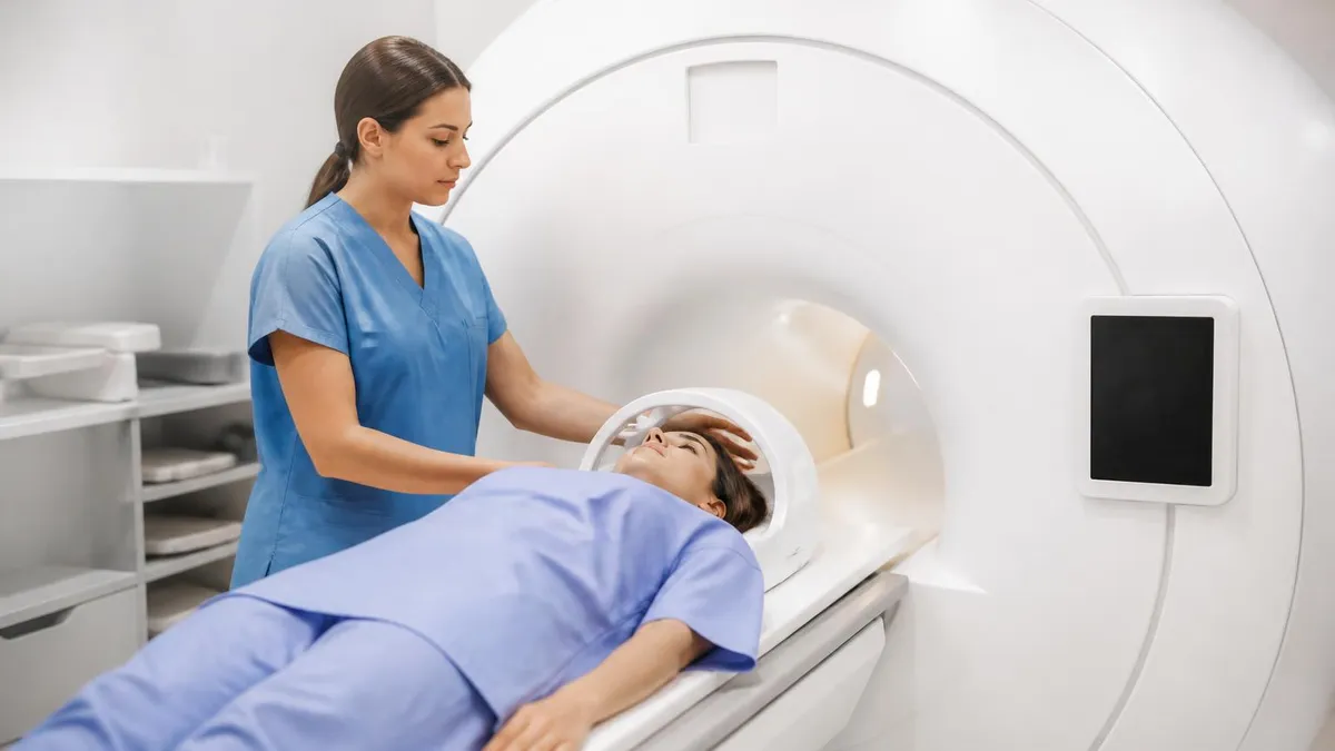
Where to Get an MRI: Facilities, Costs, and How to Choose
About the Author

Medical Laboratory Scientist & Clinical Certification Expert
Johns Hopkins UniversityDr. Sandra Kim holds a PhD in Clinical Laboratory Science from Johns Hopkins University and is certified as a Medical Technologist (MT) and Medical Laboratory Scientist (MLS) through ASCP. With 16 years of clinical laboratory experience spanning hematology, microbiology, and molecular diagnostics, she prepares candidates for ASCP board exams, MLT, MLS, and specialist certification tests.
Join the Discussion
Connect with other students preparing for this exam. Share tips, ask questions, and get advice from people who have been there.
View discussion (8 replies)