
Can an MRI Detect Scar Tissue? What the Scan Reveals and Why It Matters 2026
Can an MRI detect scar tissue? ✅ Learn how MRI identifies fibrosis, surgical adhesions, and internal scars — with contrast, sequences, and clinical limits.
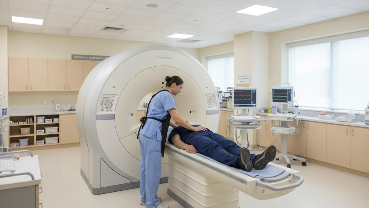
Can an MRI detect scar tissue? The short answer is yes — magnetic resonance imaging is one of the most powerful noninvasive tools available for identifying fibrotic and scar tissue anywhere in the body. Unlike X-ray or CT, which excel at visualizing bone and calcification, MRI exploits differences in soft-tissue water content and collagen density to distinguish healthy muscle, fat, and organ parenchyma from the dense, relatively dehydrated tissue that forms after injury, surgery, or chronic inflammation.
Understanding what MRI can and cannot reveal about scar tissue is essential for patients facing post-surgical follow-up, athletes recovering from tendon tears, and clinicians deciding whether additional workup is needed.
Scar tissue — known in clinical radiology as fibrosis when it affects internal organs, or as adhesions when it forms between tissue planes after surgery — has a distinctive MRI signature. On standard T1-weighted sequences, mature scar appears as a low-to-intermediate signal region, often darker than surrounding muscle. On T2-weighted sequences, acute or immature scar retains some water and can appear brighter, while mature, collagen-rich fibrosis tends to stay dark on both T1 and T2. Radiologists use this interplay of signal characteristics across multiple pulse sequences to characterize tissue age, extent, and composition with remarkable precision.
The sensitivity of MRI for scar tissue depends heavily on the region being imaged, the field strength of the magnet, and which sequences the radiologist orders. A 3 Tesla scanner equipped with dedicated surface coils and gadolinium contrast can reveal thin adhesion bands in the abdomen or pericardium that a 1.5T system might miss entirely. High-resolution protocols for the knee, shoulder, or cardiac muscle have been refined over decades, and today's MRI suites can routinely characterize scar tissue down to a few millimeters in thickness.
Gadolinium-based contrast agents play a particularly important role in scar detection. When injected intravenously, gadolinium shortens T1 relaxation time in tissues with active vascularity or inflammation. Mature scar tissue is largely avascular — meaning it has few blood vessels — so it enhances poorly or not at all on post-contrast images, while surrounding viable tissue lights up brightly. This contrast between enhancing and non-enhancing regions gives radiologists a reliable method to delineate the scar boundary and estimate how much functional tissue has been replaced.
One area where MRI scar detection has become especially clinically significant is cardiac imaging. Late gadolinium enhancement (LGE) MRI, sometimes called cardiac MRI with scar protocol, can pinpoint myocardial fibrosis caused by prior heart attack, myocarditis, or cardiomyopathy. The location and transmurality of scar burden directly influence whether a patient is a candidate for revascularization, ablation therapy, or implantable defibrillator placement. Studies consistently show that LGE-MRI outperforms echocardiography for detecting subendocardial infarcts and mid-wall fibrosis patterns invisible to ultrasound.
Outside the heart, MRI is used to evaluate post-surgical scar in the spine — distinguishing epidural fibrosis from recurrent disc herniation in a condition radiologists call failed back surgery syndrome. This distinction matters enormously because scar tissue does not benefit from repeat surgery, while a reherniated disc might.
On gadolinium-enhanced MRI, scar tissue enhances homogeneously because it contains new capillaries, whereas avascular disc material does not enhance, allowing the two conditions to be separated with about 90% accuracy in experienced hands. You can also learn more about how specialized sequences contribute to this distinction by exploring resources on can mri detect scar tissue through diffusion-weighted approaches.
The clinical takeaway is that MRI is not a single test but a flexible platform. Ordering the right protocol — the right combination of sequences, field strength, coil selection, and contrast timing — determines whether scar tissue is confidently identified or subtly missed. Patients who want the most informative study should ask their referring physician to specify a scar-dedicated protocol rather than accepting a generic routine scan of the relevant body region.
MRI: upcoming dates and official changes
- Aug 17, 2026: ARRT launched a separate entry-level Imaging Assistant, Magnetic Resonance (I.A.) credential. A reduced $75 fee applied to applications made Aug. 17–Oct. 14, 2026, for testing Oct. 15–Nov. 30, 2026. Later applicants pay $225 and can test on or after Jan. 1, 2027.[3]
- The ARRT MRI exam for postprimary-pathway candidates has 220 questions: 200 scored and 20 unscored pilot items.[1]
- Postprimary-pathway candidates get 210 minutes for the ARRT MRI exam, inside a 230-minute (3 hours, 50 minutes) appointment.[1]
- To pass the ARRT MRI exam you need a total scaled score of 75 on ARRT's 1–99 scale. This is not a percentage.[2]
- The ARRT MRI exam through the postprimary pathway costs $225, or $450 for candidates whose supporting credential is from NMTCB or ARDMS.[3]
All verified MRI facts and sources
Numbers in brackets link to the official sources listed under Sources & last verified. Dates that have passed are removed automatically.
MRI Scar Detection by the Numbers
How MRI Sequences Reveal Scar Tissue
Mature, collagen-rich scar appears dark on T1 sequences because tightly packed collagen fibers contain very little free water. This sequence helps radiologists map the extent of chronic fibrosis and differentiate it from fat, which appears bright on T1.
Acute or immature scar retains edema and fluid, producing a brighter T2 signal. Chronic, densely packed scar goes dark on T2. Comparing T1 and T2 together helps estimate tissue age and inflammation status across different injury stages.
Gadolinium contrast accumulates in the expanded interstitial space of fibrotic tissue, making scar appear bright against dark, healthy muscle on inversion-recovery sequences. This is the gold standard for cardiac scar and organ fibrosis detection worldwide.
DWI measures water molecule movement within tissue. Dense scar restricts diffusion, producing elevated signal on DWI and low apparent diffusion coefficient values, helping distinguish scar from tumor recurrence, cysts, and active inflammatory tissue.
Cardiac MRI has transformed the clinical management of patients with known or suspected myocardial scar. When a cardiologist orders a late gadolinium enhancement study, the technologist injects gadolinium contrast and then waits approximately 10 to 15 minutes before acquiring inversion-recovery images. During this delay, the contrast washes out of healthy, densely packed cardiomyocytes but accumulates in the expanded interstitial space created by scar or fibrosis. The result is a dramatic visual: dead or fibrotic myocardium appears intensely bright white while healthy muscle remains black on the inversion-recovery acquisition.
The clinical implications of myocardial scar burden are profound. Cardiologists use the transmurality of scar — measured as the percentage of wall thickness replaced by fibrosis — to predict which patients will benefit from coronary revascularization. A segment with less than 25% transmural scar is likely to recover function after bypass surgery or stenting. A segment with more than 75% transmural scar is essentially dead myocardium that will not regain contractility regardless of blood flow restoration. MRI provides this decision-critical information with spatial precision that no other noninvasive test can match.
Beyond the heart, MRI is routinely used to assess liver fibrosis in patients with chronic hepatitis, nonalcoholic fatty liver disease, and cirrhosis. Hepatic MRI with elastography sequences — a technique that measures tissue stiffness — can grade fibrosis from F0 (none) through F4 (cirrhosis) without liver biopsy. While biopsy remains the gold standard for histologic grading, MRI elastography offers the advantage of sampling the entire liver rather than just a tiny biopsy core, reducing sampling error in diseases with heterogeneous fibrosis distribution.
Pulmonary fibrosis presents a different challenge. The lungs are notoriously difficult to image with MRI because air creates susceptibility artifacts and the rapid motion of breathing degrades image quality. However, advanced techniques including ultrashort echo time sequences and free-breathing respiratory-gated protocols have made lung MRI feasible for evaluating interstitial fibrosis. In patients with idiopathic pulmonary fibrosis or connective tissue disease-related lung involvement, MRI can demonstrate the characteristic reticular pattern and honeycombing of fibrotic lung tissue, though HRCT remains the standard for lung parenchymal evaluation in most centers.
Musculoskeletal MRI is perhaps the most frequently performed scar-related application outside the heart. Post-surgical scar in the shoulder rotator cuff, the knee after ACL reconstruction, or the Achilles tendon after repair has a recognizable pattern that skilled musculoskeletal radiologists can distinguish from retear or residual tendinopathy. Mature tendon scar appears hypointense on all sequences because it lacks the organized collagen architecture and water content of normal tendon. When scar causes impingement or tethering of adjacent structures, MRI can localize the exact adhesion band and guide surgical or minimally invasive treatment planning.
Perineural fibrosis — scar tissue surrounding peripheral nerves after trauma, surgery, or radiation — is an emerging MRI application made possible by high-resolution neurography sequences. MRI neurography uses fat-suppressed T2 sequences and diffusion tensor imaging to visualize individual peripheral nerves and identify segments encased in scar. This information guides surgeons planning neurolysis procedures, helping them distinguish segments of nerve that are trapped in scar from segments that are intrinsically damaged and unlikely to benefit from release alone.
Pelvic adhesions from endometriosis, prior surgery, or pelvic inflammatory disease are another common scar-related diagnostic challenge. Endometriosis-associated fibrosis has a characteristic low T2 signal intensity reflecting dense collagen deposition. MRI is the preferred modality for mapping deep infiltrating endometriosis, identifying posterior cul-de-sac obliteration, and planning surgical resection. The ability of MRI to simultaneously assess the bowel, bladder, uterus, and ovaries in a single examination makes it uniquely suited for the complex pelvic anatomy encountered in advanced endometriosis staging.
MRI Scar Detection Across Body Regions
Musculoskeletal MRI is the most commonly ordered scar-detection study in outpatient radiology. After rotator cuff repair, ACL reconstruction, or Achilles tendon surgery, surgeons order follow-up MRI to assess healing quality and rule out retear. Mature surgical scar appears uniformly dark on T1 and T2 sequences because densely packed collagen lacks the water content of normal soft tissue. High-resolution surface coils at 3 Tesla allow radiologists to resolve scar bands as thin as 2 to 3 millimeters, providing surgeons with detailed anatomic maps before any revision procedure.
Athletes and active patients frequently develop scar from repeated micro-trauma without formal surgery. Chronic hamstring tendinopathy, for example, often involves focal fibrosis at the ischial tuberosity attachment that appears as a low-signal nodule on fat-suppressed T2 images. Distinguishing this from partial tear guides treatment decisions between conservative management, platelet-rich plasma injection, and surgical debridement. Experienced musculoskeletal radiologists correlate MRI signal patterns with patient history and clinical findings to provide the most actionable interpretation possible.
MRI for Scar Detection: Strengths and Limitations
- +No ionizing radiation — safe for repeated follow-up studies over months or years
- +Superior soft-tissue contrast compared to CT, ultrasound, or plain radiography
- +Gadolinium contrast enables reliable distinction between scar and viable tissue
- +Quantitative sequences like elastography and T1 mapping provide objective fibrosis grading
- +Whole-organ coverage in a single examination for cardiac, liver, and pelvic fibrosis
- +High spatial resolution with surface coils can detect scar bands as thin as 2–3 mm
- −Contraindicated in patients with certain pacemakers, cochlear implants, and metallic foreign bodies
- −Long scan times (45–75 minutes) require patient cooperation and breath-holding ability
- −Gadolinium contrast carries a small risk of nephrogenic systemic fibrosis in severe kidney disease
- −Lung scar remains difficult to evaluate due to motion artifact and low proton density
- −Cost and limited availability make MRI less accessible than CT in some geographic areas
- −Image quality degrades significantly in obese patients due to signal-to-noise limitations
Pros and Cons at a Glance
| Pros | Cons |
|---|---|
| No ionizing radiation — safe for repeated follow-up studies over months or years | Contraindicated in patients with certain pacemakers, cochlear implants, and metallic foreign bodies |
| Superior soft-tissue contrast compared to CT, ultrasound, or plain radiography | Gadolinium contrast carries a small risk of nephrogenic systemic fibrosis in severe kidney disease |
| Gadolinium contrast enables reliable distinction between scar and viable tissue | Lung scar remains difficult to evaluate due to motion artifact and low proton density |
| Whole-organ coverage in a single examination for cardiac, liver, and pelvic fibrosis | Cost and limited availability make MRI less accessible than CT in some geographic areas |
Preparing for an MRI Scan to Detect Scar Tissue
- ✓Tell your ordering physician about all prior surgeries so they can request the appropriate scar-detection protocol.
- ✓Inform the MRI center about any implanted devices, metal fragments, or pacemakers before scheduling.
- ✓Ask whether your study will include gadolinium contrast — scar protocols often require it for accurate delineation.
- ✓Have recent kidney function labs (creatinine, eGFR) available if contrast is planned, especially if you are over 60.
- ✓Remove all jewelry, piercings, and metal hair accessories before entering the MRI suite.
- ✓Wear comfortable, metal-free clothing or change into a provided hospital gown on scan day.
- ✓Avoid eating a heavy meal two hours before an abdominal or pelvic scar MRI to reduce bowel motion artifact.
- ✓Practice breath-holding techniques if instructed — cardiac and abdominal MRI often requires 10–20 second breath-holds.
- ✓Bring prior imaging (CT, X-ray, or previous MRI reports) so the radiologist can compare studies and document change.
- ✓Ask your physician to clearly indicate on the order whether gadolinium contrast is approved and which body region to prioritize.
The Protocol Matters as Much as the Scanner
A routine MRI ordered without a scar-specific protocol can miss clinically significant fibrosis that a properly tailored study would clearly reveal. Always ask your referring physician to specify late gadolinium enhancement, MRI elastography, or the dedicated musculoskeletal scar sequence relevant to your body region — the imaging order drives what the radiologist acquires, and a generic scan may simply not include the sequences needed to see scar tissue reliably.
Despite its many strengths, MRI for scar tissue detection has genuine limitations that clinicians and patients must understand. The most fundamental constraint is that MRI signal reflects tissue water environment and magnetic susceptibility — not cellular architecture directly. This means MRI can tell you that a region has low water content consistent with fibrosis, but it cannot replace histopathology for definitive tissue diagnosis. A densely calcified scar, a desmoid tumor, and a mature hematoma can all appear hypointense on T2, requiring the radiologist to integrate clinical history, location, and additional sequences to reach the correct interpretation.
Motion artifact is a persistent challenge across body regions. Cardiac MRI requires ECG gating to synchronize acquisition with the cardiac cycle, and arrhythmias significantly degrade image quality. Abdominal MRI requires breath-holding or respiratory triggering. Pelvic MRI can be compromised by bowel peristalsis. When a patient cannot hold still — due to pain, claustrophobia, or cognitive impairment — the resulting images may be diagnostically non-contributory despite technically adequate equipment. In these situations, CT with contrast or ultrasound may provide useful, if lower-resolution, scar-related information.
Gadolinium contrast brings its own set of considerations. Standard extracellular gadolinium agents are generally safe in patients with normal kidney function, but they carry an FDA black box warning for nephrogenic systemic fibrosis in patients with severe chronic kidney disease (eGFR below 30 mL/min/1.73 m²). For these patients, radiologists may opt for gadolinium-free protocols using T1 and T2 mapping sequences, magnetization transfer imaging, or non-contrast perfusion techniques that can still provide meaningful fibrosis characterization without contrast injection.
The interpretation of scar on MRI requires subspecialty expertise. A general radiologist reading a routine brain MRI may not be trained to characterize subtle myocardial fibrosis patterns on a cardiac study, or to distinguish post-surgical epidural scar from recurrent disc herniation on a spine scan. Patients with complex scar-related clinical questions benefit from subspecialty MRI reading — cardiac radiologists for heart studies, musculoskeletal radiologists for joint and tendon imaging, and body imaging specialists for hepatic or pelvic fibrosis assessment. Seeking interpretation from an appropriately specialized radiologist is as important as selecting the right scanner and protocol.
Artificial intelligence is beginning to change the landscape of scar detection on MRI. Deep learning algorithms trained on thousands of cardiac LGE images can now automatically segment myocardial scar and calculate scar volume with accuracy approaching that of expert human readers, in seconds rather than the 20 to 30 minutes required for manual contouring.
Similar algorithms are under development for liver fibrosis grading on MRI elastography and for musculoskeletal fibrosis mapping. These tools will not replace radiologists but will increasingly augment their workflow, flagging subtle scar, reducing inter-reader variability, and enabling quantitative longitudinal tracking of fibrosis progression or regression with therapy.
Functional consequences of scar tissue — reduced organ compliance, impaired contractility, arrhythmia susceptibility — can sometimes be assessed alongside anatomic scar detection in a single MRI session. Cardiac MRI can pair LGE scar imaging with cine sequences that measure ejection fraction and wall motion, stress perfusion sequences that identify ischemia in scar border zones, and feature-tracking strain analysis that quantifies myocardial deformation. This comprehensive functional and anatomic dataset positions MRI as the most information-dense single examination available for characterizing scar impact on organ function.
For research and clinical trials evaluating anti-fibrotic therapies — medications targeting hepatic, pulmonary, or cardiac fibrosis — MRI provides a non-invasive biomarker capable of tracking treatment response over months without repeated biopsy. Quantitative MRI parameters like extracellular volume fraction in the heart, liver stiffness on MR elastography, and T1 mapping values in the myocardium are now accepted as co-primary endpoints in pivotal trials, accelerating drug development and reducing the burden on trial participants who would otherwise require serial tissue sampling.
If your physician orders gadolinium contrast for scar detection, ensure your kidney function has been tested recently. Patients with an eGFR below 30 mL/min/1.73 m² face elevated risk of nephrogenic systemic fibrosis from certain gadolinium agents. Always disclose chronic kidney disease, dialysis status, or diabetes to both your ordering provider and the MRI facility before your appointment so the team can select the safest contrast strategy or a non-contrast alternative protocol.
Communicating effectively with your radiologist and referring physician is one of the most underappreciated aspects of getting a diagnostically useful scar MRI. Radiologists cannot tailor their protocol to clinical questions they do not know about. A requisition that reads "MRI knee, pain" will generate a standard knee protocol that may or may not include the sequences most sensitive for post-surgical scar and tendon adhesion. A requisition that reads "MRI knee with scar protocol, 14 months post-ACL reconstruction, concern for arthrofibrosis" enables the radiologist to select appropriate sequences and allocate dedicated scan time for the clinical question.
Before your appointment, prepare a brief written summary of your surgical history relevant to the area being scanned. Include the date of surgery, the procedure performed, the operating surgeon, and your current symptoms. This information transforms the radiologist from a technician reading isolated images into a clinician integrating your complete clinical picture. Bring prior MRI, CT, or ultrasound reports on a disc or through your health system's patient portal so the radiologist can compare current findings against baseline imaging and document progression, stability, or resolution of scar tissue over time.
If you receive an MRI report that mentions fibrosis, adhesions, or post-surgical scar and you do not understand the language used, you have every right to request a physician consultation to explain the findings. Radiology reports are written for clinicians and use technical terminology that can be alarming or confusing without context.
Key terms to ask about include: signal intensity on T1 and T2, enhancement pattern after contrast, transmurality of scar in cardiac reports, and stage of fibrosis in hepatic reports. Understanding these terms will help you have a more productive conversation with your specialist about what the findings mean for your treatment plan.
Serial MRI studies — repeat scans at defined intervals — are often ordered to track scar evolution over time. Mature scar typically stabilizes in signal characteristics 12 to 18 months after the initial injury or surgery, but active or progressive fibrosis will continue to expand on sequential imaging. When your physician orders follow-up MRI, it is important that the study be performed on the same scanner, with the same field strength and protocol, to ensure that observed signal changes represent true biological change rather than machine-to-machine variability. Many academic centers maintain standardized scar quantification protocols precisely to ensure longitudinal comparability.
Patients who have received radiation therapy for cancer are at particular risk for radiation-induced fibrosis, which can affect any tissue within the treatment field. Post-radiation fibrosis on MRI appears as low T2 signal, thickened, retracted tissue that may be difficult to distinguish from local tumor recurrence. Dynamic contrast-enhanced MRI, which measures the kinetics of gadolinium enhancement over time, can sometimes differentiate enhancing tumor from non-enhancing radiation fibrosis based on the rate and degree of contrast wash-in and wash-out. This distinction avoids unnecessary re-biopsy or re-treatment in patients already burdened by cancer therapy.
The relationship between MRI findings and clinical symptoms is not always straightforward. Substantial scar tissue visible on MRI may produce minimal symptoms in some patients, while modest fibrosis in a functionally critical location — such as the cardiac conduction system or the brachial plexus — can produce profound disability.
The radiologist reports what is present anatomically; the clinician integrates this with the patient's functional status, symptom severity, and treatment goals. This distinction is important for patients who receive an MRI report describing scar but are not sure what it means for their daily life — the imaging finding is one piece of a larger clinical mosaic, not an independent verdict on prognosis.
Looking ahead, MRI technology continues to advance in ways that will further enhance scar tissue detection. Photon-counting MRI, ultra-high field 7 Tesla systems, and compressed sensing reconstruction algorithms are improving spatial resolution and reducing scan times simultaneously. Artificial intelligence-powered automated scar quantification is moving from research tools to clinical software packages available at point of care. For patients and clinicians navigating the complex landscape of fibrosis imaging, staying informed about these advances — and seeking centers that have invested in state-of-the-art protocols — will remain the most reliable strategy for obtaining the most diagnostically complete evaluation of scar tissue burden.
For patients and technologists preparing for MRI board examinations, understanding scar tissue detection integrates multiple high-yield topics: pulse sequence physics, contrast agent pharmacokinetics, tissue characterization, and clinical protocol selection. Registry and board examiners frequently test candidates on why certain sequences are chosen for fibrosis detection, what the signal characteristics of scar look like on T1 and T2, and how gadolinium enhancement patterns differ between scar and viable or neoplastic tissue. Mastering these concepts requires not just memorization but a mechanistic understanding of how MRI generates image contrast from tissue properties.
The T1 relaxation time of a tissue reflects how quickly its protons return to equilibrium after an RF pulse, and this depends largely on the molecular environment. Water molecules tightly bound to large macromolecules — such as the collagen fibers that dominate scar tissue — exchange energy efficiently and relax quickly, producing a shorter T1 than free water.
However, the overall free water content of mature scar is so low that the T1-shortening effect is outweighed by the reduced proton density, resulting in low signal on both T1 and T2. This is the fundamental physics explanation for why scar appears dark: not enough free water to generate substantial MRI signal regardless of the pulse sequence used.
T2 relaxation time reflects how quickly protons lose phase coherence after excitation. Free water in a homogeneous environment has a long T2 — it appears bright on T2-weighted images. The restricted water in dense collagen has a very short T2, making scar dark. Acute scar or early fibrosis, however, contains more edematous free water because the inflammatory response has not yet fully resolved, which is why new surgical scars or recent infarcts can appear brighter on T2 than the surrounding tissue during the first weeks of healing before collagen deposition matures.
Gadolinium-based contrast agents work by shortening T1 relaxation time in their immediate environment. When gadolinium distributes into the expanded extracellular space of fibrotic tissue — which has more interstitial volume than healthy, densely packed tissue — it creates a significant T1-shortening effect that makes scar appear bright on T1-weighted post-contrast images.
This is the basis of late gadolinium enhancement: the contrast accumulates where there is more extracellular space, which corresponds anatomically to where collagen has replaced cells. The inversion time in the LGE sequence is carefully selected to null the signal from healthy myocardium, making enhancing scar stand out with maximal contrast.
Diffusion-weighted imaging adds another dimension to scar characterization. The apparent diffusion coefficient (ADC) of scar tissue is typically reduced compared to normal tissue because the dense collagen matrix physically restricts water molecule movement. This restricted diffusion produces elevated signal on high-b-value DWI images and low ADC values on the calculated map.
Importantly, tumors and abscesses also restrict diffusion, which is why DWI findings must always be interpreted in the context of morphological sequences — but in the right clinical setting, low ADC in a region of known prior surgery strongly supports scar as the explanation for restricted diffusion rather than tumor recurrence.
Magnetization transfer imaging is a less commonly used but valuable technique for scar detection that is worth understanding for advanced MRI practice. By applying off-resonance RF pulses, magnetization transfer sequences saturate the bound water protons associated with macromolecules like collagen and myelin. The degree of magnetization transfer to free water protons — expressed as the magnetization transfer ratio — is higher in collagen-rich scar than in most normal soft tissues, providing a quantitative measure of fibrotic burden that complements conventional T1 and T2 findings. This technique has been applied in cardiac imaging, hepatic fibrosis grading, and musculoskeletal fibrosis assessment.
For the MRI technologist, knowledge of scar detection protocols translates directly into better scan quality and greater clinical value. Understanding why the radiologist orders a 12-minute inversion recovery sequence after contrast injection — rather than immediately scanning post-contrast — allows the technologist to time sequences correctly and avoid the common error of imaging too early before the gadolinium distribution into extracellular scar space has reached equilibrium.
Similarly, knowing why high-resolution surface coils are preferred over body coils for musculoskeletal scar, and why specific inversion times are calculated based on the patient's heart rate during cardiac LGE, reflects the kind of protocol comprehension that distinguishes an expert MRI technologist from a button-pressing operator. This depth of understanding is exactly what MRI registry examinations test, and it is why connecting physics to clinical practice is essential for examination success.
You can continue preparing with the mri abdomen and pelvis cpt code, which includes answer explanations for every question.
You can continue preparing with the open mri near me, which includes answer explanations for every question.
Sample MRI - Magnetic Resonance Imaging Practice Questions
Try these questions from our free MRI - Magnetic Resonance Imaging practice tests. The correct answer and an explanation follow each question.
In cardiac MRI, which sequence is used to assess myocardial viability and detect scar tissue?
- A. True FISP cine imaging
- B. Late gadolinium enhancement (LGE) with inversion recovery
- C. First-pass perfusion with adenosine stress
- D. Black-blood T2 STIR
Answer: B. Late gadolinium enhancement (LGE) with inversion recovery
Late gadolinium enhancement (LGE) uses an inversion recovery sequence 10–15 minutes after GBCA injection; scarred myocardium retains gadolinium and appears bright against nulled normal myocardium.
The 'magic angle' artifact in MRI tendons occurs at approximately what angle relative to the main magnetic field?
- A. 15°
- B. 30°
- C. 45°
- D. 55°
Answer: D. 55°
Magic angle artifact occurs when a tendon is oriented at approximately 55° (the magic angle) to B0, causing artificial T1/PD signal increase that can mimic pathology.
What causes the 'zipper' artifact that appears as a line of noise across the MRI image?
- A. Patient motion during phase encoding
- B. External RF interference entering the scan room through a breach in shielding
- C. Eddy currents induced by gradient switching
- D. Incomplete fat suppression
Answer: B. External RF interference entering the scan room through a breach in shielding
A zipper artifact is caused by external RF noise at a specific frequency leaking into the Faraday-shielded room, appearing as a bright line at that frequency position.
What is the purpose of applying a saturation band in MRI?
- A. To suppress signal from specified tissue or flowing blood to reduce artifacts
- B. To increase SNR from the target anatomy
- C. To improve spatial resolution in the phase direction
- D. To shorten the overall scan time
Answer: A. To suppress signal from specified tissue or flowing blood to reduce artifacts
Saturation bands apply an RF pulse to a defined region, nulling its magnetization so it contributes no signal, which suppresses motion or flow-related artifacts.
Take the full MRI - Magnetic Resonance Imaging practice test
MRI Questions and Answers
Sources & last verified
- assets-us-01.kc-usercontent.com — number of questions, time limit (official source). “MRI Scored Items 200 Pilot Items 20 Total Items 220 Test Time 210 min” Verified Oct 15, 2026.
- The American Registry of Radiologic Technologists — passing score (official source). “Our exam scoring scale ranges from 1 to 99, and you’ll need a total score of 75 to pass the exam.” Verified Oct 15, 2026.
- The American Registry of Radiologic Technologists — exam fee, other (official source). “Postprimary (M, CT, MR, BD, CI, VI, VS, BS, R.R.A.): $225 Postprimary (CT, MR, BD, or VS—using NMTCB as supporting category): $450 Postprimary (MR, VS,…” Verified Oct 15, 2026.
How we verify: editorial & fact-checking policy. Report an error: contact us.
DWI MRI: Diffusion-Weighted Imaging Explained (2026 Guide)
MRI Lumbar Spine Without Contrast: Complete Guide to the Scan, What It Shows, and How to Prepare
MRI History: From Nuclear Resonance to Modern Scanners

MRI of Cervical Spine: Complete Guide to Imaging, Anatomy, Pathology, and Patient Preparation
Where to Get an MRI: Facilities, Costs, and How to Choose
About the Author

Medical Laboratory Scientist & Clinical Certification Expert
Johns Hopkins UniversityDr. Sandra Kim holds a PhD in Clinical Laboratory Science from Johns Hopkins University and is certified as a Medical Technologist (MT) and Medical Laboratory Scientist (MLS) through ASCP. With 16 years of clinical laboratory experience spanning hematology, microbiology, and molecular diagnostics, she prepares candidates for ASCP board exams, MLT, MLS, and specialist certification tests.