
Will MRI Show Arthritis? What the Scan Reveals and When to Use It 2026
Would arthritis show up on MRI? Yes — see cartilage loss, bone edema & synovitis in detail. Full guide to what MRI reveals. 🔎
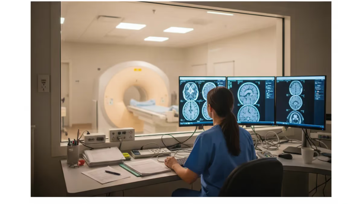
Would arthritis show up on MRI? The short answer is yes — and with considerably more detail than most patients and even some clinicians expect. MRI is uniquely capable of imaging every tissue layer affected by arthritis in a single exam: articular cartilage, subchondral bone, synovial membrane, joint fluid, ligaments, tendons, and surrounding muscles. Unlike X-ray, which can only show bone and indirect signs of cartilage loss through narrowed joint spaces, MRI directly visualizes soft-tissue pathology. That distinction matters enormously because arthritis begins in soft tissue long before bone damage becomes visible on plain film.
The two most common forms of arthritis — osteoarthritis (OA) and rheumatoid arthritis (RA) — each produce a recognizable pattern on MRI. Osteoarthritis tends to show focal cartilage thinning, subchondral bone marrow edema, osteophytes, and meniscal degeneration. Rheumatoid arthritis, by contrast, produces pannus formation (inflamed synovial tissue that erodes bone), bone marrow edema, tenosynovitis, and joint effusion. Distinguishing these patterns helps rheumatologists and radiologists tailor treatment strategies with precision that earlier imaging technologies simply could not offer.
One of the most clinically valuable aspects of MRI for arthritis is its ability to detect bone marrow edema — also called bone marrow lesions (BMLs). Bone marrow edema appears on MRI as bright signal on fluid-sensitive sequences before any structural damage is visible on X-ray or CT. Research published in major rheumatology journals has shown that BMLs in osteoarthritis strongly predict cartilage loss over the following one to two years, giving clinicians a meaningful window for early intervention. This predictive capacity makes MRI a powerful tool not just for diagnosis but for monitoring disease progression.
Synovitis — inflammation of the synovial lining — is another key arthritis finding that MRI detects with high sensitivity. On contrast-enhanced sequences, inflamed synovium lights up with gadolinium enhancement, distinguishing active inflammatory disease from chronic structural damage. This matters in clinical practice because a patient with mild X-ray changes but extensive synovitis on MRI may benefit from aggressive anti-inflammatory therapy that their plain-film findings alone would never justify. The ability to visualize synovitis has transformed how rheumatologists stage and treat early RA.
Cartilage assessment on MRI has advanced dramatically since the early days of the technique. Modern sequences such as T2 mapping, dGEMRIC (delayed gadolinium-enhanced MRI of cartilage), and T1rho imaging can quantify biochemical changes in cartilage matrix before gross morphological damage appears. These compositional MRI techniques detect proteoglycan depletion and collagen disorganization — the earliest biochemical signatures of cartilage breakdown.
While these advanced sequences are not yet routine in every clinical setting, they are increasingly used in research and at academic medical centers managing complex arthritis cases. To understand how advanced MRI sequences work in clinical settings, reading about will mri show arthritis through diffusion-weighted principles offers additional technical context.
The joints most commonly imaged for arthritis by MRI include the knee, hand, wrist, hip, shoulder, ankle, and spine. The knee is the most frequently studied joint because of the high prevalence of OA and the complex anatomy that MRI captures so well — including menisci, cruciate ligaments, and multiple cartilage surfaces. Hand and wrist MRI is especially important in rheumatoid arthritis, where early erosions at the metacarpophalangeal and wrist joints are among the first structural signs of disease and carry prognostic significance for long-term joint damage.
Patients sometimes wonder whether MRI is always necessary for arthritis evaluation. The answer depends on clinical context. When X-rays are sufficient for diagnosis and treatment planning — as in advanced OA with obvious joint space loss — MRI may not add actionable information. But when symptoms are disproportionate to plain-film findings, when inflammatory arthritis is suspected in early stages, or when surgical planning requires precise anatomical detail, MRI provides information that no other imaging modality can match. Understanding when MRI adds clinical value is key to using this powerful but costly tool appropriately.
MRI: upcoming dates and official changes
- Aug 17, 2026: ARRT launched a separate entry-level Imaging Assistant, Magnetic Resonance (I.A.) credential. A reduced $75 fee applied to applications made Aug. 17–Oct. 14, 2026, for testing Oct. 15–Nov. 30, 2026. Later applicants pay $225 and can test on or after Jan. 1, 2027.[4]
- The ARRT MRI exam for postprimary-pathway candidates has 220 questions: 200 scored and 20 unscored pilot items.[1]
- The ARRT MRI exam for primary-pathway candidates has 230 questions: 200 scored and 30 unscored pilot items.[2]
- Postprimary-pathway candidates get 210 minutes for the ARRT MRI exam, inside a 230-minute (3 hours, 50 minutes) appointment.[1]
- Primary-pathway candidates get 230 minutes for the ARRT MRI exam, inside a 250-minute (4 hours, 10 minutes) appointment that also covers the tutorial, the NDA and a survey.[2]
- To pass the ARRT MRI exam you need a total scaled score of 75 on ARRT's 1–99 scale. This is not a percentage.[3]
- The ARRT MRI exam through the postprimary pathway costs $225, or $450 for candidates whose supporting credential is from NMTCB or ARDMS.[4]
- The ARRT MRI certification exam costs $225 to apply for through the primary eligibility pathway.[4]
- ARRT MRI candidates have three attempts to pass the exam, all within three years of the date their first exam window opens.[2]
- ARRT MRI certification and registration lasts 10 years, even with annual renewal. After that, holders must complete Continuing Qualifications Requirements (CQR).[5]
Numbers in brackets link to the official sources listed under Sources & last verified. Dates that have passed are removed automatically.
MRI Exam at a Glance
| Item | Detail | Source · Verified |
|---|---|---|
| Number of questions (Postprimary pathway) | 220 questions (200 scored + 20 unscored pilot)[1] | assets-us-01.kc-usercontent.com · Oct 15, 2026 |
| Number of questions (Primary pathway) | 230 questions (200 scored + 30 unscored pilot)[2] | assets-us-01.kc-usercontent.com · Oct 15, 2026 |
| Time limit (Postprimary pathway) | 210 minutes test time (230-minute appointment)[1] | assets-us-01.kc-usercontent.com · Oct 15, 2026 |
| Time limit (Primary pathway) | 230 minutes test time (250-minute appointment)[2] | assets-us-01.kc-usercontent.com · Oct 15, 2026 |
| Passing score | Scaled score of 75 (scale 1–99)[3] | The American Registry of Radiologic Technologists · Oct 15, 2026 |
| Exam fee (Postprimary pathway) | $225 application fee ($450 with an NMTCB or ARDMS supporting credential)[4] | The American Registry of Radiologic Technologists · Oct 15, 2026 |
| Exam fee (Primary pathway) | $225 application fee[4] | The American Registry of Radiologic Technologists · Oct 15, 2026 |
| Retake policy | 3 attempts within 3 years of the first exam window[2] | assets-us-01.kc-usercontent.com · Oct 15, 2026 |
| Validity | 10 years, then Continuing Qualifications Requirements (CQR)[5] | assets-us-01.kc-usercontent.com · Oct 15, 2026 |
| Renewal | Annual renewal, $65/year; 24 CE credits every 2 years[5] | assets-us-01.kc-usercontent.com · Oct 15, 2026 |
| Delivery | Computer-based at Pearson VUE test centers[2] | assets-us-01.kc-usercontent.com · Oct 15, 2026 |
| Eligibility (Postprimary pathway) | Supporting ARRT credential (or NMTCB/ARDMS) + structured education + clinical experience[1] | assets-us-01.kc-usercontent.com · Oct 15, 2026 |
| Eligibility (Primary pathway) | ARRT-recognized MRI program + associate degree or higher + ethics[2] | assets-us-01.kc-usercontent.com · Oct 15, 2026 |
| Score release | Preliminary score at the test center; official results in about 10 business days[3] | The American Registry of Radiologic Technologists · Oct 15, 2026 |
| Testing window | One-year (365-day) exam window per application[2] | assets-us-01.kc-usercontent.com · Oct 15, 2026 |
Free MRI - Magnetic Resonance Imaging Study Tools
Interactive MRI - Magnetic Resonance Imaging resources on PracticeTestGeeks, free to start:
- MRI - Magnetic Resonance Imaging practice test — 200+ questions with instant scoring and answer explanations
- MRI - Magnetic Resonance Imaging flashcards — review key terms and concepts card by card
- MRI - Magnetic Resonance Imaging study guide — topic-by-topic overview of what the exam covers
- MRI - Magnetic Resonance Imaging cheat sheet — one-page printable summary for last-minute review
MRI and Arthritis by the Numbers
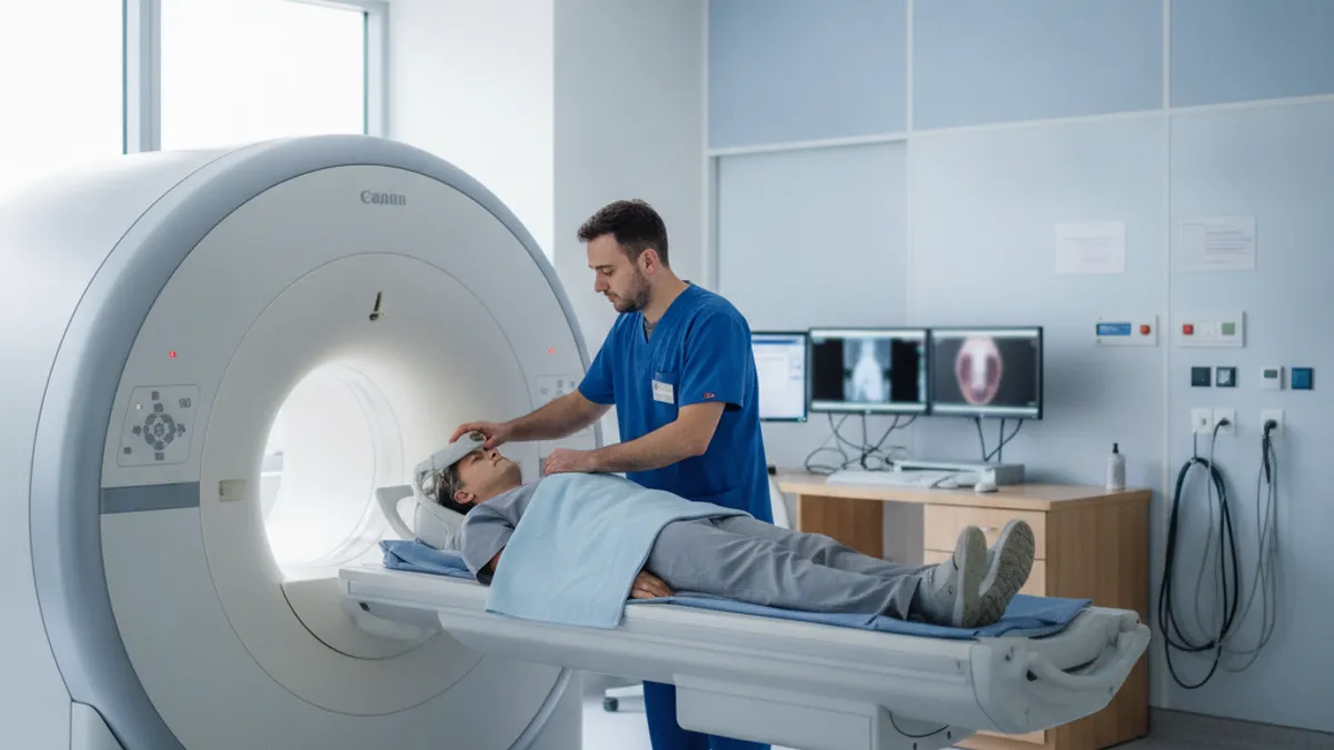
Types of Arthritis MRI Can Detect and Characterize
MRI shows focal cartilage loss, subchondral bone marrow edema, osteophytes, meniscal tears, and joint effusion. Bone marrow lesions on MRI predict cartilage loss 1–2 years in advance, making early detection clinically meaningful.
MRI detects synovitis, pannus formation, bone erosions, tenosynovitis, and joint effusion. Gadolinium contrast enhances synovial inflammation, distinguishing active RA from chronic damage and guiding treatment escalation decisions.
MRI reveals enthesitis (inflammation at tendon insertions), dactylitis, sacroiliitis, and bone erosions. It differentiates PsA from RA by the distribution and character of joint involvement across axial and peripheral joints.
MRI identifies tophi, bone erosions with overhanging edges, and joint effusion. While CT is superior for crystal detection, MRI better characterizes soft-tissue involvement and associated bone marrow edema around gouty deposits.
MRI of the sacroiliac joints detects early bone marrow edema and inflammation before plain X-rays show structural changes, enabling earlier diagnosis and treatment initiation for this chronic spinal arthritis form.
MRI findings differ substantially depending on which joint is being examined, because each joint has unique anatomy and arthritis affects different structures in each location. The knee, as the largest and most frequently imaged joint for arthritis, illustrates the full range of MRI findings. In osteoarthritis of the knee, radiologists systematically evaluate the medial and lateral compartments of the femorotibial joint as well as the patellofemoral compartment. Cartilage grading using the MOAKS (MRI Osteoarthritis Knee Score) or WORMS (Whole-Organ MRI Score) systems provides standardized assessments of cartilage loss, bone marrow lesions, meniscal damage, synovitis, and osteophytes across multiple anatomical regions.
Hand and wrist MRI in rheumatoid arthritis focuses on detecting early erosions — small cortical breaks in bone — before they are visible on X-ray. The metacarpophalangeal (MCP) joints, particularly MCP2 and MCP3, are the most common early erosion sites. The carpus is also frequently involved. Studies comparing MRI and radiography in early RA have shown that MRI detects erosions two to five years earlier than X-ray, a finding with major therapeutic implications. Early detection allows disease-modifying antirheumatic drugs (DMARDs) to be started before irreversible joint destruction occurs.
Hip arthritis presents unique MRI challenges because the hip is a deep joint surrounded by thick muscle. However, MRI excels at showing acetabular and femoral head cartilage loss, labral tears that often accompany hip OA, bone marrow edema patterns, and early osteonecrosis — a complication that mimics and can coexist with inflammatory arthritis. In patients with hip pain where X-ray shows only mild changes, MRI frequently reveals significant pathology that changes the clinical management plan, including decisions about cortisone injection versus surgical consultation.
Sacroiliac joint MRI is essential in the workup of axial spondyloarthritis, including ankylosing spondylitis. The ASAS (Assessment of SpondyloArthritis International Society) criteria specify that bone marrow edema on MRI at sacroiliac joints, when present in a characteristic distribution and above a defined threshold, constitutes a positive MRI for sacroiliitis. This criterion is now embedded in international diagnostic guidelines, meaning that MRI has moved from a supplementary tool to a diagnostic cornerstone for axial spondyloarthritis in patients who lack the classic X-ray changes of late-stage disease.
Shoulder MRI for arthritis evaluates the glenohumeral joint and acromioclavicular joint. Glenohumeral OA on MRI shows posterior cartilage loss with posterior glenoid bone erosion and biconcave glenoid deformity. Rotator cuff tears, which frequently coexist with shoulder arthritis, are definitively characterized on MRI — an important consideration because the surgical approach differs depending on whether the cuff is intact. Inflammatory arthritis of the shoulder, including RA, shows synovitis, bone erosions at the humeral head margin, and subacromial bursitis, all visible on MRI with far greater clarity than on X-ray.
Ankle and foot MRI has grown in importance for inflammatory arthritis evaluation, particularly RA and psoriatic arthritis. The metatarsophalangeal (MTP) joints of the foot are among the earliest and most common sites of RA erosions, yet plain X-rays of this region have limited sensitivity for early erosive disease. MRI of the foot in early RA can show erosions, bone marrow edema, and tenosynovitis of the peroneal and posterior tibial tendons — findings that correlate with disease activity and help guide systemic therapy decisions when clinical examination alone is insufficient.
Spinal arthritis, including facet joint osteoarthritis and inflammatory spondyloarthropathy, is well characterized on MRI. Facet joint OA on lumbar or cervical spine MRI shows cartilage loss, subchondral sclerosis, synovial cysts, and facet joint effusion. These findings help explain radicular pain patterns and guide injection therapy. In inflammatory conditions affecting the spine, MRI shows discovertebral lesions (Romanus lesions), posterior element involvement, and syndesmophyte formation at a stage when clinical symptoms may precede structural X-ray changes by years. Learning more about spinal arthritis MRI findings is an important complement to understanding peripheral joint imaging.
MRI Sequences Used to Evaluate Arthritis
The workhorse sequences for arthritis MRI are proton density (PD) fat-saturated and T2 fat-saturated sequences, which highlight fluid and edema with bright signal. These sequences are excellent for detecting joint effusion, bone marrow edema, and cartilage surface irregularities. T1-weighted sequences, without fat suppression, provide high contrast between bone marrow fat and abnormalities like erosions and bone marrow edema, making them ideal for identifying early cortical breaks at joint margins.
STIR (Short Tau Inversion Recovery) sequences offer strong fat suppression across the entire field of view and are valuable in body parts where magnetic field inhomogeneity limits spectral fat saturation. STIR is particularly useful for detecting sacroiliac joint inflammation in ankylosing spondylitis and for whole-body MRI protocols used in inflammatory arthritis staging. A standard joint arthritis protocol typically combines T1, PD fat-sat, and T2 fat-sat sequences in at least two planes — usually coronal and sagittal or axial — providing complementary views of all affected structures.
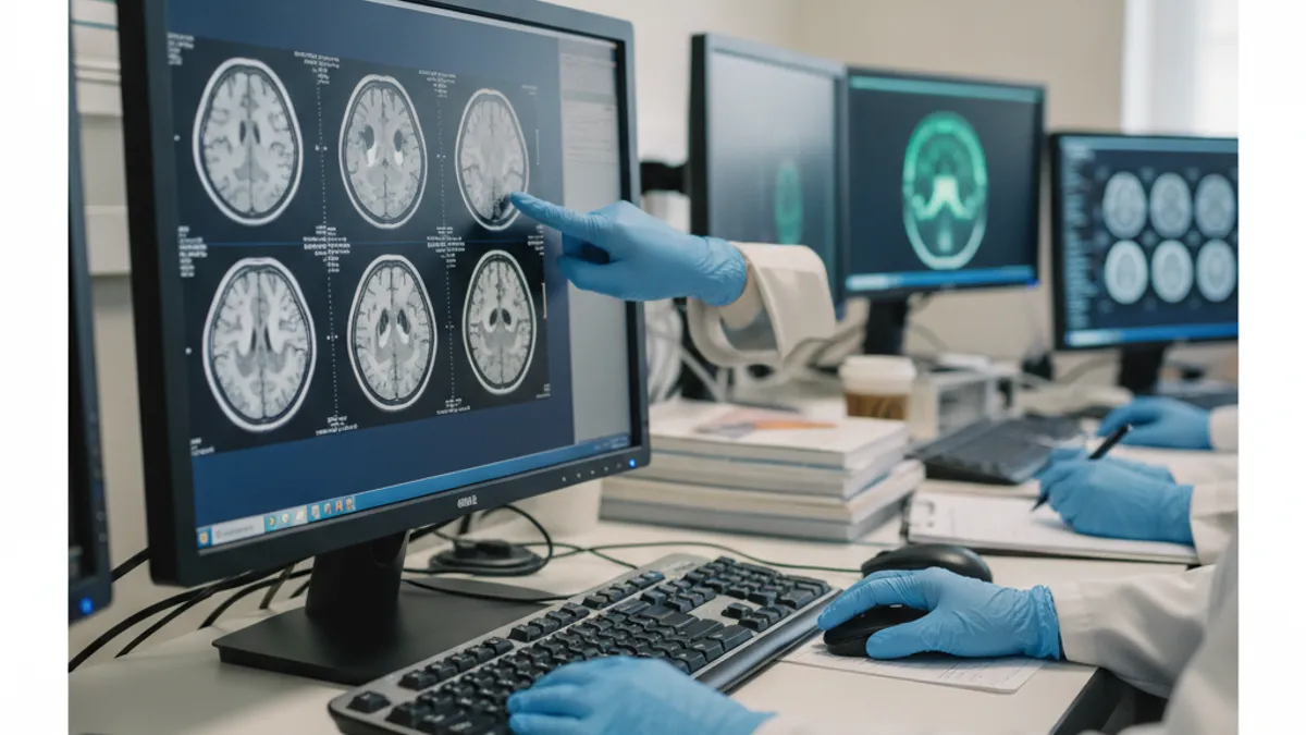
MRI for Arthritis: Advantages and Limitations
- +Detects bone marrow edema and synovitis years before X-ray shows abnormality
- +Visualizes articular cartilage directly — not just inferred from joint space width
- +No ionizing radiation, safe for repeated monitoring over time
- +Evaluates multiple tissue types (cartilage, bone, synovium, tendons, ligaments) in one exam
- +Gadolinium contrast quantifies active synovitis to guide anti-inflammatory treatment
- +Advanced sequences (T2 mapping, T1rho) detect biochemical cartilage changes pre-structurally
- −Significantly more expensive than X-ray or ultrasound ($500–$3,000 without insurance)
- −Scan duration is 45–90 minutes, which may be difficult for patients with severe joint pain
- −Contraindicated in patients with certain metallic implants, pacemakers, or cochlear devices
- −Claustrophobia affects 5–10% of patients and may require sedation or open-bore MRI
- −Gadolinium contrast carries a small risk in patients with severely impaired kidney function
- −Incidental findings (such as meniscal tears in asymptomatic knees) may lead to unnecessary procedures
Pros and Cons at a Glance
| Pros | Cons |
|---|---|
| Detects bone marrow edema and synovitis years before X-ray shows abnormality | Contraindicated in patients with certain metallic implants, pacemakers, or cochlear devices |
| Visualizes articular cartilage directly — not just inferred from joint space width | Gadolinium contrast carries a small risk in patients with severely impaired kidney function |
| No ionizing radiation, safe for repeated monitoring over time | Incidental findings (such as meniscal tears in asymptomatic knees) may lead to unnecessary procedures |
How to Prepare for an Arthritis MRI: Patient Checklist
- ✓Notify your ordering physician and imaging center of all metallic implants, including joint replacements and surgical clips.
- ✓Remove all jewelry, piercings, and hair accessories before entering the MRI suite.
- ✓Inform the technologist if you have a pacemaker, neurostimulator, cochlear implant, or insulin pump.
- ✓Tell your doctor about all medications — some affect joint fluid signal and may need to be noted for the radiologist.
- ✓If gadolinium contrast is ordered, confirm your kidney function is adequate (recent creatinine/eGFR lab values may be required).
- ✓Wear comfortable, metal-free clothing or expect to change into a hospital gown provided by the facility.
- ✓Arrive 15–20 minutes early to complete screening paperwork and answer safety screening questions.
- ✓Tell the technologist if you experience claustrophobia — anxiolytic premedication or open-bore MRI can be arranged in advance.
- ✓Avoid putting lotion, deodorant, or sunscreen on the joint being imaged, as some products contain metallic particles.
- ✓Bring your prior imaging (X-rays, previous MRIs, ultrasound reports) on CD or via electronic access for comparison.
MRI Detects Arthritis Damage 2–5 Years Before X-Ray
Studies in early rheumatoid arthritis consistently show that bone erosions visible on MRI appear two to five years before they become detectable on plain radiography. Starting disease-modifying therapy during this window — before irreversible structural damage occurs — significantly improves long-term joint function and quality of life. If your X-ray looks normal but you have persistent joint pain and swelling, an MRI of the affected joint may reveal early inflammatory changes that completely change your treatment plan.
Comparing MRI with other imaging modalities for arthritis evaluation helps clarify when each tool is most appropriate. Plain radiography remains the first-line imaging study for most arthritis workups because it is inexpensive, fast, widely available, and provides an excellent overview of joint alignment, bone density, osteophytes, and joint space width. For moderate-to-advanced osteoarthritis where the diagnosis is not in doubt and treatment decisions are primarily clinical, X-ray may be all that is needed. However, X-ray's fundamental limitation — its inability to image soft tissue — means it will always miss the earliest and most therapeutically relevant arthritis findings.
Ultrasound has emerged as an important complementary tool in inflammatory arthritis, particularly rheumatoid arthritis. Point-of-care ultrasound performed by the rheumatologist during the clinic visit can detect synovial hypertrophy, joint effusion, and power Doppler signal (indicating blood flow in inflamed synovium) in real time. Ultrasound is less expensive than MRI, involves no radiation, and allows dynamic assessment during joint movement. However, ultrasound is operator-dependent, limited by body habitus and acoustic windows, cannot evaluate bone marrow pathology, and provides an incomplete picture of joint structures compared with MRI.
CT (computed tomography) excels at visualizing cortical bone detail. In arthritis, CT is most valuable for quantifying the degree of bone erosion in gout (tophi appear as high-density deposits) and for pre-surgical planning in joints like the hip, shoulder, and wrist where precise bone geometry is needed. Dual-energy CT can actually detect uric acid crystals in gouty tophi — a capability that MRI lacks. However, CT uses ionizing radiation, and its inability to visualize cartilage and soft tissue directly limits its utility in most arthritis evaluations compared with MRI.
Nuclear medicine studies — bone scan, PET, and SPECT — detect increased metabolic activity in bone and have some role in arthritis evaluation, primarily for whole-body surveys of polyarticular disease activity. Bone scan shows increased uptake at inflamed joints but lacks specificity and anatomical resolution. PET-CT with FDG tracer can demonstrate synovitis in inflammatory arthritis and is occasionally used in research settings. However, nuclear medicine techniques involve radiation exposure, are less widely available, and cannot match MRI's structural detail for clinical arthritis management.
The practical decision between MRI and alternatives often comes down to three factors: clinical question, cost-benefit ratio, and contraindications. When the question is simply whether arthritis is present and how advanced it is structurally, X-ray is often sufficient.
When the question involves soft tissue (is the rotator cuff intact in a shoulder with arthritis?), bone marrow (is there osteonecrosis?), or synovial activity (is this RA active enough to justify adding a biologic?), MRI provides the answer. Insurance coverage is also a real consideration — most US insurance plans cover MRI for arthritis evaluation when ordered with appropriate clinical justification and after X-ray has been obtained.
Whole-body MRI (WBMRI) is an evolving technique for systemic inflammatory arthritis assessment. By imaging the entire axial and peripheral skeleton in a single session, WBMRI can map disease distribution in polyarticular RA, PsA, and AS without the cumulative radiation of multiple X-rays. WBMRI protocols have been validated for detecting active inflammatory lesions and structural damage across multiple joints simultaneously, providing a comprehensive picture of disease burden. While not yet standard of care, WBMRI is increasingly used in academic rheumatology practices and in clinical trials assessing systemic therapy for inflammatory arthritis.
MRI also plays a critical role in guiding joint procedures for arthritis management. MRI-guided or MRI-planned injections use pre-procedure imaging to precisely identify the area of maximal synovitis for targeted corticosteroid or hyaluronic acid delivery. In small joints of the hand or wrist, or in anatomically complex joints like the sacroiliac joint, MRI findings guide injection approach and needle placement, improving accuracy beyond what fluoroscopy alone can achieve. For patients considering surgery, MRI provides the anatomical roadmap that surgeons use to plan arthroscopic lavage, osteotomy, partial or total joint replacement, and cartilage restoration procedures.
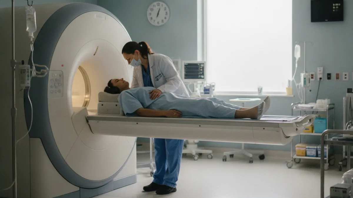
MRI is typically ordered for arthritis when X-rays are normal but symptoms persist, when inflammatory arthritis is suspected in early stages, when surgical planning requires precise anatomical detail, or when treatment response to disease-modifying therapy needs objective monitoring. Always discuss with your rheumatologist or orthopedic physician whether MRI is indicated in your specific situation — appropriate clinical ordering maximizes both diagnostic yield and insurance coverage likelihood.
Interpreting an MRI report for arthritis requires understanding the standardized language radiologists use to describe findings. Most arthritis MRI reports follow a structured format that systematically evaluates each relevant tissue compartment. For a knee OA report, you might see graded assessments of medial and lateral compartment cartilage (typically using a 0–4 scale where 0 is normal and 4 is full-thickness loss), descriptions of bone marrow lesions by location and approximate volume, meniscal grading, synovial effusion severity, and osteophyte burden. Understanding these elements helps patients engage meaningfully with their rheumatologist or orthopedic surgeon about the significance of the findings.
Bone marrow lesions (BMLs) described on an arthritis MRI report deserve particular attention. BMLs in osteoarthritis are associated with pain — large BMLs at weight-bearing surfaces correlate with joint pain severity in multiple studies — and with accelerated cartilage loss over time. When a radiology report notes prominent subchondral bone marrow edema, this finding should prompt a discussion about weight-bearing modification, physical therapy targeting joint mechanics, and potentially anti-inflammatory treatment, even if the structural cartilage damage appears mild. BMLs are not incidental findings; they are active pathological signals in the arthritis disease process.
Synovitis grading in MRI reports uses standardized scoring systems depending on the joint. In the knee, synovitis may be graded using Hoffa's fat pad signal, medial and lateral synovial recess thickening, and popliteal recess fluid. In hand and wrist RA MRI reports, the RAMRIS (RA MRI Scoring system) provides separate scores for synovitis, bone marrow edema, and erosions at defined anatomical sites. When your rheumatologist receives a RAMRIS-scored MRI report, they can track disease progression or treatment response numerically over time — a major advantage over subjective descriptive reports that make comparison difficult.
Cartilage grading nomenclature varies between reporting systems. The Outerbridge classification, originally developed for arthroscopy but adapted to MRI, grades cartilage from Grade 0 (normal) to Grade 4 (full-thickness loss with exposed subchondral bone). The ICRS (International Cartilage Repair Society) system similarly grades cartilage defects 0–4. When a report uses terms like "Grade 3 chondromalacia" or "partial thickness cartilage defect involving more than 50% of depth," these indicate significant but not complete cartilage loss — findings that typically require clinical correlation to determine whether conservative management or procedural intervention is more appropriate.
Bone erosions described in inflammatory arthritis MRI reports are defined as cortical breaks visible in at least two planes, with corresponding bone marrow signal change. Early erosions may be tiny — one or two millimeters — and easily missed on X-ray but clearly visible on high-resolution MRI sequences.
When erosions are identified, their number, size, and location relative to the joint surface are reported, because these parameters influence prognosis and the aggressiveness of antirheumatic therapy. A patient with multiple erosions at multiple MCP joints early in their RA course has a far more aggressive disease phenotype than one with synovitis but no erosions.
Incidental findings on arthritis MRI are common and require careful clinical contextualization. Meniscal tears found on knee MRI ordered for arthritis are present in the majority of adults over 50 years of age and are usually degenerative rather than traumatic.
Isolated meniscal tears in the context of knee OA do not routinely require arthroscopic surgery — a landmark finding from the METEOR and ESCAPE clinical trials demonstrated that arthroscopic partial meniscectomy provided no functional benefit over physical therapy in patients with OA and meniscal tears. Understanding which incidental findings are clinically significant versus anatomical variants is an important part of the radiologist-clinician dialogue around arthritis MRI interpretation.
Follow-up MRI timing for arthritis monitoring depends on the disease type and treatment context. In active rheumatoid arthritis being treated with biologic therapy, repeat MRI at 12 months can quantify changes in synovitis score and erosion burden, providing objective evidence of treatment response or failure.
For osteoarthritis being monitored conservatively, repeat MRI every one to two years may track disease progression in patients enrolled in research protocols, though routine surveillance MRI is not standard clinical practice. When symptoms change significantly — new severe pain, joint locking, or rapid functional decline — repeat MRI sooner than the planned interval is clinically appropriate to rule out new pathology like a displaced meniscal tear or osteonecrosis progression.
Practical tips for getting the most out of your arthritis MRI start well before the scan itself. When your physician orders the MRI, ask specifically whether the protocol is optimized for arthritis evaluation. General joint MRI protocols are designed to rule out acute injury (ligament tears, fractures) and may not include the dedicated sequences needed for detailed cartilage assessment or synovitis detection. Requesting that the ordering physician specify arthritis as the clinical indication ensures the radiologist selects the appropriate protocol — for example, adding gadolinium contrast for suspected inflammatory arthritis or specifying multi-planar cartilage sequences for suspected OA.
Choosing the right MRI facility matters more for arthritis imaging than for some other MRI indications. Higher field strength scanners (3 Tesla versus the older 1.5 Tesla standard) provide substantially better spatial resolution and signal-to-noise ratio for small joint imaging — especially hands, wrists, and feet. A 3T MRI can resolve cartilage defects and bone erosions that a 1.5T scanner may miss at small joints.
If you have inflammatory arthritis affecting small joints, asking whether a 3T scanner is available and requesting that facility is worthwhile. Dedicated extremity MRI units, which use a lower field strength but allow the affected limb to be positioned inside a smaller, open coil rather than a full-body bore, are also excellent for hand and wrist arthritis evaluation and are more comfortable for patients with severe joint pain.
Positioning and immobility during the scan significantly affect image quality. Joint motion during MRI acquisition causes blurring artifacts that degrade cartilage assessment and can obscure small erosions. Patients should be positioned comfortably with padding at pressure points to minimize the urge to move, and should be counseled to remain as still as possible throughout each sequence. For patients with severe pain that makes immobility difficult, asking about a pre-scan analgesic — a non-sedating option taken an hour before the appointment — may improve image quality by reducing pain-driven motion.
After the MRI, follow up with your ordering physician to review the results together rather than reading the radiology report alone. Radiology reports are written for clinicians, not patients, and the clinical significance of findings is highly context-dependent.
A finding described as "moderate cartilage loss in the medial compartment" may be an expected age-related finding in a 65-year-old with mild symptoms, or it may be the explanation for debilitating pain in a 45-year-old — the radiologist cannot determine clinical significance without full clinical context, which is why physician interpretation is essential. Bring your report to your rheumatologist or orthopedic physician and ask specifically how each major finding affects your treatment plan.
If your MRI shows arthritis findings that surprise you — especially if you have been told your X-rays are normal — understand that this is not unusual and reflects MRI's superior sensitivity rather than an error. Patients sometimes receive an MRI diagnosis of significant bone marrow edema or synovitis when they had assumed their pain was "just getting older." This early detection is precisely the value of MRI — it identifies pathology at a stage when intervention is most likely to slow or halt disease progression. An unexpected positive MRI is an opportunity, not a crisis.
Physical activity modifications based on MRI findings are an important practical consideration. Large bone marrow lesions at weight-bearing surfaces in knee OA are exacerbated by high-impact activity. Evidence from biomechanics research shows that lateral wedge insoles, gait retraining to reduce knee adduction moment, and water-based exercise that minimizes joint loading can reduce BML volume and associated pain. Sharing your MRI findings with a physical therapist specializing in musculoskeletal conditions allows them to tailor exercise programming to your specific joint pathology in a way that generic OA exercise programs cannot.
Finally, staying informed about evolving MRI technology for arthritis is worthwhile for patients with chronic joint disease. Artificial intelligence-assisted MRI analysis — using machine learning algorithms to automatically segment cartilage, quantify bone marrow lesion volume, and score synovitis — is entering clinical practice. These AI tools promise faster, more reproducible, and more comprehensive arthritis MRI analysis than manual radiologist assessment.
As these tools become widely available, they may enable cost-effective serial MRI monitoring of arthritis progression, transform how treatment response is measured in clinical practice, and ultimately help more patients receive timely, targeted arthritis care based on objective imaging biomarkers rather than symptom-based assessment alone.
For a fuller set of questions on this topic, see our mri abdomen and pelvis cpt code resource.
For a fuller set of questions on this topic, see our open mri near me resource.
Sample MRI - Magnetic Resonance Imaging Practice Questions
Try these questions from our free MRI - Magnetic Resonance Imaging practice tests. The correct answer and an explanation follow each question.
Which parameter, when increased, will most directly improve the signal-to-noise ratio (SNR) in an MRI image?
- A. Increasing the number of excitations (NEX/NSA)
- B. Increasing the echo train length (ETL)
- C. Decreasing the field of view (FOV)
- D. Increasing the bandwidth
Answer: A. Increasing the number of excitations (NEX/NSA)
Increasing NEX/NSA averages more signal acquisitions, which reduces noise and improves SNR proportional to the square root of the number of averages.
Which artifact appears as parallel bright and dark bands in the phase-encoding direction when the FOV is too small and the patient extends beyond it?
- A. Gibbs ringing
- B. Aliasing (wraparound)
- C. Chemical shift type 2
- D. Susceptibility artifact
Answer: B. Aliasing (wraparound)
When anatomy outside the FOV is phase-encoded, it aliases (wraps around) and appears as a superimposed copy on the opposite edge of the image, sometimes appearing as bands.
What is the primary advantage of a 3T MRI scanner compared to a 1.5T scanner?
- A. Shorter T1 relaxation times for all tissues
- B. Higher SNR that can be traded for faster scans or higher spatial resolution
- C. Elimination of susceptibility artifacts
- D. Lower SAR deposition in patients
Answer: B. Higher SNR that can be traded for faster scans or higher spatial resolution
Field strength doubling theoretically doubles SNR, which can be reinvested in thinner slices, smaller voxels, or reduced scan time.
Which of the following changes will increase spatial resolution without changing the FOV?
- A. Increasing the matrix size
- B. Decreasing the matrix size
- C. Increasing slice thickness
- D. Increasing NEX
Answer: A. Increasing the matrix size
Increasing matrix size means more phase and frequency encoding steps, reducing voxel size and improving spatial resolution within the same FOV.
Take the full MRI - Magnetic Resonance Imaging practice test
MRI Questions and Answers
Sources & last verified
- assets-us-01.kc-usercontent.com — number of questions, time limit, eligibility (official source). “MRI Scored Items 200 Pilot Items 20 Total Items 220 Test Time 210 min” Verified Oct 15, 2026.
- assets-us-01.kc-usercontent.com — number of questions, time limit, retake policy, delivery (official source). “MRI Scored Items 200 Pilot Items 30 Total Items 230 Test Time 230 min” Verified Oct 15, 2026.
- The American Registry of Radiologic Technologists — passing score, score release (official source). “Our exam scoring scale ranges from 1 to 99, and you’ll need a total score of 75 to pass the exam.” Verified Oct 15, 2026.
- The American Registry of Radiologic Technologists — exam fee, other (official source). “Postprimary (M, CT, MR, BD, CI, VI, VS, BS, R.R.A.): $225 Postprimary (CT, MR, BD, or VS—using NMTCB as supporting category): $450 Postprimary (MR, VS,…” Verified Oct 15, 2026.
- assets-us-01.kc-usercontent.com — validity, renewal (official source). “ARRT certification and registration—even with annual renewal—is limited to 10 years.” Verified Oct 15, 2026.
How we verify: editorial & fact-checking policy. Report an error: contact us.
DWI MRI: Diffusion-Weighted Imaging Explained (2026 Guide)
MRI Lumbar Spine Without Contrast: Complete Guide to the Scan, What It Shows, and How to Prepare
MRI History: From Nuclear Resonance to Modern Scanners

MRI of Cervical Spine: Complete Guide to Imaging, Anatomy, Pathology, and Patient Preparation
Where to Get an MRI: Facilities, Costs, and How to Choose
About the Author

Medical Laboratory Scientist & Clinical Certification Expert
Johns Hopkins UniversityDr. Sandra Kim holds a PhD in Clinical Laboratory Science from Johns Hopkins University and is certified as a Medical Technologist (MT) and Medical Laboratory Scientist (MLS) through ASCP. With 16 years of clinical laboratory experience spanning hematology, microbiology, and molecular diagnostics, she prepares candidates for ASCP board exams, MLT, MLS, and specialist certification tests.