
Video EEG: What It Is, How It Works, and What to Expect During Your EEG Test
Learn what a video EEG test is, how long it takes, what it costs, and what to expect. ✏️ Complete guide for patients and EEG students.
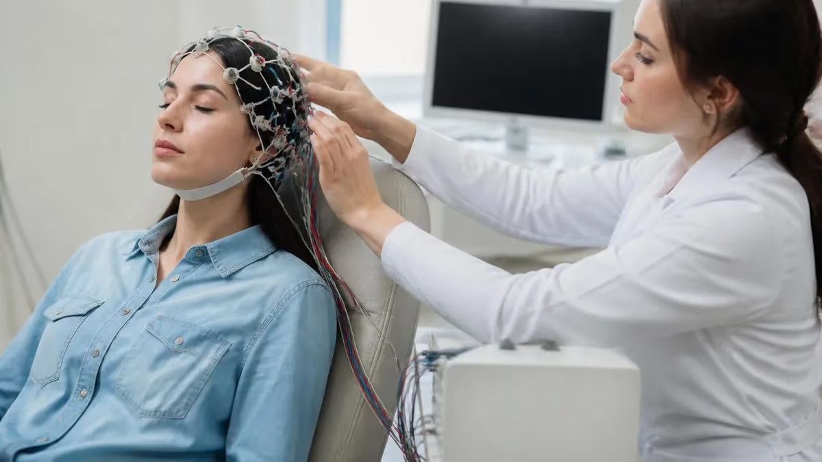
An EEG test — short for electroencephalogram — records the electrical activity of your brain through small sensors placed on the scalp. Among the most clinically powerful versions of this technology is video EEG, a specialized monitoring setup that simultaneously captures brain wave data and synchronized video footage of the patient. This dual-channel approach allows neurologists to match what the brain is doing electrically with what the body is doing physically, making it far easier to classify seizure types, rule out non-epileptic events, and determine the right course of treatment for complex neurological conditions.
Video EEG monitoring is increasingly common in epilepsy centers across the United States, and understanding what the test involves can help patients and families prepare more effectively. The EEG medical test itself is painless — electrodes conduct electricity but do not deliver any shock or stimulation. The challenge for most patients is simply the duration and the constraints of staying connected to monitoring equipment. Inpatient video EEG sessions can last anywhere from 24 hours to several weeks, depending on how long it takes to capture a representative seizure event on camera and on the electrodes simultaneously.
For EEG technologists working in clinical settings, video EEG represents one of the most technically demanding and professionally rewarding areas of practice. Technologists must maintain electrode integrity over extended periods, respond to clinical events at any hour, and document findings with precision. If you are curious about how compensation compares across these specialized roles, the video eeg technologist salary landscape varies significantly by state, certification level, and care setting.
This article explains what an EEG test is at a fundamental level, how video monitoring adds diagnostic power, what the procedure feels like from a patient perspective, how long the test typically runs, and what the test costs across different clinical environments. Whether you are a patient preparing for monitoring, a family member seeking to understand a loved one's upcoming procedure, or a student studying for EEG certification examinations, the information below is organized to answer the questions that matter most about this important diagnostic tool.
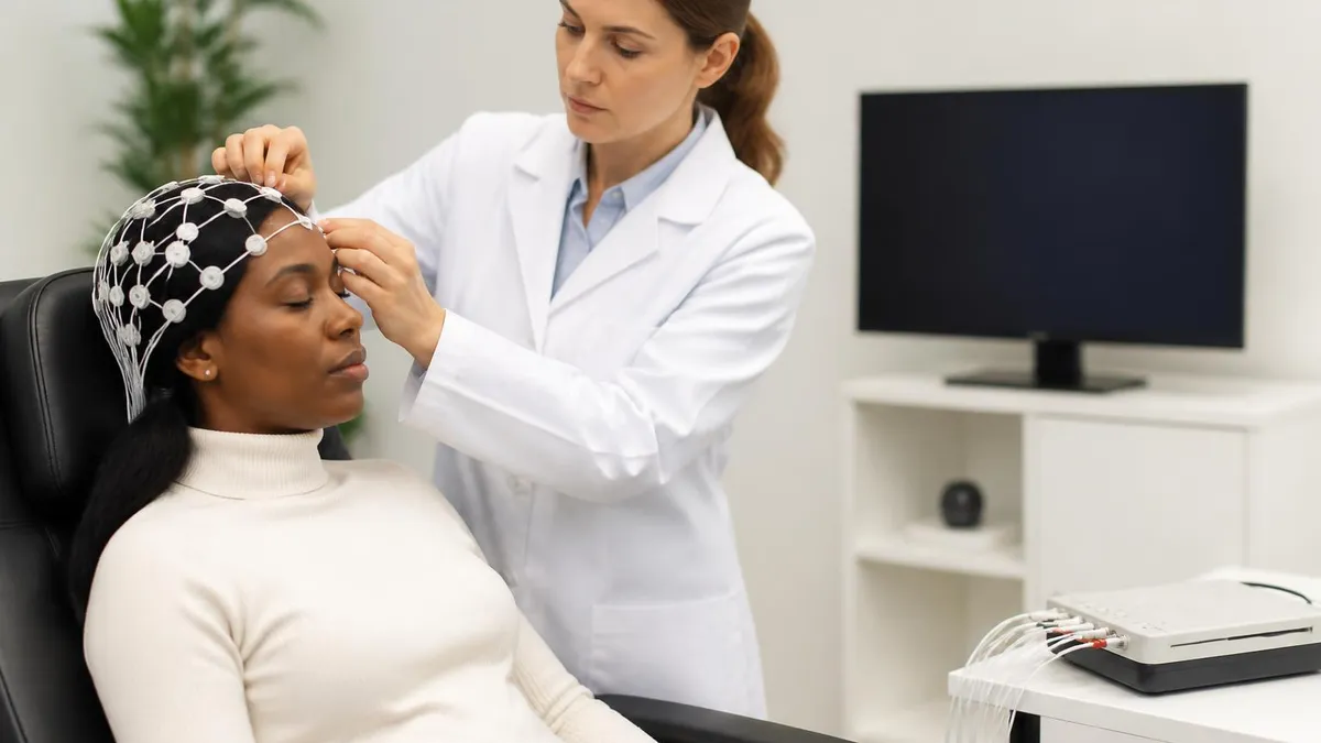
The EEG test captures brain wave patterns by measuring voltage differences between pairs of electrodes placed according to standardized maps such as the international 10-20 system. During a video EEG, those electrical signals are displayed in real time on a technologist's workstation while one or more cameras continuously record the patient.
When a clinical event occurs — a stare, a jerk, a fall, an unusual behavior — the video and the EEG tracing can be reviewed side by side, frame by frame. This level of detail allows epileptologists to make diagnostic distinctions that a routine outpatient EEG, which typically lasts only 20 to 40 minutes, simply cannot provide.
Patients are often surprised to learn how many conditions can mimic seizures without actually involving abnormal brain electrical activity. Psychogenic non-epileptic seizures, syncope, movement disorders, and even normal sleep behaviors can all look alarming and raise concern for epilepsy.
Video EEG is the gold standard test for distinguishing true epileptic seizures from these mimics because it captures both the behavioral and the electrical signatures of an event at the same moment. Without synchronized video, a technologist reviewing an EEG tracing can identify abnormal discharges but may not be able to confirm whether a clinical event actually occurred during that same window.
Understanding the full scope of the EEG test — from electrode placement to waveform interpretation to the nuances of video correlation — is also the foundation of EEG technologist certification exams. The questions on those exams frequently address normal and abnormal patterns, activation procedures, and monitoring techniques including ambulatory and video-based approaches. Reviewing practice questions across all of these domains is one of the most efficient ways to build both knowledge and confidence before sitting for a credentialing examination.
Video EEG by the Numbers
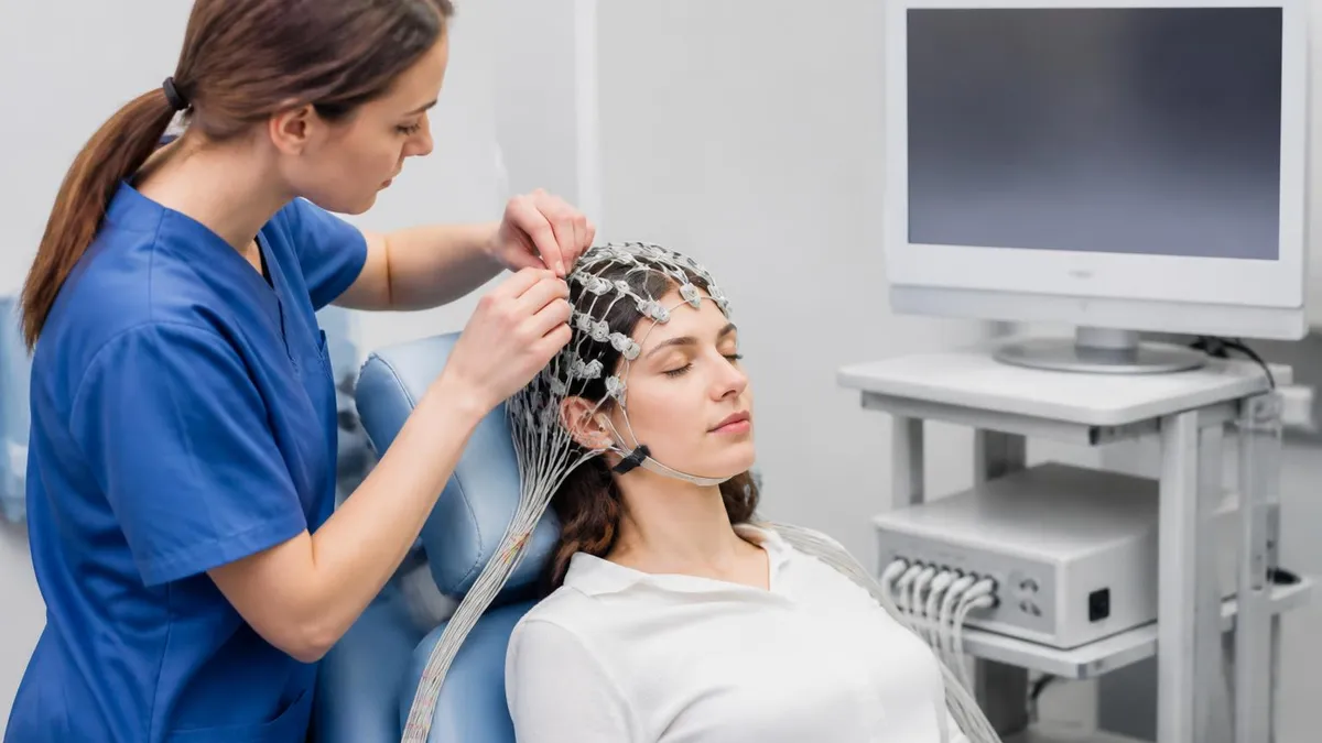
How Video EEG Works: Step by Step
Admission and Setup
Camera Positioning
Continuous Monitoring
Activation Procedures
Clinical Event Documentation
Review and Interpretation
One of the most common questions patients ask before scheduling an EEG medical test is simply: how long is an EEG test? The honest answer is that it depends enormously on the type of EEG being performed. A routine outpatient EEG — the kind ordered to evaluate a first-time seizure, assess a headache disorder, or check for encephalopathy — typically runs between 20 and 40 minutes.
The technologist applies electrodes, records resting brain activity, performs activation procedures such as hyperventilation and photic stimulation, and then removes the electrodes. From arrival to discharge, most patients spend one to two hours at the testing facility.
Sleep-deprived EEG tests are a step up in duration. Because sleep and the transitions in and out of sleep are powerful activators of epileptiform abnormalities, neurologists often order patients to stay awake for 24 hours before a routine EEG to increase the chance of capturing a sleep stage during the recording. The test itself is still only 40 to 60 minutes long, but the preparation requirement makes it a much more taxing experience. Some patients fall asleep naturally during the recording, which is exactly what the neurologist wants to see.
Ambulatory EEG testing extends the recording window to 24, 48, or 72 hours while allowing the patient to remain at home and continue their daily routine. A portable recorder attached to a belt or worn in a small backpack collects data continuously. The tradeoff is that ambulatory studies typically do not include synchronized video, which limits the ability to correlate brain activity with observed behaviors. For patients who have frequent events but may not be captured in a standard 30-minute window, ambulatory monitoring can dramatically increase diagnostic yield.
Inpatient video EEG monitoring is the longest and most intensive form of the EEG test, with sessions that routinely last three to seven days and can extend to two weeks or longer in complex cases. The goal is to capture at least two or three typical clinical events on synchronized video and EEG so that the epileptologist can classify seizure type with confidence. Because seizures are unpredictable, there is no way to guarantee capture within a set timeframe, and this uncertainty is one of the most emotionally challenging aspects of the experience for patients and families.
The length of the monitoring period also depends on the clinical question being asked. A patient being evaluated for epilepsy surgery candidacy may need longer monitoring than a patient simply being classified for medication management. When the goal is to localize the seizure onset zone for surgical planning, the team needs multiple seizures captured from multiple electrode configurations, which can extend the admission considerably. In these cases, the EMU team works closely with the patient to manage expectations and maintain comfort throughout what can become a prolonged stay.
Night shifts are particularly important during video EEG because slow-wave sleep, REM sleep, and the transitions between them are strongly associated with seizure activity in many epilepsy syndromes. Nocturnal frontal lobe epilepsy, for example, produces seizures almost exclusively out of sleep, and these events could be entirely missed by a daytime-only recording. This is why epilepsy monitoring units operate around the clock with trained technologists available to respond to clinical events at any hour and to ensure that electrode connections remain intact through the night.
For EEG students preparing for credentialing examinations, understanding the clinical indications for each monitoring modality — routine, sleep-deprived, ambulatory, and inpatient video — is fundamental. Questions about when to order which type of test, how to perform activation procedures correctly, and how to document clinical events are standard content across all major EEG certification examinations. Practicing with representative questions in each of these domains is the most reliable way to consolidate that knowledge before exam day.
EEG Practice Test Questions
Prepare for the EEG - Electroencephalography exam with our free practice test modules. Each quiz covers key topics to help you pass on your first try.
EEG Abnormal Epileptiform Patterns
EEG Exam Questions covering Abnormal Epileptiform Patterns. Master EEG Test concepts for certification prep.
EEG Activation Procedures
Free EEG Practice Test featuring Activation Procedures. Improve your EEG Exam score with mock test prep.
EEG Artifact Identification
EEG Test Prep for Artifact Identification. Practice EEG Quiz questions and boost your score.
EEG Electrode Placement and Montages
EEG Questions and Answers on Electrode Placement and Montages. Free EEG practice for exam readiness.
EEG - Electroencephalography Abnormal Epil...
EEG Mock Test covering - Electroencephalography Abnormal Epileptiform Patterns. Online EEG Test practice with instant feedback.
EEG - Electroencephalography Activation Pr...
Free EEG Quiz on - Electroencephalography Activation Procedures. EEG Exam prep questions with detailed explanations.
EEG - Electroencephalography Artifact Iden...
EEG Practice Questions for - Electroencephalography Artifact Identification. Build confidence for your EEG certification exam.
EEG - Electroencephalography Electrode Pla...
EEG Test Online for - Electroencephalography Electrode Placement and Montages. Free practice with instant results and feedback.
EEG - Electroencephalography Instrumentati...
EEG Study Material on - Electroencephalography Instrumentation and Calibration. Prepare effectively with real exam-style questions.
EEG - Electroencephalography Neuroanatomy ...
Free EEG Test covering - Electroencephalography Neuroanatomy and Physiology. Practice and track your EEG exam readiness.
EEG - Electroencephalography Non-Epileptif...
EEG Exam Questions covering - Electroencephalography Non-Epileptiform Abnormalities. Master EEG Test concepts for certification prep.
EEG - Electroencephalography Normal EEG Wa...
Free EEG Practice Test featuring - Electroencephalography Normal EEG Waveforms. Improve your EEG Exam score with mock test prep.
EEG - Electroencephalography Patient Safet...
EEG Mock Exam on - Electroencephalography Patient Safety and Ethics. EEG Study Guide questions to pass on your first try.
EEG - Electroencephalography Pediatric and...
EEG Test Prep for - Electroencephalography Pediatric and Neonatal EEG. Practice EEG Quiz questions and boost your score.
EEG ICU and Continuous EEG Monitoring
EEG Questions and Answers on ICU and Continuous EEG Monitoring. Free EEG practice for exam readiness.
EEG Instrumentation and Calibration
EEG Mock Test covering Instrumentation and Calibration. Online EEG Test practice with instant feedback.
EEG Neuroanatomy and Physiology
Free EEG Quiz on Neuroanatomy and Physiology. EEG Exam prep questions with detailed explanations.
EEG Non-Epileptiform Abnormalities
EEG Practice Questions for Non-Epileptiform Abnormalities. Build confidence for your EEG certification exam.
EEG Normal EEG Waveforms
EEG Test Online for Normal EEG Waveforms. Free practice with instant results and feedback.
EEG Patient Safety and Ethics
EEG Study Material on Patient Safety and Ethics. Prepare effectively with real exam-style questions.
What Is an EEG Medical Test? Three Perspectives
An EEG medical test is a painless, non-invasive recording of your brain's electrical activity. Small metal electrodes are attached to your scalp with a sticky gel, and they pick up the tiny voltage signals your neurons generate constantly. You will not feel any electrical current — the electrodes only listen, they never stimulate. During the test you may be asked to breathe rapidly for a few minutes or watch a flashing light, both of which can reveal abnormalities that resting brain activity might hide.
The results appear as wavy lines on a computer screen, and a neurologist reads those patterns to determine whether your brain activity is normal, shows signs of epilepsy, indicates a sleep disorder, or reflects some other neurological condition. Most patients report that the electrode application is the most uncomfortable part — particularly the scalp cleaning and gel application — but the recording itself involves simply lying still or sitting quietly. If your test includes video monitoring, a camera will record you throughout, but the footage is used only for medical purposes and reviewed only by your clinical care team.
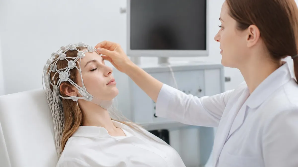
Video EEG Monitoring: Advantages and Limitations
- +Synchronizes brain wave data with video to confirm whether physical events are truly epileptic
- +Gold standard for distinguishing epileptic seizures from psychogenic non-epileptic events
- +Enables precise seizure classification to guide medication and surgical treatment decisions
- +Captures nocturnal events that outpatient tests consistently miss
- +Allows medication reduction under controlled conditions to provoke diagnostic events safely
- +Provides permanent, reviewable record that can be shared with specialists at other centers
- −Requires hospital admission lasting days to weeks, which disrupts work, school, and family life
- −Cost can reach several thousand dollars even with insurance, creating financial burden for many families
- −Seizure capture is not guaranteed, and monitoring may end without a definitive answer
- −Electrode application and maintenance over many days causes scalp discomfort and skin irritation
- −Patients must remain connected to equipment, limiting mobility and privacy significantly
- −Medication reduction to provoke seizures carries clinical risk and requires close medical supervision
What to Expect: EEG Test Preparation Checklist
- ✓Wash your hair the night before and do not apply conditioner, oil, or styling products that coat the scalp.
- ✓Confirm with your neurologist whether to take your regular medications the morning of the test.
- ✓If ordered for a sleep-deprived EEG, stay awake the entire night before your scheduled appointment.
- ✓Arrive on time — electrode application for a full 19-electrode montage takes 30 to 45 minutes.
- ✓Wear comfortable, loose-fitting clothing that is easy to change without disturbing the electrode array.
- ✓Bring entertainment for long monitoring sessions: books, tablets, headphones, and comfort items from home.
- ✓Press the event button immediately whenever you feel an aura, unusual sensation, or any possible symptom.
- ✓Ask family members or companions to stay nearby so they can observe and describe any clinical events.
- ✓Notify nursing staff promptly if any electrode feels loose, itchy, or painful during the monitoring period.
- ✓Follow all dietary and activity instructions given by the EMU team, particularly regarding caffeine and exercise.
Video EEG Changes Treatment in Up to 50% of Cases
Research published in leading epilepsy journals consistently shows that inpatient video EEG monitoring changes the diagnosis or treatment plan in approximately 40 to 50 percent of patients referred for evaluation. For patients who have been treated for epilepsy without seizure control, a significant proportion turn out to have psychogenic non-epileptic seizures, a finding that redirects care toward psychiatric treatment rather than additional anti-seizure medications. This diagnostic impact is the primary reason epilepsy monitoring units have become a standard feature of comprehensive epilepsy programs across the United States.
Understanding how to read EEG results is not a skill most patients need to develop, but having a general framework for what neurologists look for helps patients understand their reports and ask better questions during follow-up appointments. EEG results are described in terms of the dominant background rhythm, the presence or absence of abnormal waveforms, and the distribution of any abnormalities across the scalp.
A normal adult awake EEG shows a posterior dominant rhythm of 8 to 13 Hz — the alpha rhythm — that attenuates when the eyes open. Slower rhythms in regions where faster activity is expected signal dysfunction in the underlying brain tissue.
Epileptiform abnormalities are the patterns that most directly relate to seizure disorders. These include spikes, sharp waves, spike-and-slow-wave complexes, and polyspike discharges. A spike is defined as a waveform with a duration of 20 to 70 milliseconds, a sharp wave as one lasting 70 to 200 milliseconds. Both are followed by a slow wave component in classic epileptiform patterns. The distribution of these discharges — whether they are generalized across both hemispheres simultaneously or confined to one region or lobe — is critically important for diagnosis and surgical planning.
Generalized epileptiform discharges, such as the 3 Hz spike-and-wave complex seen in absence epilepsy, suggest a genetic generalized epilepsy syndrome. Focal discharges that consistently arise from one region — the left temporal lobe, for example — point toward a structural or lesional cause and raise the question of whether the patient might benefit from surgical resection of that area. When video EEG is used in surgical planning, the localization of ictal onset (where the seizure begins electrically) is compared with imaging findings and neuropsychological test results to determine whether resection is feasible and likely to be effective.
Artifacts are a major challenge in EEG interpretation and are particularly prevalent during prolonged monitoring sessions. Muscle artifact from jaw clenching, chewing, or body movement creates high-frequency noise that can obscure underlying brain activity. Eye movement artifact produces characteristic waveforms at the frontal electrodes. Electrode pop artifact — caused by a loose or degraded electrode — creates sudden, isolated high-amplitude transients that can mimic spikes if not recognized for what they are. Experienced EEG technologists and interpreting neurologists develop systematic habits for identifying and mentally subtracting these non-cerebral signals before drawing clinical conclusions.
One of the most important distinctions in EEG reading is between interictal and ictal patterns. Interictal patterns are abnormalities seen between seizures — they indicate epileptic potential but do not represent an active seizure. Ictal patterns represent ongoing seizure activity. A clinical seizure without corresponding ictal EEG activity is a strong indication that the event is non-epileptic in origin. Conversely, electrographic seizures — ictal EEG patterns without any clinical manifestation — are called subclinical or electrographic-only seizures and are important to identify, particularly in critically ill patients or those with encephalopathy.
Post-ictal changes following a seizure are also diagnostically informative. After a tonic-clonic seizure, the EEG typically shows diffuse slowing that gradually resolves over minutes to hours. Focal post-ictal slowing localized to the region of seizure onset adds further evidence for the localization hypothesis being built during a surgical evaluation. Todd's paralysis — the transient focal weakness that sometimes follows a focal motor seizure — correlates with this post-ictal focal slowing and can be visualized in the synchronized video alongside the EEG recording during video monitoring sessions.
For students preparing for EEG certification examinations, the ability to recognize and describe these patterns accurately is one of the most heavily tested competency areas. Examination questions cover normal variants that can mimic pathology, classic epileptiform patterns across different epilepsy syndromes, artifact identification and management, and the technical parameters that affect how these patterns appear on the recording. Building familiarity with a wide range of pattern examples through practice questions is the most reliable preparation strategy available for these visual recognition tasks.
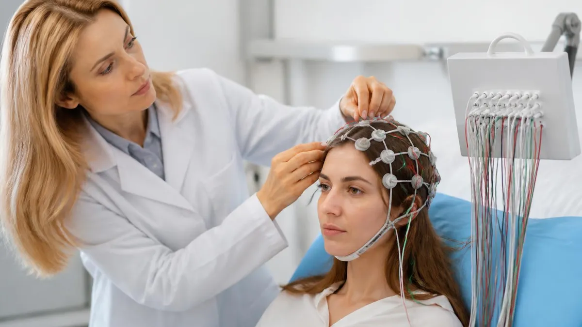
Many video EEG protocols involve supervised reduction of anti-seizure medications to increase the likelihood of capturing a seizure event during the monitoring window. This strategy is effective but not without risk — medication reduction can provoke more frequent or more severe seizures than the patient typically experiences, and in rare cases can lead to status epilepticus. Patients and families should discuss this aspect of the protocol thoroughly with the neurologist before admission, ask what safety measures are in place, and understand the criteria under which medications will be promptly reinstated.
The cost of an EEG test varies widely depending on the type of test, the clinical setting, the patient's insurance coverage, and the geographic region. A routine outpatient EEG performed at a hospital-based neurology clinic or an independent diagnostic facility typically costs between $200 and $700 before insurance adjustments. The EEG test price reflects the technical component — the electrode application, recording, and equipment — as well as the professional component, which is the neurologist's fee for interpreting the tracing. These two components are often billed separately, and patients may receive two distinct charges even for a single test.
Sleep-deprived and prolonged outpatient EEG tests cost somewhat more than routine studies because of the additional technologist time involved. Ambulatory EEG monitoring, which requires specialized portable equipment and typically includes a device rental or purchase component, runs from $400 to $1,500 depending on the provider. Some insurance plans cover ambulatory monitoring without prior authorization for patients with documented seizure history, while others require step-therapy documentation showing that a routine EEG was performed first without a definitive result.
Inpatient video EEG monitoring is the most expensive variant of the EEG medical test, with total hospital charges that can range from $5,000 to $50,000 or more for admissions lasting a week or longer. This cost reflects not just the EEG recording itself but the full inpatient hospitalization — room, nursing care, physician services, laboratory work, and any additional imaging or consultations that occur during the stay. Most major insurance plans, including Medicare and Medicaid, cover inpatient video EEG when it is ordered by a neurologist for an approved indication such as refractory epilepsy evaluation or pre-surgical seizure localization.
EEG test cost for uninsured or underinsured patients can be a serious barrier to accessing this important diagnostic tool. Some academic medical centers and federally qualified health centers offer sliding-scale payment arrangements or financial assistance programs for patients who qualify based on income. Patient advocacy organizations focused on epilepsy, including the Epilepsy Foundation, maintain resources to help patients navigate insurance coverage questions, appeal denials, and identify financial assistance programs available in their state.
The question of EEG test side effects is one that patients and families frequently ask, and the reassuring answer is that the EEG test itself carries essentially no direct side effects. The electrodes do not deliver any current into the brain, and there is no radiation exposure as there would be with CT or X-ray imaging. Some patients experience mild scalp irritation or temporary redness at electrode sites, particularly after prolonged monitoring, and the conductive gel can be difficult to wash fully out of the hair after the test. These are minor inconveniences rather than true medical side effects.
The activation procedures performed during an EEG do carry a small degree of clinical risk. Hyperventilation can provoke absence seizures in susceptible individuals, which is intentional, but can also cause light-headedness and tingling in patients without epilepsy due to the resulting drop in carbon dioxide levels. Photic stimulation with a strobe light is contraindicated in patients with known photosensitive epilepsy unless capturing a photic response is the specific clinical goal. Medication reduction during inpatient video EEG carries the most meaningful risk, as discussed above, and is always performed under direct medical supervision in an environment equipped to manage seizure emergencies.
For EEG professionals and students, understanding both the clinical and financial dimensions of the EEG test is important context for patient communication. Technologists are frequently asked by patients and families how much the test costs, how long it will take, and whether there will be any side effects. Being able to answer these questions accurately and empathetically — while directing patients to their insurance coordinator or neurologist for specific billing and coverage questions — is part of delivering high-quality patient-centered care in any EEG setting.
For EEG technologists and students preparing for credentialing examinations, video EEG represents a convergence of nearly every technical and clinical skill the field demands. The R.EEG T. examination administered by ABRET tests knowledge across domains including instrumentation and electronics, electrode application, normal and abnormal EEG patterns, special procedures including ambulatory and inpatient monitoring, and patient care. The CLTM credential, which focuses specifically on long-term monitoring, places even greater emphasis on seizure recognition, clinical event documentation, and the technical demands of continuous recording over days to weeks.
Preparing for these examinations requires a study strategy that balances breadth with depth. Candidates who only review textbook material without practicing applied questions often struggle with the case-based and pattern recognition items that appear throughout these exams. Working through practice questions that present EEG patterns, clinical scenarios, and technical troubleshooting problems in the same format as the actual examination is the most efficient way to identify knowledge gaps and build test-taking stamina at the same time.
The ambulatory EEG component of certification examinations covers the technical requirements for portable recording systems, the differences between ambulatory and inpatient video EEG in terms of diagnostic capability, the patient instructions required for successful home recording, and the common artifacts that arise when patients are unmonitored. These questions appear consistently across multiple examination administrations and reward candidates who have hands-on experience as well as theoretical knowledge of how these systems work in practice.
Activation procedures are another high-yield examination topic that connects directly to video EEG practice. Hyperventilation protocols, including the correct duration, the expected normal response, and the abnormal responses that should prompt additional evaluation, are tested in detail. Photic stimulation parameters — flash frequencies, patient positioning, contraindications, and the interpretation of photoparoxysmal responses — appear regularly. Sleep recording, including the identification of normal sleep architecture, vertex sharp waves, sleep spindles, K-complexes, and the REM-related changes that must be distinguished from pathological activity, is a substantial content area on all major EEG credentialing examinations.
Candidates preparing for EEG certification should also be comfortable with the ethical and documentation requirements of inpatient monitoring. Informed consent for video recording, the privacy considerations that govern how video footage is stored and accessed, the documentation standards for clinical event logs, and the chain of custody procedures for monitoring data that may be used in legal proceedings — such as cases involving non-epileptic events and disability determinations — are all part of professional practice in epilepsy monitoring units and may appear as examination content.
The practical value of EEG certification extends well beyond the examination itself. Certified technologists command higher salaries, have greater career mobility across clinical settings, and are often preferred candidates for positions in academic medical centers and comprehensive epilepsy programs where video EEG monitoring is a core service. Investing the time and resources to prepare thoroughly for credentialing examinations pays dividends throughout a technologist's career, and the knowledge gained in preparation directly improves the quality of care provided to patients during monitoring.
Building a consistent daily study habit — reviewing practice questions, reading explanatory feedback carefully, and tracking performance across topic areas — is more effective than marathon cramming sessions in the weeks before an examination. Most successful candidates report spending four to eight weeks in active preparation, dedicating 30 to 60 minutes per day to question practice and review. Using practice platforms that organize questions by topic and difficulty level, and that provide detailed answer explanations with references to clinical guidelines and standard textbooks, accelerates the learning process significantly compared to passive re-reading of study materials alone.
EEG Questions and Answers
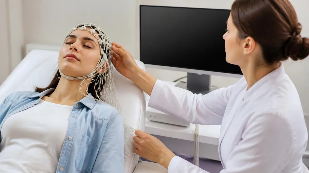
EEG Tech Salary 2026: Pay by State, Experience, and Certification
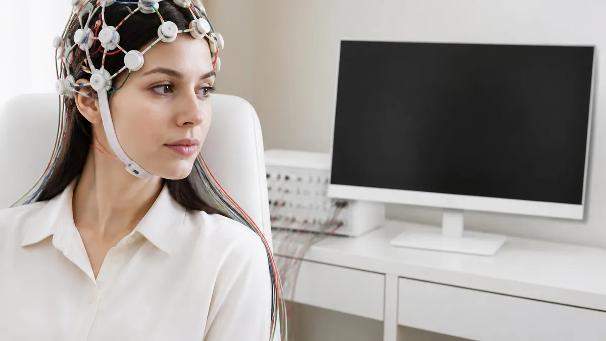
EEG Images: A Visual Guide to Brain Wave Patterns, Recordings, and Test Results
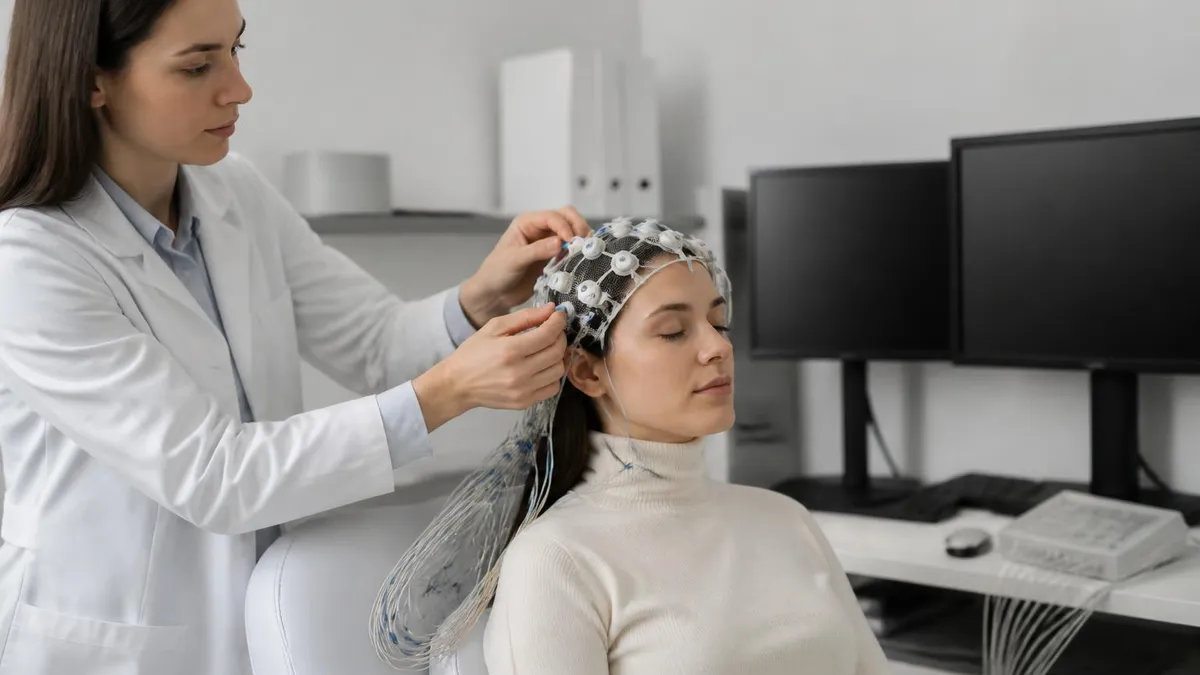
EEG for Seizures: How the Test Diagnoses Epilepsy and What to Expect
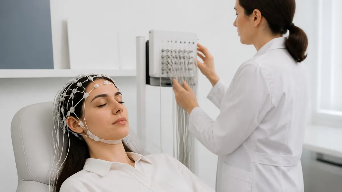
Ambulatory EEG: 24-72 Hour Home Recording for Suspected Seizures
EEG Electrode Placement: The Complete Guide to the 10-20 System, Scalp Mapping, and How an EEG Test Works
About the Author

Educational Psychologist & Academic Test Preparation Expert
Columbia University Teachers CollegeDr. Lisa Patel holds a Doctorate in Education from Columbia University Teachers College and has spent 17 years researching standardized test design and academic assessment. She has developed preparation programs for SAT, ACT, GRE, LSAT, UCAT, and numerous professional licensing exams, helping students of all backgrounds achieve their target scores.