EEG 10-20 System: Complete Guide to Electrode Placement, Brain Mapping, and What Your EEG Test Reveals
🧠 Learn how the EEG 10-20 system works, what an EEG test measures, cost, duration, and what to expect. Complete guide for patients and students.

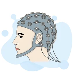
The eeg 10 20 system is the international standard that governs exactly where electrodes are placed on a patient's scalp during an EEG test — one of the most widely ordered neurological evaluations in modern medicine.
Developed in the 1950s and standardized by the International Federation of Clinical Neurophysiology, the 10-20 system gets its name from the fact that electrodes are spaced either 10% or 20% of the total front-to-back and left-to-right distances across the skull. This precise, reproducible framework ensures that brain activity recorded in a clinic in Boston is directly comparable to recordings taken in Los Angeles or anywhere else in the world.
Understanding what an EEG medical test involves starts with appreciating why standardized electrode placement matters so much. The brain generates tiny electrical signals — measured in microvolts — from millions of neurons firing in coordinated patterns. Small errors in electrode position can shift readings by enough to misidentify a region of interest, potentially leading to an incorrect diagnosis. The 10-20 system eliminates this variability by anchoring electrode positions to four anatomical landmarks: the nasion (bridge of the nose), the inion (bony bump at the back of the skull), and the two preauricular points just in front of each ear.
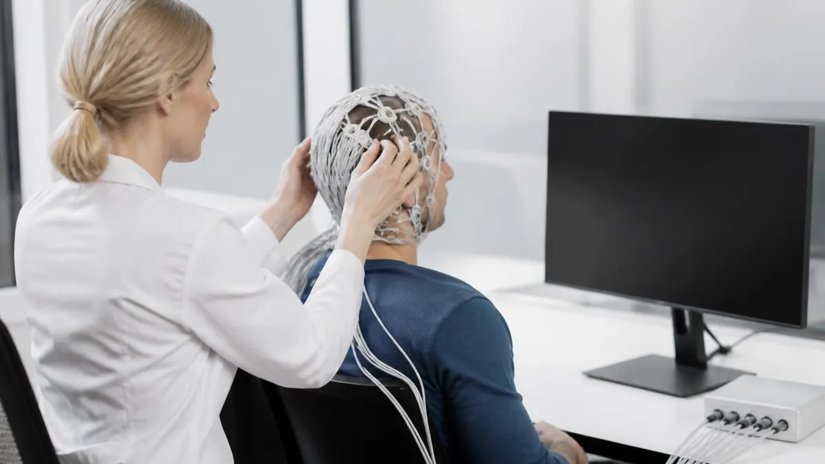
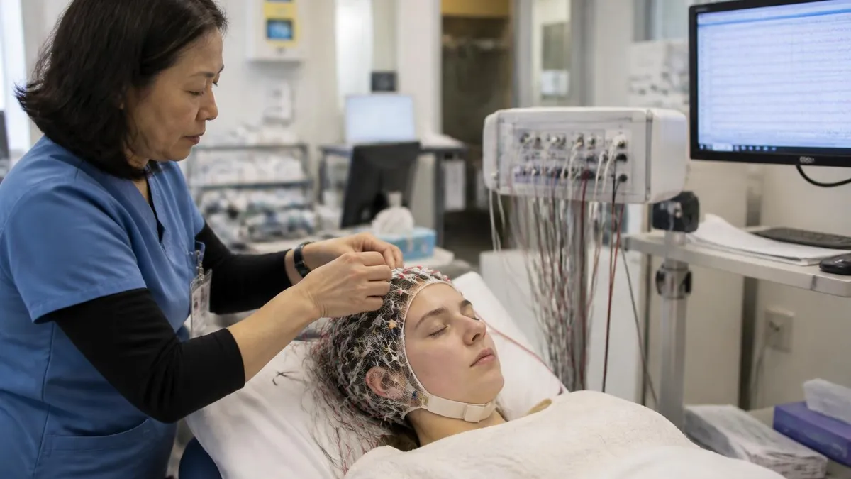
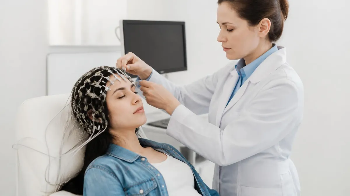
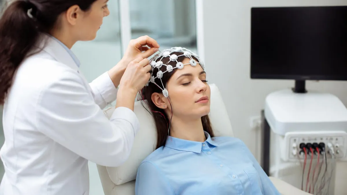
For patients wondering what is an EEG test and why their doctor ordered one, the procedure is completely painless and non-invasive. A trained technologist — called an EEG technician or neurodiagnostic technologist — measures your head, marks the electrode sites with a grease pencil or small sticker, and attaches between 19 and 256 small metal discs (electrodes) to your scalp using a conductive gel or paste. The electrodes detect voltage differences between scalp locations and feed them to an amplifier that displays the familiar wavy lines you have probably seen in medical dramas or textbooks.
The EEG test is ordered for a wide range of clinical reasons. Epilepsy is the most common indication — the test can detect abnormal electrical discharges called epileptiform activity that occur between or during seizures. Physicians also use EEGs to evaluate encephalopathy (diffuse brain dysfunction), sleep disorders, brain tumors, head trauma effects, metabolic disturbances, and to confirm brain death. In intensive care units, continuous EEG monitoring runs 24 hours a day or longer to catch subclinical seizures that produce no visible movements but still damage the brain if untreated.
The question of how long is an EEG test depends on the type ordered. A standard routine EEG in an outpatient clinic typically runs 20 to 40 minutes of actual recording time, though setup and cleanup push the total appointment to about 60 to 90 minutes.
Sleep-deprived EEGs require the patient to stay awake the night before so they fall asleep during the recording, adding another variable. Ambulatory EEGs — where a portable recorder is worn at home for 24 to 72 hours — capture brain activity across multiple days of normal life. Inpatient video-EEG monitoring for presurgical epilepsy evaluation can last a week or more.
Costs for an EEG test vary considerably depending on facility type, geographic region, and insurance coverage. The EEG test price at an outpatient hospital lab typically ranges from $200 to $700 after insurance adjustments, while the uninsured sticker price — the EEG test cost without any coverage — can range from $1,000 to $3,000 or higher for extended monitoring.
Freestanding neurophysiology labs and academic medical centers often charge differently, so calling ahead for an estimate is always worthwhile. Many insurance plans cover EEG when a physician documents a medically necessary indication such as a first seizure or evaluation of a known seizure disorder.
For students and clinicians preparing for board examinations or simply deepening their knowledge, the eeg 10 20 system is a foundational topic tested across multiple question domains — from electrode nomenclature to artifact recognition to the normal and abnormal waveforms seen in each brain region. Mastery of the 10-20 framework is not just academic trivia; it is the practical backbone that makes every EEG interpretation clinically meaningful.
EEG Test & 10-20 System by the Numbers

The 10-20 System: How Electrodes Are Named and Placed
The four reference points — nasion, inion, and bilateral preauricular points — define the skull's dimensions. Distances between them are measured in centimeters, and electrode sites are placed at 10% or 20% intervals along anterior-posterior and lateral axes.
Each electrode carries a letter indicating the brain region beneath it: Fp (frontopolar), F (frontal), C (central), P (parietal), O (occipital), T (temporal), and A (auricular/ear reference). The letter-number pairing tells you the exact location instantly.
Odd numbers are always assigned to the left hemisphere (e.g., F3, C3, P3, T3) and even numbers to the right hemisphere (F4, C4, P4, T4). Midline electrodes carry the subscript 'z' for zero — Fz, Cz, Pz — indicating they sit exactly on the sagittal plane.
High-density EEG research and source localization often use the expanded 10-10 system, which adds intermediate electrode positions to yield 74 or more channels. This provides much finer spatial resolution, useful for epilepsy surgery planning and brain-computer interface research.
For clean recordings, electrode-to-scalp impedance must typically be below 5 kΩ. The technologist checks impedance before starting, abrades the skin gently, and reapplies gel if values are too high. High impedance introduces noise and obscures genuine brain activity.
Once the electrodes are placed according to the 10-20 framework, the EEG machine records voltage differences between pairs of electrodes in configurations called montages. A referential montage measures each electrode against a common reference — often the linked ear electrodes (A1-A2) or a computed average reference.
A bipolar montage measures adjacent electrode pairs in a chain, which localizes abnormalities more precisely because a discharge appearing in only one channel of the chain indicates the boundary between abnormal and normal tissue. Switching between montages is one of the first skills an EEG reader develops, because the same brain event can look very different depending on the montage selected.
The brain rhythms captured by the EEG are grouped into frequency bands, each with distinct clinical significance. Delta waves (0.5–4 Hz) are the slowest and normally dominate only in deep sleep; their presence in an awake adult almost always indicates serious pathology such as a tumor, stroke, or encephalopathy. Theta waves (4–8 Hz) are also slow for a waking adult and may reflect drowsiness, focal structural lesions, or diffuse dysfunction, though they appear normally during childhood and adolescence. The interpretation always depends on the patient's age and state of alertness.
Alpha waves (8–13 Hz) are the hallmark of relaxed wakefulness with eyes closed, generating the prominent posterior dominant rhythm — often called the PDR — over the occipital electrodes (O1, O2). A normal adult PDR is symmetric and runs at 8.5 to 12 Hz; it attenuates (blocks) when the patient opens their eyes or performs a mental task. Asymmetry in the PDR, with one side slower or lower in amplitude than the other, can indicate ipsilateral posterior lesions such as a stroke or tumor pressing on the occipital lobe.
Beta waves (13–30 Hz) are faster rhythms associated with active thinking, anxiety, and certain medications — particularly benzodiazepines and barbiturates, which dramatically increase beta amplitude across the scalp. Focal excess beta in one region may suggest an underlying cortical lesion, while generalized beta excess is most often medication-related and benign. Gamma oscillations above 30 Hz are increasingly studied in research contexts as markers of cognitive processing and binding of perceptual information, though their role in routine clinical EEG is still emerging.
Abnormal EEG findings fall into two broad categories: epileptiform and non-epileptiform. Epileptiform abnormalities include spikes (duration 20–70 milliseconds), sharp waves (70–200 ms), and spike-and-slow-wave complexes. These patterns reflect hypersynchronous neuronal firing and are associated with a significantly elevated risk of unprovoked seizures. The location of the discharge in the 10-20 grid provides the electrographic equivalent of a GPS coordinate — a discharge maximal at F7-T3 suggests a left anterior temporal focus, pointing toward mesial temporal lobe epilepsy, the most common surgically treatable epilepsy syndrome in adults.
Non-epileptiform abnormalities include focal or generalized slowing, burst-suppression patterns in comatose patients, periodic lateralized epileptiform discharges (PLEDs), and the classic 3-Hz spike-and-wave seen in absence epilepsy, which is generalized rather than focal. Recognizing the difference between these patterns requires knowing what each electrode location represents anatomically and understanding that the 10-20 system is the common language that makes that translation possible. Without standardized placement, the geographic coordinates of any EEG finding would be meaningless or unreproducible.
For anyone preparing for EEG certification examinations or seeking deeper mastery, consistent practice with electrode nomenclature, montage switching, and waveform classification is essential. The breadth of clinical scenarios — neonatal EEG, pediatric EEG, ICU monitoring, and outpatient epilepsy follow-up — each demands familiarity with how normal brain rhythms change across the lifespan and how pathological patterns modify the baseline. The 10-20 system is the thread that ties all these scenarios together, providing a reproducible anatomical context for every tracing you will ever read or record.
What Is an EEG Medical Test? Types, Procedures, and Uses
A routine outpatient EEG is the most commonly ordered type and serves as the first-line evaluation for seizure disorders, encephalopathy, and altered mental status. The appointment lasts about 60 to 90 minutes in total, with 20 to 40 minutes of active recording. The technologist performs activation procedures — hyperventilation for three minutes and photic stimulation with a strobe light at varying frequencies — to provoke latent abnormalities that may not be visible in the resting baseline recording.
Despite its widespread use, a routine EEG captures only a brief window of brain activity, which means it can be entirely normal in a patient with epilepsy if no discharges happen to occur during that window. Studies suggest that a single routine EEG detects epileptiform activity in only about 50% of people with known epilepsy. Repeating the test, including sleep recording, or upgrading to a prolonged ambulatory EEG significantly improves the diagnostic yield, which is why physicians often order serial studies in patients with a high clinical suspicion for epilepsy but an initially normal recording.

EEG Test: Benefits vs. Limitations
- +Completely non-invasive and painless — no needles, radiation, or sedation required for most studies
- +Excellent temporal resolution — captures brain activity millisecond by millisecond, far better than MRI or CT
- +Widely available in outpatient neurology clinics, hospitals, and even some primary care settings across the US
- +Relatively affordable compared to MRI; the EEG test cost is often covered by major insurance plans with a valid clinical indication
- +Can detect abnormalities even when structural imaging (MRI, CT) is completely normal, as in many epilepsy cases
- +Portable versions allow monitoring during natural sleep and daily activities over 24–72 hours without hospitalization
- −Short routine studies capture only a brief window; a normal EEG does NOT rule out epilepsy
- −Poor spatial resolution — the 10-20 system localizes to brain regions but cannot pinpoint individual gyri the way a high-resolution MRI can
- −Susceptible to numerous artifacts — muscle, electrode, eye movement, and external electrical interference — that can mimic or obscure genuine brain activity
- −Interpretation is operator-dependent; inter-rater agreement even among experienced epileptologists can be imperfect on ambiguous patterns
- −Hair products, sweating, and head movement degrade electrode contact and increase impedance, reducing signal quality
- −EEG test side effects are rare but include scalp irritation from electrode paste and, very rarely, seizure provocation during photic stimulation in susceptible individuals
Preparing for Your EEG Test: A Step-by-Step Checklist
- ✓Wash your hair the morning of the test with regular shampoo — do not use conditioner, oils, or styling products that coat the scalp.
- ✓Ask your ordering physician whether to take or skip regular medications; most epilepsy medications are continued, but your doctor decides.
- ✓If a sleep-deprived EEG is ordered, stay awake the entire night before as instructed so you can fall asleep naturally during the recording.
- ✓Arrive at least 15 minutes early so the technologist has time to measure your head and apply all electrodes without rushing the setup.
- ✓Inform the technologist of all medications, supplements, and caffeine intake, as these all influence EEG appearance and must be documented.
- ✓Remove all hair accessories — clips, extensions, wigs, and hearing aids — before electrode application, as they cause artifacts.
- ✓Tell the technologist about any claustrophobia or anxiety so they can walk you through the flashing light portion of the test beforehand.
- ✓Breathe deeply and steadily during the hyperventilation activation procedure even if you feel light-headed; notify staff if you feel unwell.
- ✓Keep your eyes still and relax facial muscles during recording periods; blinking, chewing, and jaw clenching create eye and muscle artifacts.
- ✓Plan for a mild scalp redness or stickiness after the gel is removed; bring a comb and shampoo if you want to freshen up after the appointment.
The 10-20 System Is the Universal EEG Language
Memorizing the electrode grid is not just about passing a certification exam — it is the foundation of every EEG interpretation you will ever perform. When you can immediately picture which brain region sits beneath each electrode label, you can translate a raw tracing into a clinical localization in seconds, transforming wavy lines into actionable neurological information.
The clinical question of what is an EEG test used for extends well beyond epilepsy. In the sleep medicine world, polysomnography — the comprehensive overnight sleep study — incorporates EEG channels alongside measures of eye movements (EOG), muscle tone (EMG), heart rhythm (ECG), respiratory airflow, and oxygen saturation.
The EEG channels in a polysomnogram follow the same 10-20 placement rules and allow sleep technologists and physicians to score sleep stages: wake (W), light non-REM sleep (N1, N2), deep slow-wave sleep (N3), and REM sleep. Sleep stage scoring depends critically on recognizing the EEG signatures of each stage — vertex sharp waves and sleep spindles in N2, high-amplitude slow waves in N3, and the low-amplitude mixed-frequency activity of REM.
In neurology intensive care units, the EEG has evolved from a diagnostic tool into a real-time monitoring instrument analogous to cardiac monitoring for the heart. Quantitative EEG (qEEG) displays summarize hours of raw data into color-coded trend panels — amplitude-integrated EEG, spectral edge frequency, and seizure probability scores — allowing bedside nurses and physicians to spot deterioration at a glance without reviewing every page of raw tracing. Machine-learning algorithms are now being integrated into these platforms to flag likely seizures automatically, though human review remains essential because false-positive rates are still too high for fully automated clinical decision-making.
Neonatal EEG presents a specialized challenge because the infant brain generates rhythms that would be frankly abnormal in an adult. Normal features unique to neonates include trace alternant — alternating bursts of high-amplitude activity separated by lower-amplitude inter-burst intervals — which is normal in quiet sleep of premature infants but disappears as the brain matures toward term.
Seizures in neonates are also often subclinical and subtle, manifesting as rhythmic delta or alpha activity rather than the dramatic spike-wave complexes seen in older patients. The modified Hahn or the American Clinical Neurophysiology Society neonatal EEG guidelines specify electrode placement adapted for the smaller neonatal skull, but the 10-20 proportional system still underlies the approach.
Intraoperative neurophysiological monitoring (IONM) represents another major application of EEG in clinical practice. During surgeries near the brain or spinal cord — tumor resections, carotid endarterectomies, cardiac bypass procedures — continuous EEG monitoring alerts the surgical team to ischemia (reduced blood flow) in real time.
A sudden decrease in fast activity or a burst-suppression pattern during carotid clamping, for example, signals that the brain may not be receiving adequate perfusion from collateral circulation, prompting the surgeon to place a shunt or adjust anesthesia. The 10-20 system ensures that the monitoring technologist and the reading neurophysiologist can communicate unambiguously about which brain region is affected.
For patients wondering about EEG test side effects, the news is overwhelmingly reassuring. The test itself carries virtually no medical risk. The conductive gel used to attach electrodes may cause mild, temporary scalp redness or skin irritation in patients with sensitive skin, which resolves within hours.
The hyperventilation activation procedure commonly causes tingling in the hands and face due to transient hypocapnia (low CO2 from overbreathing) — this sensation is expected and harmless, not a sign of a problem. The photic stimulation with flashing lights can very rarely trigger a seizure in photosensitive individuals, but this occurs almost exclusively in patients who already have a photosensitive epilepsy syndrome and can often be predicted from the EEG itself before the stimulation is escalated.
The cost landscape for EEG in the United States is fragmented. The EEG test cost varies by setting: hospital outpatient departments typically bill more than freestanding neurology clinics because of facility fees. Medicare reimbursement for a routine EEG (CPT 95816 — awake and drowsy) runs approximately $100 to $200 at the physician-fee-schedule level, while the technical component billed by the facility can be several times higher.
Patients on high-deductible health plans may pay their full deductible for an EEG before insurance kicks in, making it worthwhile to call the lab and ask about self-pay discounts, which many facilities offer at 30 to 60% below the list price.
One often-overlooked aspect of the EEG medical test experience is interpreting the written report afterward. Most EEG reports follow a structured format: a description of the background activity, notation of any abnormalities, a section on activation procedures, and a final impression or clinical correlation statement.
The report will reference the 10-20 system implicitly through electrode names — if it says a sharp wave was seen at T3-T5, you now know that refers to the left mid-temporal to left posterior temporal chain, an area associated with the hippocampus and mesial temporal structures. Understanding even the basics of the 10-20 grid transforms a confusing medical report into a meaningful map of your own brain.

A normal routine EEG does not rule out epilepsy. Studies show that a single 30-minute recording misses epileptiform activity in roughly half of patients with a confirmed seizure disorder. If your doctor suspects epilepsy but your first EEG is normal, ask about a sleep-deprived study or a 24-hour ambulatory EEG to increase the chance of capturing diagnostic abnormalities during a longer recording window.
EEG certification examinations in the United States are administered by the American Board of Registration of Electroencephalographic and Evoked Potential Technologists (ABRET). The primary credential for EEG technologists is the Registered EEG Technologist (R. EEG T.) examination, which tests knowledge across domains including electrode application, patient safety, equipment troubleshooting, artifact recognition, normal and abnormal EEG patterns, and the clinical indications for various EEG study types.
The 10-20 system is explicitly tested in the electrode application domain, and examinees must demonstrate both theoretical knowledge of the placement rules and practical ability to apply electrodes accurately on a mannequin head during the hands-on component of some certification pathways.
The advanced credential, the Certified EEG Technologist (CEET), covers more complex clinical scenarios including neonatal EEG, long-term monitoring, and quantitative EEG analysis. For neurophysiology technologists who also monitor intraoperative studies, the Certified Neurophysiologic Intraoperative Monitor (CNIM) examination adds domains covering somatosensory evoked potentials, motor evoked potentials, and brainstem auditory evoked potentials in addition to EEG. In all of these certifications, the 10-20 system is a prerequisite baseline, not an advanced topic — examiners assume you have already mastered electrode nomenclature and move on to higher-order interpretation questions.
Studying for EEG certification requires more than reading textbooks. The most effective preparation combines content review with active practice interpreting sample tracings and answering question banks. Research in educational psychology consistently shows that retrieval practice — forcing yourself to recall information through questions rather than passively re-reading notes — produces significantly better long-term retention than re-reading alone. This is especially important for EEG preparation because the examination presents tracings and asks you to identify patterns, which is an active recognition task, not a passive recall of definitions.
When building a study schedule, most successful candidates recommend spending the first two to three weeks solidifying foundational knowledge: the 10-20 system electrode names and positions, normal EEG variants by age and state, artifact recognition, and the physiological basis of EEG generation. The next phase focuses on abnormal patterns — epileptiform discharges, focal and generalized slowing, encephalopathic patterns, and the EEG correlates of specific syndromes like West syndrome (hypsarrhythmia), Lennox-Gastaut syndrome (slow spike-and-wave), and juvenile myoclonic epilepsy (polyspike-wave). The final study phase should be dedicated almost entirely to practice questions and timed mock examinations under realistic testing conditions.
Time management during the actual certification examination is a practical skill that many candidates underestimate. The R. EEG T. examination includes a substantial number of tracing-based questions where you must identify a pattern and choose the correct answer from four options. Spending too long on any single tracing can leave you rushing through the final portion of the exam.
A useful strategy is to flag difficult tracing questions on the first pass, answer every question you can complete confidently in under 45 seconds, then return to flagged items with whatever time remains. This approach ensures you collect every easy point before investing time in the harder ones.
For comprehensive study resources, combining official ABRET examination blueprints with high-quality practice question banks gives you both the content map and the retrieval practice needed for strong performance. Practice questions that mirror the clinical scenarios on the real examination — asking you to identify the electrode at a given 10-20 position, recognize an epileptiform pattern in a described tracing, or choose the most appropriate montage for a specific clinical scenario — build the mental agility needed for test-day success.
Pairing these resources with peer study groups or online communities of EEG professionals adds accountability and exposes you to the real-world clinical experience of others preparing for the same credential.
Taking a systematic and evidence-based approach to EEG study — grounded in the 10-20 system, reinforced by active question practice, and guided by a realistic timeline — gives any candidate the best possible chance of earning their certification and building a clinical career in neurodiagnostics. The field continues to grow as continuous monitoring expands into new settings and as digital EEG technology makes recording more accessible across the full range of clinical environments, from rural outpatient clinics to cutting-edge academic medical centers.
Practical mastery of the 10-20 system begins with a simple mnemonic: memorize the midline chain first (Fpz, Fz, Cz, Pz, Oz), then learn the symmetrical lateral chains on each side. Once you can recite the midline electrodes from front to back without hesitation, adding the lateral electrodes — Fp1/Fp2, F3/F4, F7/F8, C3/C4, T3/T4, T5/T6, P3/P4, O1/O2 — becomes a matter of recognizing the left-odd/right-even number rule and the anatomical letter codes.
Drawing the 10-20 grid from memory on blank paper until you can produce it accurately in under two minutes is a classic and highly effective self-test that most experienced technologists and readers did during their training.
When beginning to interpret actual EEG tracings, always follow a systematic approach rather than jumping immediately to the most dramatic-looking deflections. Start by identifying the study parameters: paper speed (usually 30 mm/second), sensitivity (typically 7 µV/mm for adults), and filter settings (low-frequency filter 1 Hz, high-frequency filter 70 Hz). Then assess the background — what is the dominant rhythm in the posterior regions, what is its frequency and amplitude, and is it symmetric?
Only after characterizing the baseline should you look for abnormalities, because the background tells you what is normal for this particular patient and state, giving you the context needed to recognize what is truly deviant.
Artifact recognition is one of the most practically important EEG skills and one of the areas where students most commonly struggle.
The most frequent artifacts in routine EEG are electrode artifact (a single channel showing high-amplitude noisy activity due to poor contact), electromyographic artifact (muscle activity creating fast, irregular high-frequency spikes especially over the temporal and frontal electrodes), eye blink artifact (large slow deflections maximal at Fp1 and Fp2 that propagate posteriorly with a predictable polarity field), and cardiac artifact (a rhythmic pulse-synchronous artifact often best seen at ear reference electrodes). Learning to identify these confidently prevents misidentifying a frontopolar electrode artifact as a pathological frontal sharp wave — a critical distinction with real diagnostic consequences.
Hyperventilation and photic stimulation findings also require systematic interpretation. Hyperventilation normally produces bilateral generalized slowing in the delta and theta range, which is a physiological response to hypocapnia and is entirely normal — particularly prominent in children and young adults. Persistent focal slowing or an asymmetric buildup during hyperventilation, however, raises concern for an underlying focal lesion on the side that slows more.
Photic stimulation at frequencies between 6 and 18 Hz is most likely to drive photoparoxysmal responses in photosensitive patients; a true photoparoxysmal discharge extends beyond the occipital electrodes and outlasts the stimulus by more than 100 ms, distinguishing it from the harmless occipital photic driving response seen in normal individuals.
Long-term monitoring skills become critical for technologists working in epilepsy monitoring units or ICUs. During prolonged recordings, patient movement, nursing care, and equipment changes create recurrent artifacts that must be flagged in the recording log so reviewers know what was happening at any given moment.
The event marker — a button the patient or technologist presses to time-stamp clinical events — is one of the most important tools in long-term EEG because it links the electrical record to the patient's experience. Teaching patients and family members to use the event marker reliably is a practical skill that directly improves diagnostic yield.
Keeping up with the evolving EEG literature is also part of professional development for anyone working in neurodiagnostics. Organizations such as the American Clinical Neurophysiology Society (ACNS) publish standardized terminology and updated clinical practice guidelines that define how specific EEG patterns should be named, interpreted, and reported. The ACNS critical care EEG terminology, for example, provides a structured vocabulary for ICU patterns that allows clinicians at different hospitals to communicate unambiguously — building on the same principles of standardization that make the 10-20 system valuable in the first place.
Whether you are a patient trying to understand what your ordered EEG test involves, a student beginning your neurodiagnostics training, or an experienced technologist preparing for an advanced certification, the 10-20 electrode system is the indispensable foundation beneath everything.
It transforms the scalp into a reproducible coordinate map, gives every brain region an addressable electrode, and makes the global language of EEG interpretable regardless of where or by whom the recording was made. Investing time to master it thoroughly — not just to pass a test but to internalize it — pays clinical dividends for the entire span of a career in neurophysiology.
EEG Questions and Answers
EEG Electroencephalography Practice Test PDF (Free Printable 2026)
What Does an EEG Measure? Brain Waves, Electrical Activity, and What Your Results Reveal
How to Read an EEG Test: A Complete Guide to Brain Wave Patterns and Results
EEG Waves Explained: Delta, Theta, Alpha, Beta, and Gamma Brain Rhythms
EEG Monitoring: What Is an EEG Test, How It Works, Cost, and What to Expect
About the Author
Educational Psychologist & Academic Test Preparation Expert
Columbia University Teachers CollegeDr. Lisa Patel holds a Doctorate in Education from Columbia University Teachers College and has spent 17 years researching standardized test design and academic assessment. She has developed preparation programs for SAT, ACT, GRE, LSAT, UCAT, and numerous professional licensing exams, helping students of all backgrounds achieve their target scores.