
EEG 10-10 System: Complete Guide to Electrode Placement, the EEG Test, and What to Expect
🧠 Learn how the EEG 10-10 system works, what an EEG test costs, how long it takes, and what to expect before and after.

The EEG 10-10 system is the internationally standardized method for placing electrodes on the scalp during an electroencephalography recording. Whether you are preparing for a clinical EEG test, studying for a neurodiagnostic technology certification, or simply trying to understand what happens when electrodes are affixed to your head, understanding this electrode grid is essential. The 10-10 system expanded the older 10-20 system to include more electrode sites, giving clinicians a far more detailed map of brain electrical activity across frontal, temporal, parietal, and occipital regions.
An EEG medical test measures the electrical impulses produced by neurons communicating with each other. Those impulses are tiny — measured in microvolts — yet they carry enormous diagnostic information about seizure disorders, sleep abnormalities, encephalopathies, and brain death determination. The electrodes of the 10-10 system act as listening posts distributed evenly across the scalp, with each position named by a letter indicating its brain region and a number indicating its lateral position. Odd numbers sit on the left hemisphere; even numbers sit on the right; the letter z marks the midline.
People commonly ask what is an EEG test before their appointment and are often surprised to learn how non-invasive the procedure is. No needles pierce the scalp. No electricity enters your body. The electrodes only record signals; they never transmit them. A typical routine EEG uses 19 to 21 electrodes placed according to the 10-20 subset of the 10-10 system, while research-grade or high-density recordings can use 64, 128, or even 256 electrode sites that fill in the remaining positions defined by the full 10-10 grid.
The designation 10-10 refers to the spacing rules that govern electrode placement. Each electrode is positioned either 10 percent or 20 percent of a measured skull distance away from its neighbors, ensuring proportional coverage regardless of head size. A large adult and a small child will have electrodes in the same relative cortical positions even though the absolute centimeter distances differ considerably. This proportionality is what makes the system universal and allows EEG findings from clinics around the world to be compared meaningfully in research and in clinical practice.
Understanding the eeg 10 10 system is also important for anyone taking a credentialing examination in neurodiagnostic technology. The American Board of Registration of Electroencephalographic and Evoked Potential Technologists (ABRET) tests candidates extensively on electrode placement accuracy, measuring conventions, and the rationale behind the grid structure.
Misplaced electrodes can mimic or mask pathological findings, so precision in following the system is not an academic exercise — it has real clinical consequences for patients whose diagnoses depend on accurate recordings. For a broader look at how the EEG compares to other cardiac and brain monitoring tests, see our overview of the eeg 10 10 system.
The cost of an EEG test varies widely depending on setting, insurance coverage, and test duration. A routine outpatient EEG at a hospital-based neurology clinic typically runs between $200 and $900 before insurance adjustments, while ambulatory EEGs that record continuously for 24 to 72 hours can cost $1,000 to $3,000 or more. Understanding these figures helps patients prepare financially and advocate for appropriate testing. Throughout this guide we will cover the full picture: what the 10-10 system looks like in practice, how the EEG test is performed, what it costs, how long it takes, and what your results mean.
This article is structured to serve two audiences simultaneously. For patients and families, it demystifies an often anxiety-provoking procedure and explains exactly what will happen before, during, and after the test. For students and technologists, it provides a thorough review of the 10-10 electrode naming conventions, measurement techniques, and clinical applications that appear on board examinations. Both groups will find concrete numbers, practical checklists, and evidence-based explanations throughout every section.
EEG Test & 10-10 System by the Numbers
How the 10-10 Electrode Grid Is Measured and Applied
Identify Nasion and Inion
Mark the Midline Positions
Measure Left-Right Distance
Place Temporal Chain Electrodes
Verify Impedances and Apply Gel
Confirm Montage and Begin Recording
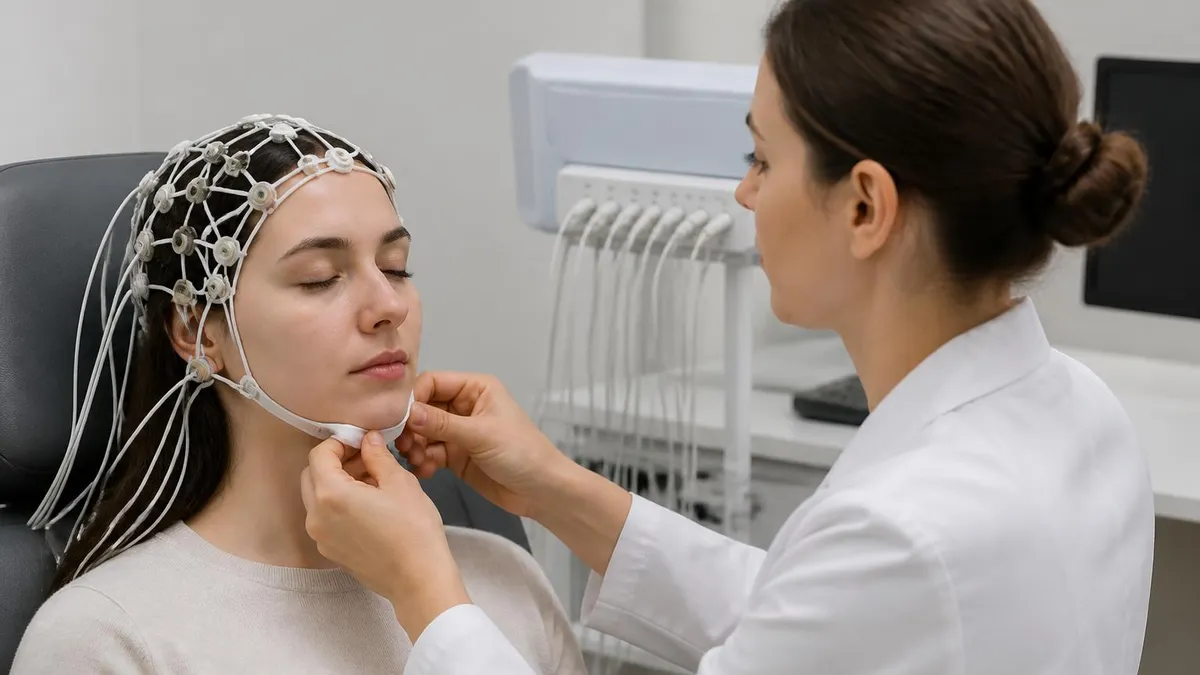
When patients ask what is an EEG test in practical terms, the best answer is that it is a passive, painless recording session that captures the brain's spontaneous electrical rhythms over a defined period. The patient reclines in a chair or lies on a table while the EEG technologist applies electrodes using conductive paste or a gel-filled cap. The recording typically lasts 20 to 40 minutes for a routine study, though some protocols extend to several hours or even days in an inpatient monitoring setting.
Before the electrodes are applied, the technologist will measure the patient's head according to the 10-10 or 10-20 convention, marking each site with a wax pencil or erasable marker. The scalp is lightly abraded at each mark to reduce the insulating layer of dead skin cells, which would otherwise increase impedance and degrade signal quality. This light abrasion feels similar to a gentle scratching sensation and leaves no lasting marks. The electrode paste or gel is then applied, and the electrode cup is pressed into position and secured with collodion glue or a fabric cap.
During the recording, the technologist will ask the patient to perform several activation procedures designed to elicit abnormal activity that may not appear during a resting baseline. Hyperventilation for three minutes causes cerebral vasoconstriction and can provoke absence seizures or other paroxysmal discharges. Photic stimulation involves a strobe light flashing at frequencies ranging from 1 to 30 Hz, which can trigger photoparoxysmal responses in susceptible individuals. Sleep deprivation before the test is another common activation method, as the drowsy and early sleep states lower the seizure threshold and unmask epileptiform discharges that are suppressed during wakefulness.
The raw EEG signal is amplified approximately one million times before being displayed on the recording system. Modern digital EEG systems sample at 256 to 512 Hz per channel and store the data in formats that allow technologists and neurologists to review recordings at any montage after acquisition. The neurologist who interprets the study will switch between multiple montages, adjust filter settings, and examine individual waveform morphologies to reach a clinical conclusion. This interpretive process typically takes 15 to 30 minutes for a routine study and longer for complex or lengthy recordings.
The EEG medical test is ordered for a wide range of clinical indications. Epilepsy diagnosis and management account for the majority of outpatient EEGs, but the test is also used to evaluate encephalopathy (diffuse brain dysfunction), assess the depth of sedation or coma in ICU patients, investigate spells of altered awareness, and monitor for subclinical seizures in patients who have had a known seizure and are being treated with anticonvulsants. In brain death evaluation, the EEG serves as one of the ancillary tests confirming electrocerebral silence when clinical examination alone is insufficient.
Patients and families frequently worry about what they will experience during the EEG. It is worth emphasizing clearly: the electrodes only detect electrical signals from your brain; they do not send any electricity into your body. You cannot receive a shock from an EEG. The most uncomfortable part of the procedure for most people is the application and removal of the electrode paste, which can pull slightly on hair.
Some patients find the hyperventilation activation procedure mildly dizzy-making, but this resolves within seconds of stopping the breathing exercise. Photic stimulation can cause mild discomfort in people sensitive to flickering light, and the technologist can modify or skip this step if needed.
After the EEG is complete, the technologist removes the electrodes and helps the patient wash out any remaining paste from their hair. Most people drive themselves home without difficulty. A full written report from a board-certified neurologist is typically available within one to three business days, and the ordering physician will discuss the results and any clinical implications during a follow-up appointment. In urgent inpatient settings, a preliminary verbal report may be communicated to the treating team within hours of recording completion.
EEG Test Duration, Cost, and Side Effects: What to Expect
A routine outpatient EEG typically takes 20 to 40 minutes of actual recording time, but patients should plan for a total appointment of 60 to 90 minutes to allow for check-in, electrode application, post-recording cleanup, and checkout. Ambulatory EEG studies, which record continuously while the patient goes about daily activities, run for 24 to 72 hours. Inpatient video-EEG monitoring for presurgical epilepsy evaluation can last several days to weeks, depending on how quickly the clinical team captures a sufficient number of representative seizures.
Sleep-deprived EEGs add roughly 30 extra minutes to the recording protocol to ensure that drowsiness and stage 1 sleep are captured on the trace. Neonatal EEGs, which require specialized electrode caps and careful waveform interpretation, typically run 60 minutes or longer to observe multiple sleep-wake cycles. Intraoperative EEG monitoring during neurosurgery or carotid endarterectomy runs for the entire duration of the surgical procedure, which can range from two to eight hours or more depending on case complexity.

Advantages and Limitations of the EEG 10-10 System
- +Proportional spacing ensures accurate cortical coverage on any head size, from neonates to large adults
- +Standardized naming convention allows EEG findings to be compared and replicated across labs worldwide
- +Higher electrode density than the older 10-20 system captures activity from more cortical regions
- +Non-invasive placement with no needles, radiation, or injected contrast — safe for all age groups
- +Supports millisecond-level temporal resolution, far exceeding the time precision of MRI or PET scanning
- +Flexible: the same grid supports routine clinical EEG, high-density research EEG, and source localization
- −Electrode application is time-consuming — a full 10-10 setup can take 30 to 45 minutes for experienced technologists
- −Electrode paste residue in hair is uncomfortable and inconvenient, particularly for patients with thick or curly hair
- −Scalp EEG has poor spatial resolution compared to intracranial recording; cannot resolve sources smaller than a few centimeters
- −Volume conduction spreads signals across multiple electrodes, making precise source localization difficult without computational modeling
- −Movement and muscle artifacts degrade signal quality; prolonged recordings in uncooperative patients yield limited interpretable data
- −Electrode impedance rises over time as paste dries, potentially compromising data quality in very long recordings without re-gelling
Pre-EEG Test Preparation Checklist
- ✓Wash and dry your hair the morning of the test — do not apply conditioner, oil, or styling products, which increase impedance
- ✓Ask your doctor whether to continue, reduce, or hold antiepileptic medications before the test
- ✓Follow any sleep deprivation instructions: for a sleep-deprived EEG, stay awake until midnight and rise by 4 AM
- ✓Eat a full meal before your appointment — low blood sugar can alter EEG patterns and make you feel faint during hyperventilation
- ✓Avoid caffeine for at least 8 hours before the test, as it reduces drowsiness needed to capture sleep transitions
- ✓Arrive at least 15 minutes early to complete paperwork and allow the technologist adequate setup time
- ✓Inform the technologist of all medications, supplements, and any known scalp conditions or adhesive allergies
- ✓Remove hair extensions, wigs, or tight braids that would prevent electrode access to the scalp
- ✓Plan for 60 to 90 minutes total appointment time — routine recording is 20 to 40 minutes but setup and cleanup add time
- ✓Arrange a ride home if you are unusually fatigued from sleep deprivation, even though no sedation is used in the EEG itself
Cz Is the Most Critical Electrode for Vertex Wave Detection
The Cz electrode, positioned exactly at the midpoint of the skull according to the 10-10 system, is uniquely important for detecting vertex sharp waves during drowsiness and stage 1 sleep. These normal waveforms appear as high-amplitude sharp transients maximal at Cz and must not be mistaken for pathological epileptiform discharges. Accurate Cz placement — confirmed by equal measurements from nasion to inion and from left to right preauricular point — is therefore essential for correct EEG interpretation in every clinical setting.
Reading an EEG report requires understanding a specific vocabulary that neurologists use to describe brain wave patterns. The report will typically describe the background rhythm — the dominant frequency and amplitude of activity seen during the majority of the recording — and then comment on any focal or generalized abnormalities.
A normal adult background shows a posterior dominant rhythm of 8 to 13 Hz (alpha range) that attenuates with eye opening, plus lower-amplitude beta activity frontally. Slower frequencies, called theta (4 to 7 Hz) and delta (0.5 to 3 Hz), are abnormal in a wakeful adult and suggest cortical dysfunction when they appear in excess.
Epileptiform discharges are the findings most patients are anxious to hear about. These include spikes (less than 70 milliseconds in duration), sharp waves (70 to 200 milliseconds), and spike-and-slow-wave complexes. The term epileptiform does not mean the patient is having a seizure at the moment of recording — it means the EEG shows waveform morphologies associated with an increased risk of spontaneous seizures. A single isolated sharp wave may be clinically insignificant; frequent bilateral synchronous spike-wave discharges at 3 Hz are highly specific for idiopathic generalized epilepsy syndromes like childhood absence epilepsy.
Focal slowing refers to theta or delta activity restricted to one brain region, typically indicating focal cortical injury, ischemia, or postictal suppression in that area. The 10-10 electrode system is particularly valuable for localizing focal abnormalities because the additional electrode sites in the extended grid allow clinicians to map the precise field distribution of a discharge or area of slowing.
A lesion in the left temporal lobe, for example, will produce focal delta activity at T7, FT7, and TP7 — electrode designations that exist in the 10-10 system but not in the more limited 10-20 system used by some older facilities.
Generalized abnormalities affect the entire scalp simultaneously and are typically caused by metabolic disturbances, toxic exposures, diffuse encephalitis, or genetic epilepsy syndromes. Triphasic waves are a pattern of generalized slow waves with a distinctive three-phase morphology that is classically associated with hepatic encephalopathy, though the same pattern can appear in other metabolic encephalopathies and in certain medication toxicities. Burst-suppression is a severe pattern characterized by alternating periods of very-high-amplitude activity and electrocerebral silence, seen in deep anesthesia, severe anoxic brain injury, and some refractory status epilepticus cases treated with barbiturate coma.
The concept of normal variants is critically important for avoiding misdiagnosis. Several benign waveform patterns closely resemble epileptiform activity and must be recognized as normal to prevent unnecessary anticonvulsant therapy. These include wicket spikes (temporal arc-shaped discharges in adults), small sharp spikes (low-amplitude brief discharges in sleep), 14-and-6 Hz positive bursts (posterior head region in adolescents), and SREDA (subclinical rhythmic electrographic discharge of adults). Mastery of these normal variants requires extensive pattern recognition experience, which is why board examinations test them rigorously.
EEG source localization is an advanced application that uses the electrode density of the 10-10 system to mathematically estimate where inside the brain a recorded surface discharge originates. Techniques such as LORETA (low-resolution electromagnetic tomography) and equivalent dipole modeling take the simultaneously recorded voltages at dozens of electrode sites and work backward to estimate the cortical or subcortical generator. The accuracy of these models improves substantially with more electrode channels, which is why research and presurgical planning protocols routinely use 64 to 256 electrode high-density montages that far exceed the standard 10-10 grid in spatial sampling.
For patients preparing for a clinical EEG, the most important take-home message about results is to wait for a formal written interpretation by a qualified neurologist rather than trying to interpret the raw waveforms from the recording screen. Technologists are trained to acquire a technically excellent recording and to note obvious abnormalities, but clinical interpretation — including the integration of EEG findings with the patient's history, examination, and other test results — requires the expertise of a physician. Preliminary impressions from technologists or from online image searches are frequently misleading and can cause unnecessary anxiety.

Some ordering physicians request that patients reduce or hold antiepileptic medications before an EEG to increase the likelihood of capturing epileptiform activity. This should ONLY be done under direct physician supervision. Abruptly stopping anticonvulsants without medical guidance can trigger prolonged or dangerous seizures, including status epilepticus. Always confirm with your neurologist exactly which medications to hold, at what dose reduction, and for how many days before your scheduled EEG test.
Preparing for the ABRET R.EEG.T. (Registered EEG Technologist) examination requires a thorough command of the 10-10 system, including not only the electrode names and measurement conventions but also the underlying anatomy those electrode sites overlie. Fp1 and Fp2 overlie the orbitofrontal cortex; F7 and F8 overlie the inferior frontal gyri; C3 and C4 overlie the primary motor and somatosensory cortices at the hand representation area; O1 and O2 overlie the primary visual cortex. Knowing these relationships allows technologists to predict which electrodes should show abnormalities based on a patient's clinical presentation and imaging findings.
The examination also tests measurement technique in detail. For the nasion-to-inion measurement, candidates must know that Fpz is at 10% of the total distance from the nasion, Fz at 30%, Cz at 50%, Pz at 70%, and Oz at 90%. The remaining 10% from Oz to the inion is not occupied by a standard 10-10 electrode.
For the transverse measurement from left preauricular point to right preauricular point through Cz, the positions at 10%, 30%, 50%, 70%, and 90% correspond to T7 (or T3 in older nomenclature), C3, Cz, C4, and T8 (T4) respectively. The circumferential measurement running from nasion over the temporal regions to the inion places the T7 and T8 electrodes at the correct ear-level position.
One of the most commonly confused aspects of the 10-10 system for students is the nomenclature change that occurred when the system was updated in 2001 by the American Clinical Neurophysiology Society. In the revised system, what was formerly called T3 became T7, T4 became T8, T5 became P7, and T6 became P8.
The new names better reflect the anatomical position of these electrodes — T7 and T8 sit over the temporal lobes, while P7 and P8 are posterior enough to be considered parieto-temporal. Both naming conventions still appear in clinical practice and in published literature, so technologists and students must be fluent in both systems and recognize the cross-references instantly during examination and clinical work.
For students studying the eeg test content domain for certification, the electrode placement questions are among the highest-yield topics on the ABRET examination. Practice questions frequently present a head diagram and ask which electrode is misplaced, or present a description of a cortical region and ask which electrode overlies it.
The best preparation strategy combines memorization of the measurement percentages with a clear three-dimensional mental model of how the skull landmarks relate to the underlying cortical anatomy. Drawing the system repeatedly from memory, without reference materials, is one of the most effective study techniques reported by candidates who have passed the examination on their first attempt.
Clinical applications of the 10-10 system extend beyond routine epilepsy monitoring. Intraoperative neurophysiological monitoring for spine and brain surgery uses subset montages selected to monitor the specific cortical territories at risk during the procedure. Neonatal EEG uses a modified electrode placement system that adapts the proportional spacing rules to the much smaller and differently shaped neonatal skull.
Polysomnography, or overnight sleep study, incorporates a subset of EEG electrodes — typically F3, F4, C3, C4, O1, O2 — to stage sleep according to the American Academy of Sleep Medicine scoring rules, with each electrode placed according to 10-10 conventions for consistency across laboratories.
Research applications of high-density EEG have opened entirely new fields of cognitive neuroscience. Event-related potential (ERP) studies use 64 to 256 electrode caps placed according to the 10-10 system and its extensions to measure brain responses to sensory, cognitive, and motor events with millisecond precision.
Components like the P300 (a positive deflection at centroparietal sites 300 milliseconds after an infrequent target stimulus) and the N400 (a negative deflection at temporal sites following semantically incongruent words) have become foundational tools in cognitive neuroscience, neurological assessment, and brain-computer interface development. None of this research would be possible without the standardized electrode positioning framework that the 10-10 system provides.
For technologists and students who want to deepen their understanding of how the EEG relates to other neurodiagnostic and cardiac monitoring modalities, understanding the distinctions and overlaps between electrophysiological testing methods is essential professional knowledge. The broader landscape of electrical recording technologies — from scalp EEG to invasive intracranial monitoring to cardiac rhythm analysis — shares fundamental principles of electrode placement, signal amplification, and artifact recognition that transfer across specialties and strengthen overall clinical competency.
Practical preparation for your EEG test — whether as a patient or as a student technologist performing one for the first time — benefits enormously from understanding what can go wrong and how to prevent it. The most common source of recording failure in clinical EEG is high electrode impedance, which introduces noise that obscures the underlying brain signals.
Impedance rises when electrode paste dries, when the scalp is not adequately prepped, when there is excessive hair between the electrode cup and scalp surface, or when a patient has a thick layer of sebum from not washing their hair before the appointment. Checking impedance before starting the recording and re-applying paste to any site above 5 kilohms is always faster than trying to salvage a noisy recording during post-processing.
Muscle artifact is the second most common technical problem in outpatient EEG, particularly in anxious or uncomfortable patients who unconsciously tense their scalp, temporalis, or frontalis muscles. The resulting high-frequency electromyographic activity can completely obscure focal or generalized cortical abnormalities in the underlying EEG.
Skilled technologists manage this by speaking calmly to patients throughout the recording, adjusting the patient's position for maximum comfort, and using high-frequency filters appropriately when reviewing artifact-contaminated sections. For examinations, candidates should be able to identify muscle artifact by its high-frequency morphology and scalp distribution limited to electrode sites overlying muscles rather than showing the broad field characteristic of genuine cortical activity.
Movement artifact appears when electrode leads shift position, when the patient adjusts in their chair, or when cables brush against surfaces during ambulatory recordings. Proper cable management — securing leads together, using strain relief loops near each electrode, and routing cable bundles away from the patient's arms and collar — dramatically reduces movement artifact in both laboratory and ambulatory settings. In inpatient video-EEG monitoring, simultaneous video recording allows the reviewing neurologist to correlate any EEG change with the corresponding patient behavior, distinguishing true ictal activity from movement or arousal artifact with far greater confidence than EEG alone.
Electrode pop artifact is a brief, high-amplitude, sharply contoured discharge limited to a single electrode that can be mistaken for an epileptiform sharp wave by inexperienced reviewers. The diagnostic distinction is distribution: a genuine epileptiform discharge has an electrical field that spreads across multiple adjacent electrodes in a physically plausible pattern consistent with the underlying dipole orientation, while an electrode pop is visible only at one channel and often shows a square or biphasic morphology unlike the smooth asymmetric contour of a true sharp wave.
The 10-10 system's denser electrode grid makes field analysis more reliable, because more surrounding channels are available to confirm or refute the presence of a plausible cortical field.
Environmental 60 Hz interference from AC power sources is another common artifact that beginning technologists must learn to manage. Modern differential amplifiers with high common-mode rejection ratios suppress this noise effectively when electrode impedances are balanced and low. Imbalanced impedances — one electrode with 2 kilohms and an adjacent one with 10 kilohms — cause the amplifier's common-mode rejection to fail, allowing 60 Hz noise to enter the recording.
The solution is always to identify and fix the high-impedance electrode rather than increasing the low-frequency filter cutoff, which would distort the genuine low-frequency brain signals that carry important clinical information about delta slowing and encephalopathy.
For students approaching their ABRET examination, the best final preparation strategy combines timed practice question sessions with active recall of the electrode naming and measurement rules. Writing out the full 10-10 grid from memory — naming every electrode position, its measurement percentage, and the cortical region beneath it — reinforces the interconnected knowledge structure that the examination tests from multiple angles.
Reviewing normal variants versus epileptiform patterns using published atlas images, with immediate feedback from answer keys, builds the pattern recognition skills that cannot be acquired from text alone. Spaced repetition over four to six weeks before the examination produces substantially better retention than cramming in the days immediately before the test date.
Finally, remember that the EEG examination is not just a test of technical knowledge — it also evaluates patient interaction and safety skills. A technically perfect electrode application performed on an anxious or uncomfortable patient who has not been adequately prepared and reassured is not a complete success. The best EEG technologists combine precise measurement technique with clear communication, empathy, and the ability to adapt their approach to patients ranging from frightened children to critically ill adults. These human skills are as central to the profession as knowing that Cz sits at the 50 percent mark of the nasion-to-inion measurement.
EEG Questions and Answers
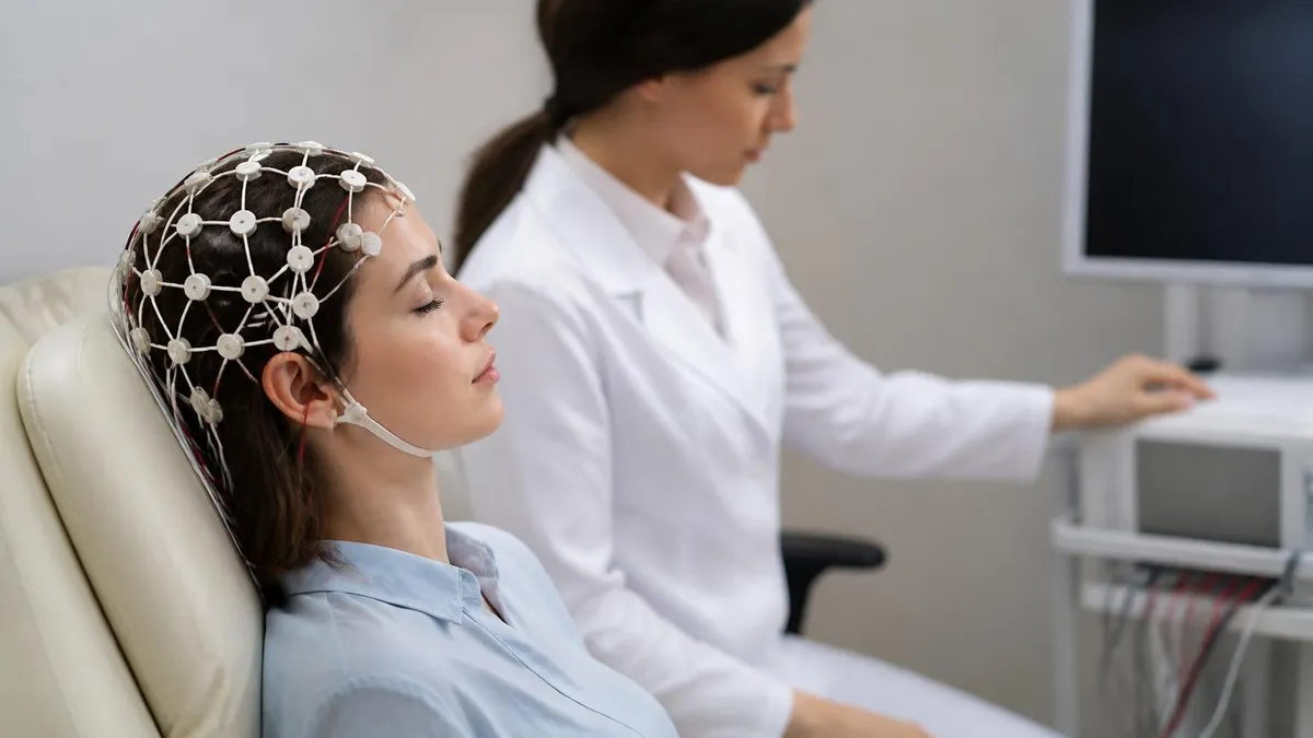
EEG vs ECG vs EKG: What Each Test Measures, How They Differ, and What to Expect
EEG Electroencephalography Practice Test PDF (Free Printable 2026)

Seizure EEG: What the EEG Test Shows, How It Works, and What to Expect
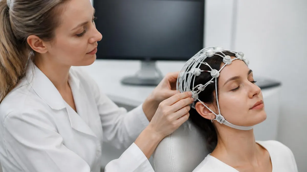
Sleep EEG: How Overnight Brain Wave Testing Works and What Your Results Mean
EEG Test Cost in 2026: Prices, Insurance Coverage, and How to Save
About the Author

Educational Psychologist & Academic Test Preparation Expert
Columbia University Teachers CollegeDr. Lisa Patel holds a Doctorate in Education from Columbia University Teachers College and has spent 17 years researching standardized test design and academic assessment. She has developed preparation programs for SAT, ACT, GRE, LSAT, UCAT, and numerous professional licensing exams, helping students of all backgrounds achieve their target scores.