Abnormal EEG Waves: What Your EEG Test Results Really Mean 2026 June
What do abnormal EEG waves mean? Learn EEG test results, wave patterns, cost, and next steps. 🧠 Clear guide for patients and students.

An EEG test — short for electroencephalogram — records the electrical activity of your brain through small sensors attached to your scalp. When neurologists review the results, they are looking for patterns that fall outside the normal range. These irregular patterns, collectively known as abnormal eeg waves, can point to a wide range of neurological conditions, from epilepsy and sleep disorders to metabolic disturbances and brain injuries. Understanding what these patterns mean is the first step toward an accurate diagnosis and an effective treatment plan.
The EEG medical test has been used clinically since the 1930s, and it remains one of the most valuable tools in modern neurology. Unlike imaging studies such as MRI or CT scans, which capture the physical structure of the brain, an EEG captures function — the real-time rhythm of neurons firing together. This distinction matters enormously when a patient has normal-looking brain anatomy but abnormal electrical behavior. In those cases, the EEG is often the only test that reveals what is actually going wrong.
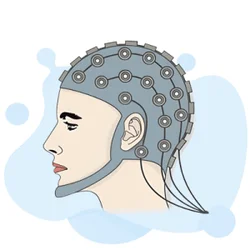
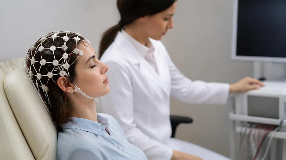
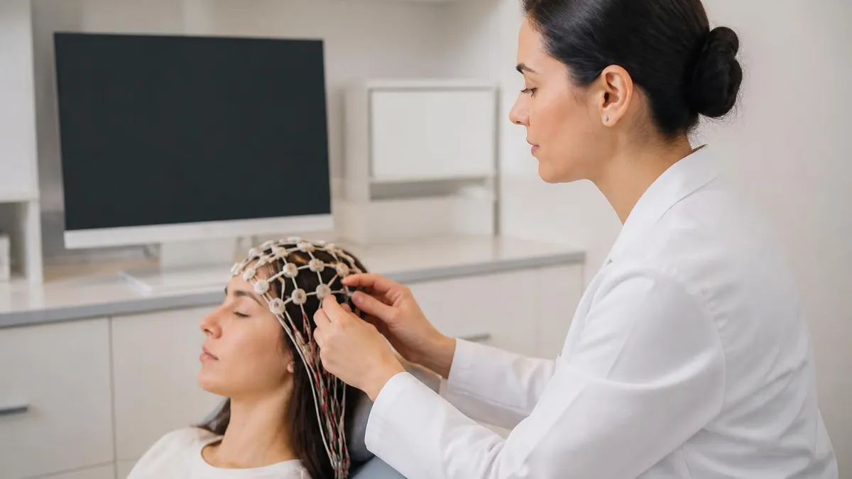
Many patients wonder what is an EEG test before they walk through the clinic door. In simple terms, it is a painless, noninvasive recording session in which a technician places 19 to 25 electrodes on specific scalp locations defined by the international 10-20 system. The electrodes detect tiny voltage fluctuations — measured in microvolts — and amplifiers turn those signals into the familiar wave tracings that neurologists read. The entire setup process typically takes 20 to 45 minutes, and the actual recording lasts anywhere from 20 minutes to several hours depending on the clinical question.
How long is an EEG test? A routine outpatient EEG usually runs about 20 to 40 minutes of actual recording time after setup. A prolonged video-EEG monitoring session, used to capture seizures on camera while recording brain waves simultaneously, can run for several days in a hospital setting. Ambulatory EEGs, which patients wear at home, typically record for 24 to 72 hours. The length of the study is chosen based on how frequently the abnormality is expected to appear — rare events require longer recording windows to be caught.
Before the test, patients are usually asked to wash their hair, avoid caffeine, and sometimes to sleep-deprive themselves slightly so they are more likely to fall asleep during the recording. Sleep is clinically useful because certain abnormal wave patterns appear only during drowsiness or light sleep. Technicians may also perform activation procedures — having the patient breathe rapidly (hyperventilation) or flashing a strobe light (photic stimulation) — to provoke latent abnormalities that would not appear during quiet wakefulness alone.
The EEG test cost in the United States varies significantly depending on the type of study and the facility. A routine outpatient EEG typically ranges from $200 to $700 before insurance adjustments. Prolonged video-EEG monitoring can cost $5,000 to $15,000 or more per day in a hospital epilepsy monitoring unit. Most commercial insurance plans and Medicare cover medically indicated EEGs, though prior authorization may be required. Patients without insurance can often negotiate cash-pay discounts or access community health centers that offer the test at reduced rates.
EEG test side effects are minimal because the procedure is entirely noninvasive. The gel used to secure electrodes can temporarily make your hair feel sticky, and some patients experience mild scalp discomfort from electrode placement. The hyperventilation activation procedure can cause temporary lightheadedness or tingling. In rare cases, photic stimulation triggers a seizure in photosensitive individuals — but this happens in a controlled clinical setting where trained staff are present and prepared to respond immediately.
EEG Test by the Numbers
Types of Abnormal EEG Wave Patterns
Sharp waves, spikes, and spike-and-slow-wave complexes that strongly correlate with seizure disorders. These waveforms have a characteristic pointed morphology and are the most clinically significant abnormal EEG findings neurologists look for when evaluating epilepsy.
Abnormally slow delta or theta activity confined to one brain region. This pattern typically indicates a localized structural problem such as a stroke, tumor, or cortical contusion, and the location of the slowing can guide further imaging.
Generalized excess of slow frequencies (delta or theta) replacing normal alpha and beta rhythms. This nonspecific pattern appears in encephalopathy caused by metabolic disturbances, toxic exposures, systemic illness, or severe global brain dysfunction.
Alternating bursts of high-amplitude activity and periods of near-electrical silence. This pattern indicates severely impaired brain function and is seen in deep anesthesia, hypoxic-ischemic encephalopathy, or certain catastrophic brain injuries.
Repeating waveforms at regular intervals, including lateralized periodic discharges (LPDs) and generalized periodic discharges (GPDs). These patterns are often seen in critically ill patients and can be associated with nonconvulsive seizure activity.
Understanding what causes abnormal EEG results requires a working knowledge of how different diseases affect the brain's electrical environment. The brain is an electrochemical organ, and any process that disrupts the delicate balance of ion channels, neurotransmitters, or synaptic connections can alter the EEG. Neurologists use the pattern of abnormality — its frequency, distribution, morphology, and relationship to clinical state — to narrow the differential diagnosis before ordering additional tests. The EEG is rarely the final word, but it is often the test that points the team in the right direction.
Epilepsy is the condition most commonly associated with abnormal EEG findings, particularly epileptiform discharges such as spikes and sharp waves. However, it is critical to understand that a single abnormal EEG does not diagnose epilepsy on its own, and a single normal EEG does not rule it out.
Epilepsy is a clinical diagnosis based on the history of unprovoked, recurrent seizures. The EEG provides supportive evidence. Approximately 20 percent of people with confirmed epilepsy will have a normal routine EEG, especially if the recording does not capture an event or if the seizure focus is deep within brain structures not easily detected by surface electrodes.
Metabolic and toxic encephalopathies produce diffuse EEG slowing that reflects widespread neuronal dysfunction rather than a focal structural lesion. Common causes include hepatic encephalopathy from liver failure, uremic encephalopathy from kidney failure, hypoglycemia, hypoxia, hyperammonemia, drug toxicity, and sepsis-associated encephalopathy. The degree of EEG slowing often correlates with the severity of the encephalopathy, making the EEG a useful tool for tracking a patient's neurological trajectory in the ICU even when clinical assessment is limited by sedation or intubation.
Herpes simplex encephalitis is one of the most important infectious conditions that produces a distinctive EEG pattern — periodic lateralized discharges over the temporal lobes, often with a characteristic one- to two-per-second periodicity. This pattern, in the right clinical context (fever, acute confusion, temporal lobe symptoms), should prompt immediate antiviral treatment even before MRI or CSF results return, because early treatment dramatically improves outcomes. The EEG's ability to rapidly identify this pattern has saved lives by shortening the time to acyclovir administration.
Sleep disorders also produce recognizable EEG abnormalities. Narcolepsy is associated with sleep-onset REM periods — meaning patients enter REM sleep within 15 minutes of falling asleep rather than the normal 60 to 90 minutes. A sleep EEG or polysomnography can capture this finding and help differentiate narcolepsy from other causes of excessive daytime sleepiness. Parasomnias such as REM sleep behavior disorder show characteristic loss of the normal muscle atonia that should accompany REM sleep, a finding visible only on a study that includes both EEG and EMG channels.
Brain tumors, strokes, and traumatic brain injuries produce focal EEG abnormalities that correspond anatomically to the lesion site. In stroke, acute infarcts suppress the fast frequencies (alpha and beta) and replace them with slow delta or theta activity over the affected hemisphere. As the stroke matures and brain tissue reorganizes over weeks to months, the EEG slowing often improves, even when structural imaging shows persistent damage. This recovery of electrical activity can be a more sensitive marker of functional recovery than imaging alone, which is one reason neurologists sometimes use serial EEGs to monitor stroke patients.
Autoimmune encephalitis — a growing and increasingly recognized category of neurological disease — produces EEG patterns that range from diffuse slowing to a distinctive pattern called extreme delta brush, which is highly specific for anti-NMDA receptor encephalitis in young patients. The EEG finding of delta brushes in the right clinical context (subacute psychiatric symptoms, movement disorder, autonomic instability in a young woman) should prompt urgent autoimmune antibody testing and immunotherapy. Early recognition and treatment can result in dramatic recovery even after prolonged severe illness.
What Is an EEG Test: Procedure, Cost, and Side Effects
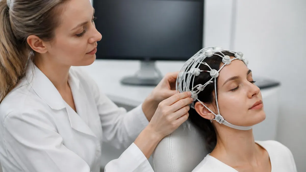
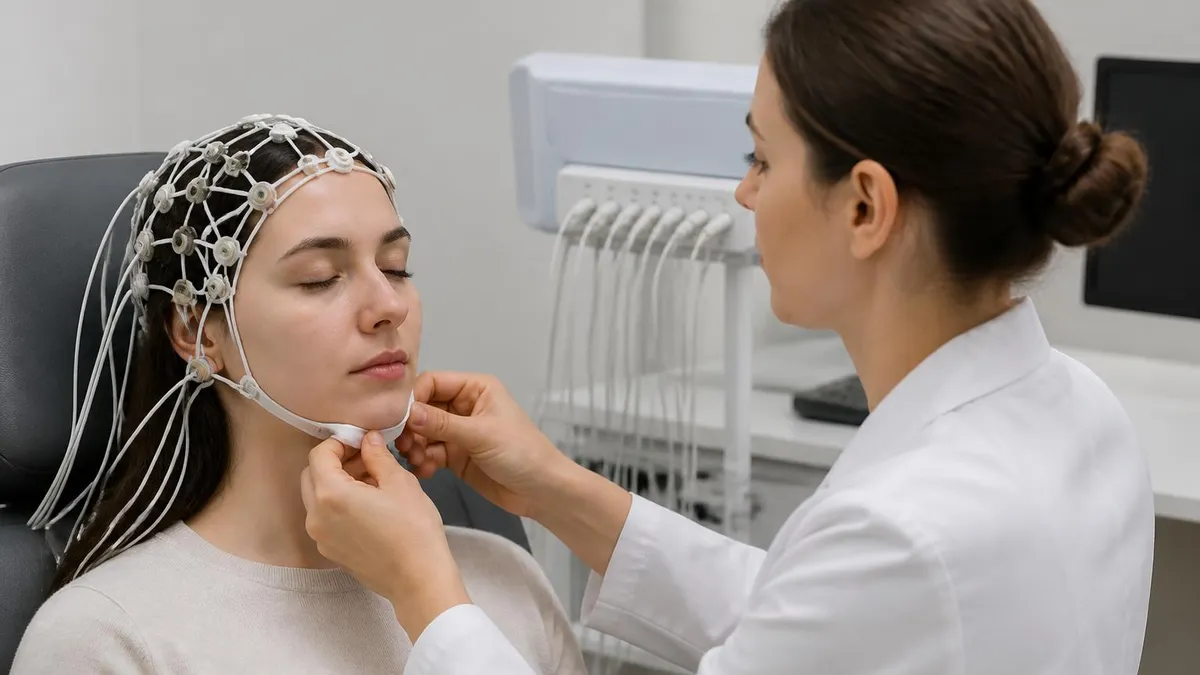
During an EEG test, a trained EEG technologist applies 19 to 25 electrodes to your scalp using a conductive gel or paste, following the standardized international 10-20 placement system. Each electrode detects the electrical potential at its location relative to a reference. You will lie back in a reclining chair or bed, close your eyes, and rest quietly while the machine records your brain's activity. The technologist will ask you to breathe deeply for several minutes and may briefly flash a strobe light as part of standard activation procedures.
Most patients describe the experience as relaxing rather than stressful. Nothing penetrates the skin — the electrodes simply sit on the surface of your scalp. If you are scheduled for a sleep-deprived EEG, you will have been asked to stay awake for part of the night beforehand so that you drift into drowsiness or light sleep during the recording, which helps the technologist capture activity patterns that only emerge in transitional sleep states. After the recording ends, the gel is wiped away and you can return to normal activities immediately.

EEG Test: Benefits and Limitations
- +Noninvasive and painless — no needles, injections, or radiation exposure
- +Provides real-time functional brain data that MRI and CT cannot capture
- +Essential for epilepsy diagnosis, seizure classification, and surgical planning
- +Relatively affordable compared to MRI or inpatient monitoring in routine form
- +Can identify emergent patterns like burst-suppression or periodic discharges in ICU patients
- +Useful for monitoring sedation depth, detecting nonconvulsive status epilepticus, and tracking encephalopathy
- −A single routine EEG misses abnormalities in up to 20% of epilepsy patients
- −Cannot pinpoint the exact anatomical location of deep seizure foci without invasive electrodes
- −Results require expert interpretation — errors occur in inexperienced hands
- −Artifacts from muscle activity, eye movement, and electrode movement can mimic or mask abnormalities
- −Does not directly image brain structure — must be combined with MRI for complete evaluation
- −Prolonged monitoring studies are expensive and logistically demanding for patients
Preparing for Your EEG Test: 10-Step Checklist
- ✓Wash your hair the morning of the test but skip conditioner, which can interfere with electrode adhesion.
- ✓Avoid caffeine for at least 8 hours before the appointment, as it can alter wave patterns.
- ✓Take all prescribed medications as usual unless your neurologist specifically instructs otherwise.
- ✓If ordered a sleep-deprived EEG, stay awake for 4 to 6 extra hours before the test.
- ✓Eat a light meal beforehand — hypoglycemia can cause EEG changes unrelated to your condition.
- ✓Wear comfortable, loose clothing and leave hair accessories like braids or extensions at home.
- ✓Arrive 10 to 15 minutes early to complete paperwork and allow time for electrode application.
- ✓Tell the technologist about all medications, supplements, and any history of photosensitivity.
- ✓Plan to relax during the recording — closing your eyes and breathing slowly improves signal quality.
- ✓Arrange a ride home if you were sleep-deprived, as drowsiness after the test can affect driving safety.
Normal EEG Does Not Rule Out Epilepsy
Up to 20 percent of people with confirmed epilepsy will have a normal routine EEG. If your first study is negative but your clinical symptoms strongly suggest seizures, your neurologist may order a prolonged ambulatory EEG, a sleep-deprived study, or inpatient video-EEG monitoring to increase the diagnostic yield. Never interpret a normal EEG as definitive proof that seizures are not occurring.
Interpreting EEG results is a specialized skill that takes neurologists and neurophysiologists years to develop. When your physician reviews an EEG report with you, the language can seem technical and intimidating. Understanding the basic vocabulary — terms like focal versus diffuse, epileptiform versus nonepileptiform, and reactive versus nonreactive — helps you participate meaningfully in that conversation and ask the right follow-up questions. A good neurologist will translate the findings into plain language, but coming to that appointment already familiar with the concepts puts you in a far stronger position as an advocate for your own health.
The EEG report will typically describe the background activity first — the dominant frequency and amplitude of the brain's resting rhythm. In a healthy awake adult, this is usually an alpha rhythm of 8 to 12 Hz, most prominent over the occipital (back of head) regions when eyes are closed. This background rhythm slows during drowsiness and disappears during deeper sleep stages. If the background is diffusely slow — dominated by theta (4–8 Hz) or delta (0.5–4 Hz) frequencies during wakefulness — the report will note this as abnormal background slowing, which indicates some degree of global cerebral dysfunction.
After describing the background, the report addresses specific abnormalities. A common finding that confuses patients is the description of a sharp wave or spike in a single hemisphere. Patients often assume that any sharp-looking wave on the tracing is abnormal. In reality, EEG readers must carefully distinguish true epileptiform discharges — which have very specific morphological criteria including a sharp contour, a defined field of spread across electrodes, and appropriate after-going slow wave — from normal benign variants and artifacts that can superficially resemble abnormal findings but have no clinical significance.
Benign EEG variants are one of the most important concepts for patients and clinicians to understand. Patterns such as small sharp spikes (SSS), rhythmic mid-temporal theta of drowsiness (RMTD), and wicket spikes are normal variants that are occasionally mistaken for epileptiform activity by less experienced readers. Misidentifying these patterns as abnormal can lead to unnecessary epilepsy diagnoses, inappropriate antiseizure medications, and the profound personal and professional consequences that come with an incorrect epilepsy label — including driving restrictions and occupational limitations. This is precisely why EEG interpretation should be performed by board-certified clinical neurophysiologists.
When the EEG report identifies a focal abnormality, the next question is always whether the clinical presentation matches the anatomy. An EEG showing right temporal sharp waves in a patient with episodes of déjà vu, lip smacking, and hand automatisms makes perfect clinical sense — the right temporal lobe is the classic location for that semiology of focal aware seizures.
On the other hand, right temporal sharp waves in a patient with exclusively generalized tonic-clonic seizures and a family history of juvenile myoclonic epilepsy would raise questions about whether the EEG finding is driving the clinical picture or whether it is an incidental variant.
The concept of EEG reactivity is particularly important in the ICU setting. A reactive EEG is one in which the background activity changes in response to external stimulation — such as a loud noise or sternal rub — even if those changes are subtle. Reactivity indicates that some level of corticothalamic connectivity is preserved, which is a marker of less severe brain dysfunction and carries a relatively better prognosis. A nonreactive EEG, in which the background pattern does not change with any stimulation, indicates severely disrupted cortical function and is associated with worse outcomes in comatose patients.
After receiving your EEG results, it is reasonable to ask your neurologist several specific questions: What is the overall background activity, and is it appropriate for my age and level of alertness? Were any epileptiform discharges seen, and if so, where were they located? Are there any patterns of clinical significance that need immediate action?
Does the EEG finding change my diagnosis or medication plan? And finally — is a repeat study or more prolonged monitoring recommended? These questions help ensure you leave the appointment with a clear understanding of where you stand and what comes next in your care pathway.

If you have an abnormal EEG and begin experiencing new or worsening seizures, prolonged confusion lasting more than a few minutes, sudden inability to speak or understand language, or weakness on one side of your body, seek emergency care immediately. These symptoms may indicate a new neurological event — such as seizure escalation, stroke, or encephalitis — that requires urgent evaluation. Do not wait for an outpatient appointment.
For EEG technologists and students preparing for board certification through organizations such as ABRET (American Board of Registration of Electroencephalographic and Evoked Potential Technologists), a strong command of abnormal EEG wave patterns is essential. The R. EEG T. examination tests candidates on waveform recognition, artifact identification, montage selection, patient preparation, and clinical correlation across a wide range of neurological conditions. Mastery of the material covered in this article — from epileptiform discharges to periodic patterns to encephalopathic slowing — forms the core of what the exam expects candidates to know.
Epileptiform patterns on the EEG examination can be divided into those associated with focal (partial) epilepsies and those associated with generalized epilepsies. Focal epileptiform patterns include centrotemporal spikes (the hallmark of self-limited epilepsy with centrotemporal spikes, formerly called benign rolandic epilepsy), temporal lobe sharp waves associated with mesial temporal lobe epilepsy, and frontal lobe epileptiform discharges that can be subtle and require careful review of multiple montages to identify. Generalized epileptiform patterns include the 3-Hz spike-and-wave of childhood absence epilepsy, the polyspike-and-wave of juvenile myoclonic epilepsy, and the irregular spike-and-wave seen in Lennox-Gastaut syndrome.
Activation procedures are a critical component of both clinical EEG practice and board examination content. Hyperventilation lowers carbon dioxide levels in the blood, causing cerebral vasoconstriction and a slight reduction in cerebral blood flow. This metabolic change can activate epileptiform discharges, particularly the 3-Hz spike-and-wave of absence epilepsy. In children, hyperventilation reliably provokes absence seizures in untreated patients, making it one of the most diagnostically useful activation procedures available. Photic stimulation activates photoparoxysmal responses in photosensitive individuals, most commonly over the posterior head regions at flash frequencies between 10 and 20 Hz.
Sleep activation is perhaps the most powerful tool in the EEG technologist's arsenal for increasing epileptiform yield. Both focal and generalized epileptiform discharges increase in frequency during non-REM sleep — particularly in the transition from wakefulness to drowsiness (stage N1) and during deeper slow-wave sleep (stage N3). This is why sleep deprivation before a routine EEG increases the diagnostic yield — it makes patients more likely to fall asleep during the recording, providing a window into sleep-activated abnormalities that a fully alert recording would miss entirely.
Artifact recognition is an equally important skill that distinguishes a competent EEG reader from an expert one.
Physiological artifacts — produced by the patient's own body — include electrocardiographic artifact (from the heart's electrical signal picked up by scalp electrodes), electromyographic artifact (from scalp and facial muscle activity), eye movement artifact (producing large deflections in frontal channels), and pulse artifact (from electrode movement over a scalp artery). Technical artifacts include electrode pop artifact from poor electrode contact, 60-Hz electrical interference from nearby equipment, and movement artifact from patient repositioning. Misidentifying any of these as a brain wave pattern leads to errors in interpretation.
Continuous EEG monitoring (cEEG) in the neurological ICU has emerged as a standard of care at major academic medical centers for critically ill patients with altered consciousness, known or suspected seizures, or conditions at high risk for nonconvulsive status epilepticus.
Studies have shown that 8 to 48 percent of comatose ICU patients have nonconvulsive seizures detectable only by EEG — events that are invisible to bedside clinical observation but that contribute to secondary brain injury if untreated. The ability to recognize ictal and interictal patterns on continuous EEG tracings is now a critical competency for intensive care neurologists and advanced practice providers working in neurocritical care settings.
For students and technologists who want to deepen their understanding of EEG patterns through structured practice, working through categorized question banks focused on abnormal EEG waves provides an efficient way to build pattern recognition. The more tracings you read — even simulated ones in an exam-style format — the faster and more accurately you will recognize key patterns in the clinical environment. Practice with immediate feedback trains the eye and the analytical mind simultaneously, accelerating the kind of expertise that would otherwise require years of unstructured clinical exposure.
If you or a family member has just received abnormal EEG results, knowing how to navigate the next steps makes a significant difference in outcomes. The most important first step is scheduling a follow-up appointment with a board-certified neurologist — ideally one who specializes in epilepsy or clinical neurophysiology — rather than relying solely on the interpretation of a general practitioner who may be less familiar with the nuances of EEG findings.
An epileptologist or clinical neurophysiologist can review the raw EEG data, not just the written report, and provide a more detailed interpretation in the context of your specific history and symptoms.
During your follow-up, bring a detailed seizure diary if you have been having episodic events. Note the date, time, duration, what you were doing when the event started, what the event looked and felt like from your perspective, any warning symptoms (aura) that preceded it, and how you felt afterward (the postictal period). This information dramatically improves the clinician's ability to classify seizure type and localize the likely seizure focus, which in turn guides medication selection and further evaluation.
The EEG result tells the neurologist what your brain looks like electrically; your seizure diary tells them what your seizures look like clinically. Together, these data points build the complete picture.
If your neurologist recommends antiseizure medication based on abnormal EEG findings, it is important to understand what the medication is targeting, what the common side effects are, and when you should expect to see a clinical response. Most antiseizure medications take several weeks to reach steady-state blood levels, and dose adjustments may be required over time. Do not stop antiseizure medications abruptly without medical guidance — sudden discontinuation can trigger rebound seizures, including status epilepticus, even in patients who have been seizure-free for extended periods. Always taper under physician supervision.
Patients with abnormal EEG results are often concerned about driving restrictions. In the United States, each state independently sets the seizure-free period required before a person with epilepsy may legally drive. Most states require 3 to 12 months of seizure freedom, though regulations differ for commercial versus personal vehicle licenses. An abnormal EEG alone — without a history of unprovoked seizures — generally does not trigger a legal driving restriction, but your physician may still recommend caution depending on the pattern found and your overall clinical situation. Always discuss driving specifically with your neurologist.
Lifestyle modifications can complement medical treatment for patients with seizure disorders identified through EEG. Sleep deprivation is one of the most potent seizure triggers for many individuals — even a single night of poor sleep can lower the seizure threshold significantly. Alcohol consumption, especially in excess or during withdrawal, is another major trigger. Stress management, regular sleep schedules, and avoiding flickering lights in photosensitive patients are practical, evidence-based strategies that many patients find meaningfully reduce their seizure frequency alongside medication. These are not substitutes for medical treatment but powerful adjuncts that give patients agency over their condition.
Advances in EEG technology continue to expand the diagnostic possibilities available to clinicians. High-density EEG systems with 256 electrodes can provide much more precise source localization than conventional 19-electrode setups. Intracranial EEG — placing electrodes directly on or within the brain cortex — offers the ultimate spatial resolution for surgical planning in drug-resistant epilepsy.
Wearable EEG devices are under active development for home-based long-term monitoring with consumer-grade hardware. Machine learning algorithms trained on thousands of EEG recordings are beginning to outperform human readers at detecting specific patterns, though clinical deployment of fully automated interpretation tools remains a work in progress that will require rigorous validation before replacing human oversight.
The field of EEG continues to evolve rapidly, and patients who stay engaged with the latest developments — by reading reputable neurology resources, participating in epilepsy patient communities, and asking informed questions at their clinical appointments — are better positioned to benefit from emerging diagnostic and therapeutic options. If you are a student or technologist building your career in neurophysiology, the same principle applies: staying current with the EEG literature, pursuing continuing education, and practicing systematic waveform reading are the habits that separate competent technologists from truly excellent ones who provide meaningful value to their clinical teams.
EEG Questions and Answers
EEG Electroencephalography Practice Test PDF (Free Printable 2026)
Sleep EEG: How Overnight Brain Wave Testing Works and What Your Results Mean
EEG Test Cost in 2026: Prices, Insurance Coverage, and How to Save
EEG vs ECG vs EKG: Brain and Heart Test Differences
Abnormal EEG Results: What They Mean, Common Patterns, and Next Steps
About the Author
Educational Psychologist & Academic Test Preparation Expert
Columbia University Teachers CollegeDr. Lisa Patel holds a Doctorate in Education from Columbia University Teachers College and has spent 17 years researching standardized test design and academic assessment. She has developed preparation programs for SAT, ACT, GRE, LSAT, UCAT, and numerous professional licensing exams, helping students of all backgrounds achieve their target scores.