
Wearable EEG Devices: What They Are, How They Work, and What to Expect from Your EEG Test
What is an EEG test? 🧠 Learn how wearable EEG devices work, what they cost, side effects, and how long the test takes.

An EEG test — short for electroencephalogram — is one of the most important diagnostic tools in modern neurology, and the rise of wearable EEG devices has fundamentally changed how and where this test can be performed.
Traditionally confined to hospital settings, the EEG medical test now reaches patients in their homes, workplaces, and everyday environments thanks to compact, portable hardware that records the brain's electrical activity continuously over hours or even days. Understanding what these devices do, how they differ from clinical systems, and what the data actually means is increasingly essential knowledge for EEG technologists preparing for board certification.
The standard EEG test works by placing small metal electrodes on the scalp according to the internationally recognized 10-20 electrode placement system. Each electrode detects tiny voltage fluctuations produced by synchronized postsynaptic potentials in cortical neurons. These signals — measured in microvolts — are amplified, filtered, and displayed as a series of waveform channels that clinicians read to identify normal rhythms, abnormal slowing, epileptiform discharges, and other neurological signatures. The entire signal chain must maintain exceptionally high fidelity because even minor artifacts from muscle movement or electrode pop can mimic or obscure clinically meaningful patterns.
Wearable EEG devices apply the same fundamental electrochemical principles but engineer them into form factors small enough to clip behind the ear, embed in a headband, or integrate into a soft cap. The engineering tradeoffs are substantial: consumer-grade wearables may use only 2 to 14 electrodes and rely on dry electrode contacts that sacrifice some signal quality for convenience, whereas medical-grade ambulatory systems designed for long-term monitoring still use the full 10-20 array with impedance-checked, gel-based contacts. Knowing these distinctions matters enormously when interpreting recordings, because channel count and electrode contact quality directly affect the diagnostic yield of the study.
From a clinical perspective, the EEG medical test ordered by a physician serves purposes far beyond the curiosity-driven applications of consumer brain-computer interfaces. Neurologists use EEG to diagnose epilepsy and classify seizure types, evaluate loss of consciousness, investigate encephalopathy, confirm brain death, monitor depth of anesthesia during surgery, and support the workup of sleep disorders. Each of these applications has specific technical requirements — bandwidth, electrode density, synchronization with video — that influence whether a wearable or stationary system is appropriate for the study.
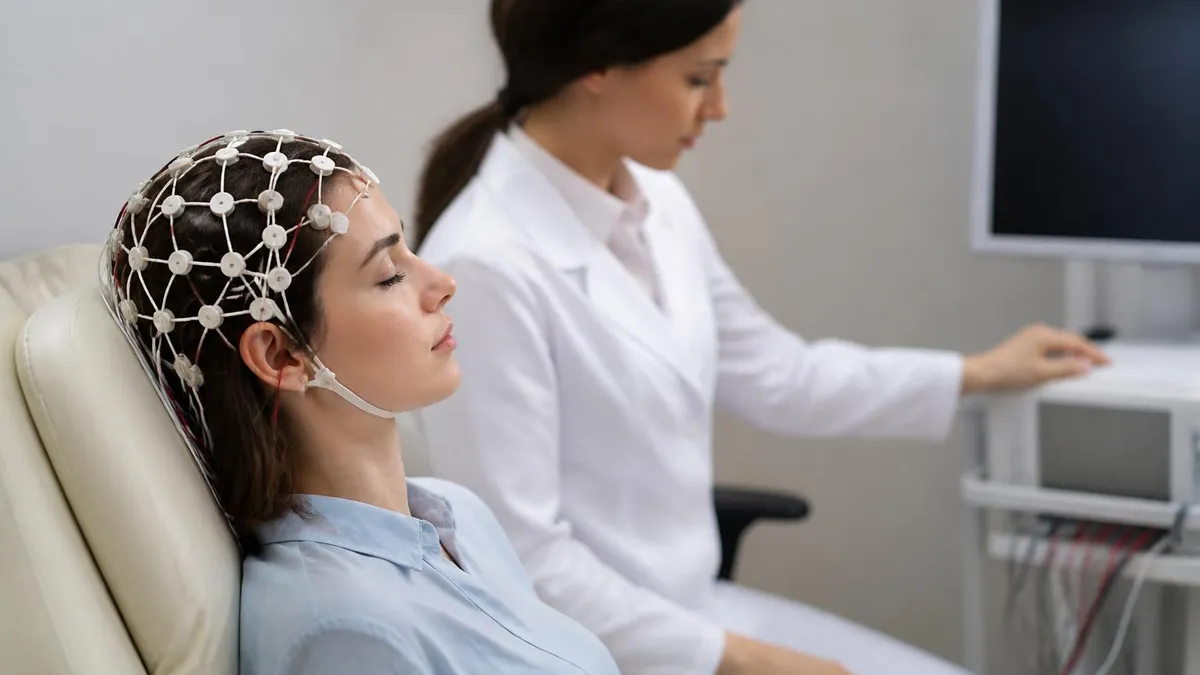
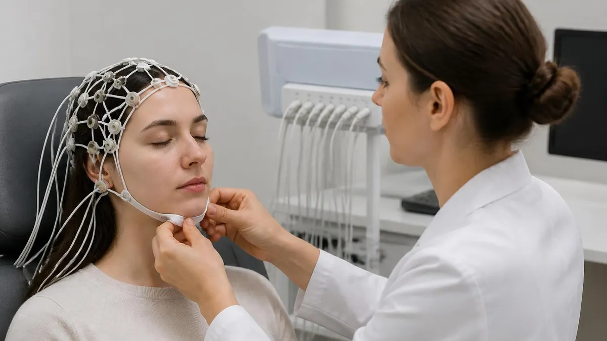
Many patients wonder what is an EEG test like in terms of physical experience. The procedure is painless and non-invasive. A trained EEG technologist measures the patient's head, marks scalp locations with a grease pencil, and applies electrodes using conductive gel or paste. In wearable ambulatory setups, a pre-fitted cap or adhesive electrode array replaces the individual placement step, dramatically shortening setup time.
Once the recording begins, patients are typically asked to relax with eyes closed, then open, and may be asked to breathe deeply for three minutes or look at a flashing light — these are activation procedures designed to provoke latent abnormalities that might not appear during a resting baseline.
The question of EEG test cost is a practical concern for many patients, particularly those without comprehensive insurance. A routine outpatient EEG typically costs between $200 and $900 before insurance adjustments in the United States, while prolonged ambulatory studies can range from $500 to $3,000 depending on the duration and the facility's technical fees.
Wearable systems intended for home use are increasingly available through equipment rental programs or as physician-prescribed devices billed through durable medical equipment channels. EEG test price varies significantly by region, provider type, and whether the recording is interpreted by a board-certified neurologist or an EEG reader in a remote reading center.
For those studying toward the ABRET R. EEG T. or CLTM credentials, mastering the technical and clinical dimensions of both traditional and wearable EEG formats is non-negotiable. Exam questions frequently test knowledge of ambulatory recording indications, artifact recognition in mobile environments, and the appropriate use of activation procedures in long-term monitoring contexts. This article walks through every major dimension of the topic — device types, clinical use cases, pros and cons, costs, and exam preparation strategies — to give technologists and students a comprehensive foundation.
Wearable EEG Devices by the Numbers

Types of Wearable EEG Devices Used in Clinical Practice
Medical-grade portable recorders using the full 10-20 electrode array with conductive gel. These systems capture 24- to 72-hour recordings outside the hospital, ideal for capturing infrequent seizures or characterizing events that occur during daily activities.
Consumer and research-grade devices using spring-loaded or foam dry electrodes, typically 8 to 14 channels. Faster to apply and more comfortable for extended wear, but generally produce higher impedance and more motion artifact than gel-based clinical systems.
Soft fabric caps with embedded electrodes distributed across the scalp. Used in research settings, neonatal monitoring, and some clinical ICU applications. Reduce setup time dramatically and allow repeated consistent placement across sessions.
Minimalist form-factor devices with only 2 to 4 electrodes positioned at temporal and mastoid sites. Limited in diagnostic resolution but useful for sleep staging, medication adherence monitoring, and consumer wellness applications including focus and stress tracking.
Understanding what is an EEG test from a technical standpoint requires appreciation of the signal processing chain that connects scalp electrodes to the final waveform display. Raw EEG signals arriving at the amplifier inputs range from 5 to 300 microvolts in amplitude. The differential amplifier rejects common-mode noise — electrical interference that appears equally on both inputs — and amplifies only the voltage difference between electrode pairs. This common-mode rejection ratio, or CMRR, is one of the most important specifications of any EEG amplifier, whether built into a desktop console or a wearable recording device clipped to a patient's belt.
After amplification, signals pass through analog or digital filters that define the frequency bandwidth of the recording. High-frequency filters, set typically between 35 and 70 Hz in routine clinical work, remove high-frequency muscle artifact and electrical line noise. Low-frequency filters, usually set at 0.1 Hz, eliminate slow baseline drifts caused by sweat, electrode polarization, or patient movement. Wearable EEG devices often apply these filters digitally in firmware, which means the technologist reviewing the recording remotely has less flexibility to retrospectively adjust filter settings than they would with raw digital data captured at full bandwidth.
Montage selection is another critical technical concept for any EEG technologist. A montage defines which pairs of electrodes are compared to display each channel on the recording. Referential montages compare each electrode to a common reference point, while bipolar montages chain adjacent electrodes together to highlight local voltage gradients. Most modern wearable systems store raw data referenced to a single electrode and allow clinicians to re-reference and re-montage the recording during review — a major advantage over older analog systems where the montage was baked into the recorded signal at acquisition time.
For those studying for the ABRET examination, understanding the physics and engineering of EEG acquisition is tested heavily alongside pattern recognition. The R. EEG T. exam blueprint specifically includes amplifier characteristics, electrode impedance requirements, filter settings, and calibration procedures. In the context of wearable eeg devices, questions may ask candidates to identify which artifact patterns are more prevalent in ambulatory versus routine recordings, or to explain why higher electrode impedance in dry-contact systems increases susceptibility to 60 Hz interference.
The question of how long is an EEG test is straightforward for routine outpatient studies but becomes more nuanced for wearable ambulatory formats. A standard routine EEG including setup, recording, and electrode removal typically lasts between 45 minutes and 90 minutes total, with the actual recording phase running 20 to 40 minutes.
Prolonged outpatient EEGs — sometimes called extended routine EEGs — run for 1 to 2 hours. Ambulatory wearable studies are prescribed for 24, 48, or 72 hours, though the recording is interrupted periodically when the patient charges the device or swims. Video-EEG monitoring in an epilepsy monitoring unit may extend for days or weeks.
Activation procedures are a defining feature of EEG methodology that every technologist must master regardless of whether the recording is being done on a fixed system or a portable wearable device. Hyperventilation — asking the patient to breathe rapidly and deeply for 3 minutes — decreases arterial CO2, producing cerebral vasoconstriction and a mild hypoxic effect that can activate generalized spike-wave discharges in patients with idiopathic generalized epilepsies.
Photic stimulation, using a stroboscope flashing at frequencies between 1 and 30 Hz, tests for photoparoxysmal responses. Sleep deprivation before the recording and sleep recording itself are additional activation methods that increase diagnostic yield for epilepsy evaluation.
Wearable ambulatory devices capture activation procedures differently than lab-based systems. The patient carries a push-button event marker to annotate the recording during hyperventilation attempts at home. Some advanced ambulatory systems include integrated accelerometers that automatically flag periods of patient movement or gross motor activity, helping reviewers correlate waveform changes with posture and activity. These engineering solutions attempt to replicate the controlled activation environment of a clinical lab in the patient's natural setting, though they introduce new challenges in artifact discrimination and event annotation accuracy.
What Is an EEG Medical Test Used For?
The most common indication for an EEG test is the evaluation of suspected epilepsy. Interictal epileptiform discharges — spikes, sharp waves, and spike-wave complexes recorded between clinical seizures — appear in roughly 50% of patients with epilepsy on a routine 20-minute recording. Extending the recording to 24 to 72 hours using an ambulatory wearable system increases sensitivity to approximately 80 to 90%, making long-term wearable EEG an essential tool in the epilepsy diagnostic workup for patients whose routine study was non-diagnostic.
Wearable ambulatory devices also allow neurologists to capture ictal recordings — EEG patterns during actual clinical events — which are essential for confirming seizure type and determining whether events are epileptic or non-epileptic in origin. Psychogenic non-epileptic seizures, which can closely mimic convulsive epileptic events clinically, produce no ictal EEG change, making the capture of a habitual event on wearable EEG diagnostically definitive in many cases. ABRET exam questions frequently test the candidate's ability to distinguish ictal from interictal patterns and to identify artifacts that can mimic epileptiform activity.

Wearable EEG Devices: Advantages and Limitations
- +Captures brain activity during real-life activities, not just in a controlled lab environment
- +Dramatically increases seizure capture rate compared to routine 20-40 minute recordings
- +Allows patients to sleep in their own beds during sleep-related EEG studies
- +Reduces hospital visits and associated costs for patients requiring prolonged monitoring
- +Newer dry-electrode systems eliminate the mess of conductive gel and reduce setup time
- +Remote monitoring capability allows a single technologist to oversee multiple patients simultaneously
- −Dry electrode contact typically produces higher impedance and more artifact than gel-based systems
- −Reduced electrode count in consumer devices limits spatial resolution for localizing focal abnormalities
- −Motion artifact from daily activities is substantially higher than in a controlled recording environment
- −Patients must manage device charging, electrode maintenance, and event logging without direct technologist supervision
- −Data transmission and cloud storage raise patient privacy and HIPAA compliance considerations
- −EEG test cost for ambulatory studies is higher than routine outpatient EEG before insurance adjustments
How to Prepare for Your EEG Test: 10-Step Checklist
- ✓Wash your hair the night before and do not apply conditioner, oil, or styling products that increase scalp impedance
- ✓Take all regularly prescribed medications on schedule unless your neurologist specifically instructs you to hold them
- ✓Avoid caffeine for at least 8 hours before the test, as it can suppress slow-wave activity
- ✓If ordered for a sleep-deprived EEG, stay awake all night before the morning appointment as instructed
- ✓Arrive 15 minutes early to allow time for paperwork and electrode setup without rushing the technologist
- ✓Inform the technologist of all medications, supplements, and any recent illnesses before recording begins
- ✓Relax during the recording and try not to chew, clench your jaw, or tense your neck muscles
- ✓Perform the hyperventilation activation procedure as directed — breathe deeply and rapidly for the full 3 minutes
- ✓Keep your eyes open and fixed on the specified target during photic stimulation to ensure consistent responses
- ✓For ambulatory studies, press the event button every time you experience any unusual symptom or sensation
Ambulatory EEG Increases Seizure Capture Rate by Up to 4×
Studies comparing 24-hour ambulatory wearable EEG to routine 20-minute recordings consistently show a 3- to 4-fold increase in the rate of capturing interictal epileptiform discharges in patients with known or suspected epilepsy. For patients with infrequent events, extending to 72 hours increases yield further. This is why neurologists routinely escalate to ambulatory EEG when an initial routine study is negative but clinical suspicion for epilepsy remains high.
EEG test cost is one of the most searched questions among patients referred for neurological evaluation, and the answer depends on several intersecting factors that technologists and patients alike should understand. The technical component of an EEG — covering electrode application, recording, and data transmission — is billed separately from the professional component, which covers physician interpretation. On an Explanation of Benefits statement, patients may see two separate charges for a single EEG study: one from the hospital or outpatient facility and one from the reading neurologist's practice group.
For routine outpatient EEG, facility technical fees typically range from $150 to $500, while professional interpretation adds another $50 to $200. In aggregate, EEG test cost before insurance often falls between $200 and $900 at an outpatient neurology clinic or hospital.
Ambulatory wearable EEG studies that run for 24 to 72 hours carry higher costs reflecting the longer recording duration, the equipment, and the increased time required for data review: total costs typically range from $500 to $3,000 depending on duration and provider. Video-EEG monitoring in a hospital epilepsy monitoring unit is billed at inpatient rates and can reach $10,000 to $30,000 for a multi-day admission.
Insurance coverage for medically indicated EEG studies is generally robust under most commercial health plans and Medicare when the study is ordered by a physician and supported by an appropriate diagnosis code. The most commonly used ICD-10 codes for EEG include G40.x series codes for epilepsy and seizure disorders, G47.x codes for sleep disorders, and R56.x codes for convulsions. Patients with Medicare Part B typically owe 20% of the Medicare-approved amount after meeting their deductible. Medicaid coverage varies by state but generally covers medically necessary EEG at rates substantially below commercial insurance reimbursement.
EEG test price for self-pay patients can sometimes be negotiated directly with the facility's billing department. Many hospital systems offer charity care programs or sliding-scale fees based on income. Some independent outpatient EEG labs offer self-pay rates that are significantly lower than hospital facility fees — sometimes $150 to $250 all-inclusive for a routine study — making it worth calling ahead to compare pricing if out-of-pocket cost is a concern. Patients should request an itemized cost estimate before scheduling and confirm whether the interpreting physician is in-network if their plan uses a narrow network.
For EEG technologists, understanding reimbursement is relevant beyond patient counseling — it directly influences which studies get ordered and how testing is structured at the institutional level. The increasing availability of affordable wearable ambulatory EEG systems has expanded access to prolonged recording for patients who previously could only access routine EEG at a local clinic. This democratization of diagnostic EEG access has clinical implications: more patients receive the longer recordings needed to achieve diagnosis, reducing the time to appropriate treatment for seizure disorders and sleep conditions.
The cost of the wearable hardware itself is a separate consideration for facilities evaluating whether to implement an ambulatory EEG program. Medical-grade ambulatory EEG recorders from manufacturers such as Natus, Nihon Kohden, and Compumedics range from approximately $5,000 to $25,000 per unit depending on channel count and feature set. EEG machine cost for full hospital-grade diagnostic systems is considerably higher, often ranging from $30,000 to $150,000. Facilities typically depreciate this equipment over 5 to 7 years and factor it into technical fee calculations when setting self-pay and contractual rates.
For ABRET exam candidates, questions about EEG reimbursement and documentation are a relatively small but present part of the exam blueprint, particularly in the area of professional standards, laboratory accreditation through ASET and the ABRET organization, and documentation requirements for studies submitted for board review. Understanding the difference between technical and professional components of EEG billing, and knowing which Current Procedural Terminology codes apply to routine, extended, ambulatory, and video-EEG monitoring, reflects the comprehensive practice knowledge expected of a credentialed technologist.

Some patients mistakenly believe they should stop anti-seizure medications before an EEG to increase the chance of recording a seizure. This can be extremely dangerous and should never be done without explicit instructions from your neurologist. Abrupt withdrawal of anti-seizure medications can precipitate prolonged seizures or status epilepticus. If a medication taper is clinically indicated for a diagnostic admission, it must be supervised in a monitored inpatient epilepsy unit — never at home.
EEG test side effects are a topic that frequently concerns patients before their first recording, and the reassuring truth is that a standard diagnostic EEG carries essentially no direct physiological risk. The electrodes record electrical activity passively — no electricity is sent into the brain or scalp during a standard EEG. The most common discomforts patients report are scalp tenderness at electrode sites, particularly if the technologist used abrasive skin prep paste to reduce impedance, and a mild headache after the hyperventilation activation procedure, which resolves within minutes of returning to normal breathing.
Photic stimulation carries a small but real risk of provoking a photoparoxysmal response or, in rare cases, a clinical seizure in patients with photosensitive epilepsy. For this reason, photic stimulation is contraindicated in patients with a known strong photosensitive response unless the clinical indication specifically requires testing the photosensitive threshold. Technologists performing photic stimulation should always have seizure safety protocols in place — side rails up on the bed, call system within reach, and rescue medication accessible — even though clinical events during routine outpatient photic stimulation are uncommon.
For wearable ambulatory EEG devices worn over 24 to 72 hours, the most commonly reported adverse experiences are scalp irritation or contact dermatitis beneath long-wearing electrodes, particularly at occipital electrode sites where hair makes maintaining good contact challenging. Patients with sensitive skin or latex allergy should inform the technologist before electrode application, as some electrode pastes and adhesives contain latex or skin-sensitizing preservatives. Modern medical-grade electrode pastes are generally well-tolerated, and severe allergic reactions are rare.
From a radiation and electromagnetic safety standpoint, EEG produces zero ionizing radiation, making it categorically different from CT scanning or X-ray procedures. EEG is safe during pregnancy, safe for neonates and premature infants, safe for patients with pacemakers and implanted cardiac defibrillators, and safe for patients with cochlear implants. The radiofrequency signals used by wireless wearable EEG transmitters to send data to a base station or smartphone are at extremely low power levels, comparable to a smartwatch, and have not been associated with adverse biological effects at diagnostic exposure durations.
For patients asking specifically about EEG test side effects related to the gel or paste used to attach electrodes, the primary concern is removal. Electrode paste can be difficult to wash out of hair, particularly if the patient has thick or long hair. Technologists typically recommend washing with warm water and a standard shampoo immediately after the study, and some facilities provide patients with a small packet of electrode paste remover. Dry-electrode wearable systems eliminate this inconvenience entirely, which is one practical advantage driving patient preference for ambulatory studies using dry-contact technology.
The psychological experience of wearing an ambulatory EEG device in public is worth acknowledging as a real patient concern, even though it is not a physical side effect. Patients required to wear visible electrode leads, a cap, or a recording device attached to their clothing for 24 to 72 hours may feel conspicuous or anxious.
Modern ambulatory systems are becoming increasingly discreet — some devices are designed to look like hearing aids or fitness trackers — but patients should be counseled realistically about the appearance of the equipment they will be wearing and given strategies for explaining it to family, coworkers, and others they encounter during the recording period.
When comparing EEG to other neurological tests, its safety profile is one of its most compelling advantages. Unlike MRI, EEG has no contraindications related to metallic implants or claustrophobia. Unlike CT or PET scanning, it involves no radiation. Unlike lumbar puncture or cerebral angiography, it is entirely non-invasive. This combination of safety, low cost relative to MRI, and unique ability to record real-time brain electrical activity second-by-second makes EEG — including its modern wearable forms — an indispensable and irreplaceable tool in the neurological diagnostic armamentarium for the foreseeable future.
Preparing effectively for the ABRET R. EEG T. examination requires understanding not just the clinical applications of EEG but the technical operational details that differentiate a competent technologist from a merely adequate one. The examination blueprint, publicly available through the ABRET organization, allocates approximately 20 to 25% of questions to equipment operation and troubleshooting — a domain that now explicitly includes ambulatory and wearable EEG systems. Candidates who study only pattern recognition and clinical interpretation without mastering the engineering and operational side of EEG will find themselves underprepared for a significant proportion of exam questions.
For wearable and ambulatory EEG specifically, exam candidates should be able to identify the indications that distinguish ambulatory outpatient EEG from video-EEG monitoring unit admission, explain the impedance requirements for ambulatory versus routine recordings, list the information that must be included in patient instructions for a home ambulatory study, and describe how to troubleshoot common ambulatory artifact patterns including electrode pop, chewing artifact, and movement-related myogenic artifact that is dramatically more prevalent in out-of-lab recordings.
The ability to recognize when artifact mimics pathology — and when pathology might be dismissed as artifact — is one of the highest-order skills tested on the credentialing examination.
Study resources for the ABRET examination have expanded considerably in recent years, with online practice question banks, video tutorial libraries, and digital atlas collections replacing the traditional text-heavy study guides that dominated EEG education for decades. Practice questions that emphasize clinical vignettes with waveform images are particularly valuable because they train the visual pattern recognition skills that are central to real-world EEG interpretation. Timed practice sessions that simulate the 3-hour examination format build the cognitive endurance needed to maintain accuracy on the later sections of the test when mental fatigue is a factor.
One practical study strategy that many successful ABRET candidates endorse is organizing study content around the major ABRET content domains — patient care and safety, technical skills, EEG data acquisition and processing, and EEG interpretation — rather than studying topics in isolation. This domain-based approach mirrors the examination structure and helps candidates identify their weakest areas early so they can allocate study time proportionally.
Candidates who score poorly on practice questions related to activation procedures, for example, should dive deep into the hyperventilation physiology, the normal HV response, the pathological HV response, and the contraindications to HV before moving on to other content areas.
Hands-on experience with actual EEG equipment — including ambulatory wearable devices if available at the candidate's facility — cannot be replaced by reading alone. ABRET requires a minimum number of supervised procedures before a candidate becomes eligible to sit the examination, precisely because procedural competence is a prerequisite for clinical credibility. Candidates should seek every opportunity to perform electrode application on patients with varying hair types and scalp conditions, troubleshoot impedance problems in real time, apply activation procedures with proper timing and patient instructions, and review recordings with a supervising EEG physician to build interpretive judgment.
Time management during the examination is a skill that deserves explicit preparation. The ABRET R. EEG T. examination contains 225 questions to be completed in approximately 3 hours, leaving an average of just under 48 seconds per question.
Most candidates find that straightforward recognition questions can be answered in 20 to 30 seconds, leaving additional time for the more complex clinical scenario questions that require multi-step reasoning. Practicing with timed question sets that mirror this pace helps candidates develop an internal sense of when to commit to an answer versus when to flag a question and return to it after completing the rest of the section.
Finally, candidates should approach the ABRET examination with confidence grounded in systematic preparation rather than anxiety about the breadth of the content domain. The examination tests a defined body of knowledge that is thoroughly mapped in the ABRET content outline, and candidates who have completed an accredited EEG technology program, accumulated the required clinical hours, and studied systematically with quality practice materials are well-positioned for success.
Every question answered correctly on a practice test is a reinforcement of knowledge that will be available on examination day — and the technologists who pass this examination go on to provide diagnostic clarity for patients whose neurological conditions depend on the quality of their recordings.
EEG Questions and Answers
EEG vs ECG vs EKG: What Each Test Measures, How They Differ, and What to Expect
EEG Electroencephalography Practice Test PDF (Free Printable 2026)

Seizure EEG: What the EEG Test Shows, How It Works, and What to Expect
EEG Test Cost in 2026: Prices, Insurance Coverage, and How to Save

Cost of EEG Machine: What Hospitals, Clinics, and Labs Actually Pay in 2026
About the Author

Educational Psychologist & Academic Test Preparation Expert
Columbia University Teachers CollegeDr. Lisa Patel holds a Doctorate in Education from Columbia University Teachers College and has spent 17 years researching standardized test design and academic assessment. She has developed preparation programs for SAT, ACT, GRE, LSAT, UCAT, and numerous professional licensing exams, helping students of all backgrounds achieve their target scores.