EEG Systems: A Complete Guide to the EEG Test, How It Works, and What to Expect
Learn what an EEG test is, how EEG systems work, test duration, cost, and side effects. 🧠 Your complete US patient and student guide.

An EEG test — short for electroencephalogram — is one of the most important diagnostic tools in modern neurology, and understanding how eeg systems work is essential for both patients preparing for the procedure and technologists studying for certification. The test measures the brain's electrical activity by recording voltage fluctuations produced by ionic current flows within neurons.
Physicians order an EEG medical test to investigate a wide range of neurological conditions, from epilepsy and seizure disorders to sleep disturbances, encephalopathy, and even brain death confirmation in critical care settings. For the millions of Americans who undergo this test each year, knowing what to expect makes the entire experience far less intimidating.
The history of EEG stretches back to 1924, when German psychiatrist Hans Berger first recorded human brain electrical activity. Since then, EEG systems have evolved from bulky analog machines with ink-pen recorders into highly sophisticated digital platforms capable of capturing hundreds of data channels simultaneously. Modern hospital-grade EEG systems sample electrical signals at rates of 256 Hz to 2,000 Hz or more, preserving even the most subtle high-frequency oscillations that older machines would have completely missed. This technological leap has dramatically improved diagnostic accuracy for complex epilepsy syndromes and subtle encephalopathies.
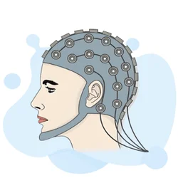
Patients are often curious about what is an EEG test before they arrive at the clinic. In straightforward terms, a trained EEG technologist places a cap or individual electrodes onto the scalp using a conductive gel. These electrodes pick up the tiny electrical signals generated by the brain, amplify them roughly one million times, and display them as waves on a computer screen.
The entire setup process typically takes 20 to 45 minutes, after which the actual recording phase begins. The patient is asked to relax, sometimes breathe rapidly, and may be exposed to flashing lights — all designed to provoke any abnormal electrical patterns that might not appear during rest alone.
One of the most frequently asked questions is how long is an EEG test. For a standard outpatient routine EEG, the total appointment time — including electrode placement, recording, and electrode removal — typically runs between 60 and 90 minutes. The recording phase itself is usually 20 to 40 minutes.
However, if a physician suspects nocturnal seizures or needs to capture a seizure event on video, a prolonged or ambulatory EEG may be ordered, which can last anywhere from 24 hours to several days. Sleep-deprived EEGs add another variable, requiring patients to arrive having slept only a few hours the night before.
The EEG medical test is completely non-invasive and painless. No needles penetrate the skin, no electrical current is sent into the brain, and no sedation is typically required for cooperative adult patients. The electrodes simply sit on the surface of the scalp, acting as passive sensors. This safety profile makes EEG uniquely valuable across the entire lifespan — it can be performed on premature neonates in intensive care units, on elderly patients with cognitive decline, and on patients who are sedated or unconscious in the ICU. Very few medical diagnostic tests can claim such broad applicability with so little risk.
For students and technologists working toward ABRET certification or other EEG credentialing, developing a thorough understanding of EEG system components is not optional — it is foundational. The R. EEG T. and CLTM examinations both test knowledge of amplifier specifications, electrode impedance standards, digital sampling theory, filter settings, and artifact recognition. These technical domains separate a skilled technologist from someone who can merely apply electrodes and press record. Mastery of EEG system fundamentals also improves the quality of recordings, reducing artifact contamination and ensuring that the neurologist reading the study has the clearest possible data to interpret.
Whether you are a patient trying to understand what awaits you at your next neurology appointment or a student building your knowledge base for a high-stakes certification exam, this comprehensive guide covers everything you need to know about EEG systems, the EEG test process, costs, side effects, and practical preparation strategies that make a real difference in outcomes.
EEG Test by the Numbers

Core Components of Modern EEG Systems
Silver-silver chloride disc or cup electrodes placed according to the International 10-20 system capture microvolt-level electrical signals from the scalp surface. Impedance must be below 5 kΩ for clean recordings.
EEG amplifiers boost brain signals roughly one million times while rejecting common-mode noise through high common-mode rejection ratios (CMRR ≥ 100 dB). Without amplification, the brain's microvolts would be invisible to any recording system.
ADCs sample continuous electrical signals at 256–2,000 Hz, converting them into digital data streams. Higher sampling rates preserve high-frequency activity (HFOs) important for locating seizure onset zones in epilepsy surgery candidates.
Low-frequency filters (high-pass, typically 0.1–1 Hz) remove slow drift artifacts. High-frequency filters (low-pass, typically 70 Hz) eliminate muscle noise. Notch filters at 60 Hz cancel US power-line interference from recordings.
Digital EEG workstations allow neurologists to adjust montages, change time scales, apply retrospective filters, and add annotations. HIPAA-compliant cloud platforms now enable remote reading from off-site locations nationwide.
Understanding exactly how an EEG test unfolds from the moment you arrive at the clinic helps reduce anxiety and ensures you cooperate with the technologist in ways that maximize recording quality.
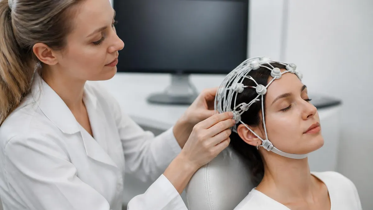
The first step is a brief pre-procedure interview in which the EEG technologist reviews your medical history, documents current medications, and asks whether you followed any preparation instructions — such as washing your hair the night before and avoiding caffeine on the morning of the test. Clean hair free of conditioner, oils, or styling products dramatically improves electrode contact and lowers impedance, which directly translates to cleaner brain wave recordings.
Next, the technologist measures your head and marks electrode placement sites using a grease pencil and a flexible tape measure. This step follows the International 10-20 System, a standardized method that defines electrode positions as percentages of the distance between skull landmarks — specifically the nasion, inion, and pre-auricular points. The system places electrodes at Fp1, Fp2, F3, F4, C3, C4, P3, P4, O1, O2, and additional sites depending on the clinical question. Standardization ensures that recordings made at different hospitals are directly comparable, which is critical when a patient transfers between facilities or participates in multi-site research.
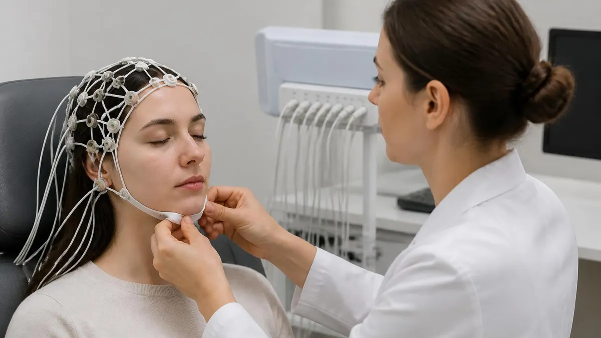
Once the sites are marked, the technologist applies a small amount of abrasive gel to each location using a blunt needle or cotton-tipped applicator, gently scrubbing the skin to further reduce impedance. Conductive electrode gel is then applied before each electrode is secured in place. For routine outpatient studies, a paste like Ten20 or Elefix holds individual electrodes snugly against the scalp. Some facilities use pre-gelled electrode caps, which speed up application significantly and are especially useful for pediatric patients or high-volume clinical operations where throughput matters.
The recording phase begins once all electrodes are secured and impedances verified. The technologist instructs the patient to lie still, close their eyes, and relax. Technologists monitor recordings in real time, watching for artifacts from eye blinking, muscle movement, and electrode pops, annotating the record with patient state changes.
Most routine EEG protocols include at least two activation procedures: hyperventilation, during which the patient breathes rapidly for three minutes, and photic stimulation, where a strobe light flashes at frequencies ranging from 1 to 30 Hz. These procedures activate the brain in ways that can unmask epileptiform abnormalities invisible during rest, making them standard components of virtually every routine adult EEG protocol.
After the recording phase concludes, the technologist removes the electrodes and helps the patient clean residual gel from their hair. This process takes 10 to 20 minutes and typically requires a second shampooing at home to fully remove any remaining conductive paste. The raw EEG data is then submitted to a board-certified neurologist or neurophysiologist for interpretation, a process called EEG reading.
The physician reviews multiple montage displays — referential, bipolar longitudinal, and bipolar transverse — before dictating a formal report that addresses whether the background activity is normal, whether epileptiform discharges are present, and whether the findings are consistent with the clinical question.
Results turnaround varies considerably by setting. In hospital inpatient or ICU environments, EEG results are often needed urgently — a neurologist may read a study within 30 to 60 minutes of recording completion. Outpatient routine studies typically have a turnaround of one to five business days before the ordering physician receives the report and discusses findings with the patient. Ambulatory and video-EEG studies, which generate many hours of data, may take a week or longer to fully review, particularly if a clinical event was captured and requires detailed analysis and clinical correlation.
For students preparing for credentialing examinations, understanding every technical step of this workflow is essential. Exam questions routinely test knowledge of why each step matters — why high impedance creates noise, why hyperventilation induces hypocapnia that can provoke absence seizures, and why filter settings must be documented in every study. Technologists who understand the reasoning behind each protocol step make better clinical decisions when unusual situations arise, such as a patient who cannot cooperate with hyperventilation or a study contaminated by artifact from a pacemaker or deep brain stimulator.
EEG Test Cost, Price, and Insurance Coverage
The EEG test price without insurance in the United States typically ranges from $200 to $700 for a standard outpatient routine study. Facility type plays a major role: hospital outpatient departments usually charge $400 to $700, while independent neurology clinics or freestanding diagnostic centers may charge as little as $150 to $350 for the same study. Geographic location also matters — studies performed in major metropolitan areas like New York or San Francisco tend to cost more than those in rural Midwest or Southern states.
Patients paying out of pocket can often negotiate significant discounts by asking the billing department for a self-pay rate before scheduling. Many facilities discount bills by 20 to 40 percent for cash-paying patients. Additionally, ambulatory EEG test cost without insurance can climb to $1,000 to $3,000 or more depending on duration, because the equipment rental and extended technologist time increase the total charge substantially. Always request an itemized estimate in advance and ask whether the interpreting neurologist bills separately from the technical facility fee.

Pros and Cons of the EEG Test
- +Completely non-invasive — no needles, no incisions, no radiation exposure of any kind
- +Provides real-time measurement of brain electrical activity that no imaging test can replicate
- +Safe for all ages from premature neonates to elderly patients, including those who are unconscious
- +Relatively affordable compared to MRI or CT scanning, especially with insurance coverage
- +Can detect seizure activity even when the patient has no obvious clinical symptoms during the test
- +Ambulatory systems allow multi-day recordings at home, capturing events that brief clinic studies miss
- −Cannot definitively identify the structural cause of abnormalities — MRI is needed for that
- −A normal EEG does not rule out epilepsy, because interictal recordings can appear completely normal
- −Electrode gel is messy and requires thorough hair washing after the procedure is complete
- −Prolonged or video-EEG studies require hospital admission that can be expensive and inconvenient
- −Interpretation requires specialized neurophysiology training — misreads can occur at facilities without dedicated expertise
- −Some patients experience anxiety during the photic stimulation phase, particularly those with photosensitivity
How to Prepare for Your EEG Test
- ✓Wash your hair the night before with regular shampoo — avoid conditioner, oils, or any styling products.
- ✓Ask your physician whether to take or skip your usual medications on the morning of the test.
- ✓Avoid caffeine for at least 8 hours before the appointment to ensure accurate resting brain wave recordings.
- ✓Get a full night of sleep unless your physician specifically ordered a sleep-deprived EEG.
- ✓Eat a normal meal before arriving — low blood sugar can affect brain wave patterns and cause artifacts.
- ✓Arrive at least 15 minutes early to complete paperwork and allow time for electrode preparation.
- ✓Wear comfortable, loose-fitting clothing — you will be lying or sitting still for up to 90 minutes.
- ✓Inform the technologist about all current medications, supplements, and any history of seizures or fainting.
- ✓Remove any hair accessories, barrettes, or wigs before the technologist begins electrode placement.
- ✓Plan for someone to drive you home if your physician ordered a sedated or sleep-deprived study.
A Normal EEG Does Not Rule Out Epilepsy
Up to 50 percent of patients with confirmed epilepsy have a normal routine EEG on first recording, because seizure-generating neurons may not fire during a brief outpatient session. If your neurologist suspects epilepsy despite a normal EEG, ask about prolonged ambulatory monitoring or a sleep-deprived study — these significantly increase diagnostic yield and should not be skipped simply because an initial test appeared normal.
One of the questions patients ask most frequently is whether the EEG test has side effects or poses any health risks. The reassuring answer is that a properly performed EEG is one of the safest diagnostic tests in all of medicine.
Because no electrical current is sent into the brain — the electrodes only receive signals, never transmit them — there is no risk of electrical shock, burn injury, or neural stimulation from a standard scalp EEG. The conductive gel used to improve electrode contact is water-soluble, non-toxic, and washes out of hair with normal shampoo, though it may require two wash cycles to fully remove.
The most commonly reported EEG test side effects are mild and transient. Hyperventilation, which is a standard activation procedure in most routine EEG protocols, can cause lightheadedness, tingling in the hands and feet, and occasionally brief perioral numbness due to the hypocapnia it induces. These symptoms resolve within a minute or two of returning to normal breathing. In rare cases, particularly in patients with undiagnosed absence epilepsy, hyperventilation can trigger a brief seizure — but this is actually a diagnostically useful event that helps confirm the diagnosis rather than a harmful complication.
Photic stimulation — the flashing strobe light used to test for photosensitive epilepsy — carries a small risk of triggering a seizure in the approximately 3 to 5 percent of epilepsy patients who have photosensitive responses. For this reason, technologists performing photic stimulation monitor recordings in real time and are trained to stop the stimulus immediately if the patient begins showing clinical signs of a seizure or if the recording shows generalized epileptiform discharges that threaten to evolve into a clinical event. The protocol is designed with safety stops at every stimulation frequency.
Patients with known photosensitive epilepsy sometimes express concern about whether photic stimulation should be performed at all. The American Clinical Neurophysiology Society (ACNS) guidelines recommend that photic stimulation be performed on virtually all routine adult EEGs, including in patients with known photosensitive epilepsy, unless there is a specific clinical reason to exclude it. The diagnostic value of documenting a photosensitive response — which can guide medication choice and lifestyle counseling — outweighs the very small risk of inducing a brief, self-limited seizure in a monitored medical setting where staff are prepared to intervene.
Allergic reactions to electrode gel are theoretically possible but extraordinarily rare in practice. If you have a known sensitivity to topical products or a history of contact dermatitis, inform the technologist before the procedure so that alternative gel formulations can be considered. Some patients with very sensitive scalps find that the abrasive skin preparation step causes mild redness that resolves within a few hours. No long-term skin effects from routine EEG preparation have been documented in the medical literature, and the procedure leaves no marks, scars, or permanent changes to the scalp or hair.
For patients who are claustrophobic, it is worth knowing that EEG is nothing like an MRI scan — there is no enclosed tunnel, no loud banging noise, and no requirement to hold completely still inside a confined space. You lie on a comfortable exam table or recline in a chair while the technologist sits nearby monitoring the recordings.
Many patients find the experience relaxing, particularly during the eyes-closed resting phase. Some patients even fall asleep during the study, which is entirely acceptable and actually provides additional useful diagnostic information about sleep architecture and the presence or absence of sleep-activated epileptiform discharges.
Invasive EEG monitoring — used in presurgical epilepsy evaluation when scalp EEG fails to localize the seizure onset zone — is an entirely different procedure with its own distinct risk profile. Intracranial EEG involves placing electrode grids or depth electrodes directly on or within brain tissue through a surgical approach.
Risks include infection, hemorrhage, and neurological deficits related to electrode placement. However, this invasive procedure should not be confused with the standard scalp EEG that most patients encounter. When someone asks what is an EEG test in general neurology practice, they almost always mean the non-invasive scalp-based version described throughout this guide.

While serious adverse events from routine scalp EEG are extremely rare, contact your ordering physician if you experience a prolonged seizure lasting more than five minutes, severe headache, skin blistering at electrode sites, or any neurological symptom that is new or worsening after the procedure. These events are uncommon but warrant prompt medical evaluation to rule out an unrelated cause.
For aspiring EEG technologists and current students pursuing ABRET certification, building a career in electroencephalography begins with understanding that this field sits at a unique intersection of neuroscience, electronics, and patient care. The primary credential in the field is the R. EEG T. (Registered Electroencephalographic Technologist), awarded by ABRET Neurodiagnostic Credentialing and Accreditation after candidates pass a rigorous written examination covering anatomy, neurophysiology, instrumentation, recording techniques, waveform recognition, and patient care. A solid grasp of eeg systems hardware and software is tested extensively across multiple examination domains.
The educational pathway to becoming a credentialed EEG technologist typically begins with an associate degree or certificate program in neurodiagnostic technology, offered by community colleges and allied health schools across the United States. These programs usually take one to two years to complete for students entering with a high school diploma or GED. Curricula cover neuroanatomy and neurophysiology, EEG equipment operation and troubleshooting, electrode placement and preparation, artifact identification, basic waveform interpretation, patient interaction and safety, and clinical practicum hours in affiliated hospitals or clinics.
The ABRET R. EEG T. examination itself consists of 200 multiple-choice questions and must be completed within a four-hour testing window. Content is divided into five domains: patient care and safety (15%), instrumentation and data acquisition (20%), recording techniques (25%), waveform recognition and interpretation (30%), and professional responsibilities (10%). The waveform recognition domain is where most candidates spend the bulk of their preparation time, as it requires not just memorization of waveform names but true pattern recognition skill developed through extensive practice with real EEG data.
Beyond the entry-level R. EEG T., the field offers additional credentials for specialization. The CLTM (Certified Long-Term Monitoring Technologist) credential focuses on extended monitoring applications including video-EEG, neonatal EEG, and ICU monitoring. The CNIM (Certified Neurophysiologic Intraoperative Monitoring) credential qualifies technologists to monitor brain and spinal cord function during neurosurgical procedures. Each of these advanced credentials requires additional clinical experience and a separate examination, representing meaningful career progression opportunities with corresponding salary increases.
Salary for certified EEG technologists in the United States varies by credential level, geographic location, and clinical setting. Entry-level R. EEG T. holders in outpatient clinic settings typically earn between $45,000 and $58,000 annually. Technologists with CLTM credentials working in hospital inpatient or ICU settings commonly earn $58,000 to $72,000.
Those holding the CNIM credential, particularly those employed by dedicated intraoperative monitoring companies, frequently earn $80,000 to $120,000 or more, especially when compensation includes on-call premiums and case-based bonuses. Travel EEG technologist positions, where credentialed techs fill short-term contract roles at facilities with staffing shortages, can yield total compensation packages exceeding $100,000 annually including housing and travel stipends.
Professional development in EEG does not stop at initial credentialing. ABRET requires credential holders to complete continuing education credits for renewal, ensuring that practicing technologists stay current with evolving technology and clinical guidelines. The American Clinical Neurophysiology Society (ACNS) publishes guideline updates that directly affect how EEG studies are performed and documented. Technologists who actively engage with professional literature, attend annual meetings, and participate in peer review processes within their departments tend to advance more quickly and contribute more meaningfully to the clinical teams they support.
For students currently in EEG training programs or self-studying for certification, consistent practice with question banks and case-based scenarios is the most efficient way to build examination readiness. Reviewing both normal variants and pathological patterns is important — the examination tests ability to distinguish normal theta slowing of drowsiness from pathological temporal theta in an adult, to recognize benign epileptiform transients of sleep (BETS) as normal rather than epileptiform, and to identify artifacts from their characteristic morphology and distribution. These distinctions require repeated exposure and active recall practice, not passive reading alone.
Practical preparation strategies make a measurable difference in EEG examination performance, and the most effective approach combines conceptual understanding with heavy volume practice. Begin your study plan by mapping the five ABRET examination domains to your own knowledge gaps. Most candidates discover that instrumentation and waveform recognition require the most remediation, while patient care content feels more intuitive. Prioritize time accordingly — spending equal hours on all domains when your mastery is unequal is one of the most common and costly preparation mistakes.
When studying EEG waveform recognition, work with actual EEG tracings rather than stylized textbook illustrations whenever possible. The difference between a real spike-wave complex and a textbook drawing of one is substantial. Many online EEG libraries and academic medical center teaching files offer free access to annotated case studies. Review at least five to ten new cases per week during your preparation period, actively attempting to identify all waveforms before reading the provided interpretation. This active retrieval approach builds pattern recognition far more effectively than passive review of annotated studies.
For the instrumentation domain, build a solid mental model of how a signal travels from the neuron to the computer screen. Start at the electrode-scalp interface, understand impedance and why it matters, trace the signal through the differential amplifier stage, understand how the ADC discretizes the continuous voltage, and then study how filter settings shape the final waveform appearance. Understanding why a high-pass filter at 1 Hz rather than 0.1 Hz will eliminate slow cortical potentials helps you answer filter-setting questions correctly even when the specific numbers in the question differ from any scenario you memorized.
Artifact recognition deserves dedicated study time because artifacts contaminate real EEG data constantly, and misidentifying an artifact as a pathological waveform is a significant error. Build a personal reference library of common artifacts: eye blink artifact (bifrontal, time-locked to eye movement), ECG artifact (rhythmic, QRS-morphology discharges with periodicity matching cardiac rate), muscle artifact (high-frequency, widespread, typically worst in temporal regions), chewing artifact (rhythmic, 1–2 Hz, temporal predominance), and electrode pop artifact (single-channel high-amplitude brief discharge with abrupt onset and offset). Each artifact has characteristic features that distinguish it from true cortical activity.
Time management during the actual ABRET examination is a skill that requires deliberate practice. With 200 questions in four hours, you have approximately 72 seconds per question. Many candidates report spending too long on difficult waveform identification questions early in the examination, creating time pressure that leads to rushed answers on later questions. Practice timed question sets under realistic conditions — use a timer, do not look up answers mid-session, and review your performance afterward to identify patterns in your errors. Consistent timing errors on a specific content domain signal the need for additional focused review.
Study groups and peer learning accelerate preparation significantly. Explaining why a particular waveform is abnormal — teaching it to a fellow student — forces you to articulate reasoning that passive reading never demands. Online EEG communities, professional society forums, and dedicated EEG study groups on social platforms connect students across the country who are preparing for the same examinations. These networks also provide moral support during what can be a demanding preparation period, particularly for students working full-time clinical positions while studying for their credentials.
Finally, pay close attention to the ACNS guidelines on EEG recording parameters, as these are frequently tested in the instrumentation domain. Electrode placement according to the 10-20 system, minimum electrode requirements for routine studies, recommended filter settings, minimum sampling rates for digital EEG, and electrode impedance standards are all fair game on the examination. These technical standards exist for excellent clinical reasons — they ensure that EEG recordings are of sufficient quality to support valid diagnostic conclusions — and understanding those reasons makes the standards far easier to remember and apply under examination conditions.
EEG Questions and Answers
EEG Electroencephalography Practice Test PDF (Free Printable 2026)
Sleep EEG: How Overnight Brain Wave Testing Works and What Your Results Mean
EEG Test Cost in 2026: Prices, Insurance Coverage, and How to Save

Travel EEG Tech Jobs: The Complete Guide to Working as a Mobile Electroencephalography Technologist

EEG Pronunciation, Meaning, and Everything You Need to Know About the EEG Test
About the Author
Educational Psychologist & Academic Test Preparation Expert
Columbia University Teachers CollegeDr. Lisa Patel holds a Doctorate in Education from Columbia University Teachers College and has spent 17 years researching standardized test design and academic assessment. She has developed preparation programs for SAT, ACT, GRE, LSAT, UCAT, and numerous professional licensing exams, helping students of all backgrounds achieve their target scores.