What Does MRI Stand For? Meaning, Uses, and How It Works 2026 June
📗 MRI stands for Magnetic Resonance Imaging. Learn how MRI scans work, what they detect, preparation tips, and what results mean.
MRI stands for Magnetic Resonance Imaging. It's a medical imaging technology that uses powerful magnetic fields and radio waves to produce detailed images of the body's internal structures — organs, soft tissues, bones, and the brain — without using ionizing radiation like X-rays or CT scans. The images MRI produces are typically far more detailed for soft tissue than what X-rays or CT scans provide, which is why MRI is the preferred diagnostic tool for a wide range of conditions affecting the brain, spinal cord, joints, and abdominal organs.
The word "magnetic" refers to the strong magnetic field the machine generates — typically 1.5 to 3 Tesla (the unit of magnetic field strength), which is roughly 30,000 to 60,000 times stronger than Earth's magnetic field. "Resonance" refers to the way hydrogen atoms in your body respond when briefly excited by radio wave pulses while inside this strong magnetic field. "Imaging" refers to the output — the detailed cross-sectional pictures that a computer assembles from those atomic responses.
Together, these three processes produce images that reveal anatomy and tissue characteristics with a level of detail that changed how medicine diagnoses and monitors dozens of conditions.
MRI was developed in the 1970s and became clinically available in the early 1980s. It represented a major advance over existing imaging technologies because it could visualize the brain and spinal cord in a way that X-rays couldn't, and it could differentiate between tissue types — healthy tissue versus tumor, inflamed tissue versus healthy — in a way that earlier imaging couldn't. The field has continued to evolve, with functional MRI (fMRI) mapping brain activity, diffusion MRI tracking nerve fiber pathways, and cardiac MRI providing detailed views of the heart.
For background on what MRI technologists do and the professional standards around MRI, the what does mri stand for study guide provides context on the clinical and certification side.
Understanding what MRI stands for and how it works is relevant not just for patients preparing for their first scan, but for anyone studying for a healthcare career or the MRI certification exam. MRI technologists — also called MRI technicians or MRT — are responsible for operating MRI equipment, positioning patients, selecting imaging protocols, and ensuring image quality. The MRI registry examination administered by the American Registry of Radiologic Technologists (ARRT) and the American Registry of Magnetic Resonance Imaging Technologists (ARMRIT) tests detailed knowledge of MRI physics, safety, anatomy, and patient care.
The abbreviation MRI has become so embedded in everyday healthcare language that most patients encounter it before they fully understand what it involves. Knowing the meaning behind the acronym helps set accurate expectations for what the scan does, how it works, and what it can and cannot detect.
How MRI Works
Inside an MRI machine, a powerful magnetic field aligns the hydrogen atoms in your body's water molecules. Hydrogen atoms are ideal for MRI because the human body is roughly 60% water, making hydrogen the most abundant atom with detectable MRI properties. When the magnetic field is applied, the protons in hydrogen atoms align with the field — like compass needles pointing north.
Next, the machine emits brief pulses of radio waves at a specific frequency that temporarily disrupts this alignment, causing the hydrogen protons to absorb energy. When the radio wave pulse stops, the protons return to their original alignment and release the absorbed energy as radio signals. Different tissue types — fat, muscle, brain matter, tumors, fluid — release these signals at different rates and intensities. The MRI machine detects these tiny radio signals and a computer processes them to construct detailed cross-sectional images.
The contrast between tissue types on an MRI image depends partly on the "relaxation time" — how quickly different tissues return to their baseline magnetic alignment after each radio wave pulse. T1-weighted and T2-weighted images are the two most common image types in MRI, each sensitive to different tissue properties. T1 imaging shows anatomy clearly, with fat appearing bright. T2 imaging highlights fluid-containing structures, making it useful for detecting inflammation, edema, and lesions. Radiologists and technologists select imaging sequences based on what they're looking for and what area of the body is being examined.
Contrast agents — typically gadolinium-based substances injected intravenously — are sometimes used to enhance MRI images. Gadolinium behaves differently from surrounding tissue in the magnetic field, making blood vessels and areas of abnormal vascular permeability (like tumors or active inflammation) appear brighter and more distinct.
Contrast-enhanced MRI is commonly used for tumor staging, vascular imaging, and assessing blood-brain barrier disruption. Patients with certain kidney conditions may have contraindications to gadolinium contrast — this is assessed before every contrast-enhanced scan. Information on the MRI certification exam and what imaging knowledge is tested is available in the what does mri stand for practice test resources.
Advanced MRI techniques continue to expand what the technology can do. Diffusion tensor imaging (DTI) maps the pathways of white matter tracts in the brain, enabling visualization of neural connections that are otherwise invisible. This has transformed neurosurgical planning — surgeons can now see which fiber tracts are adjacent to a tumor before operating.
Perfusion MRI measures blood flow through tissue and is used to assess stroke and tumor vascularity. Magnetic resonance spectroscopy (MRS) measures chemical concentrations in tissue, providing a metabolic fingerprint that helps distinguish between tissue types. Each of these techniques builds on the same fundamental physics principle as standard MRI, extended through different pulse sequences and acquisition strategies.
What MRI Scans Are Used For
Brain tumors, stroke, MS, dementia, traumatic brain injury, seizure disorders. MRI can detect lesions, hemorrhages, and structural abnormalities not visible on CT. Gold standard for most neurological diagnoses.
Herniated discs, spinal stenosis, joint injuries, ligament tears, bone marrow disease. Preferred over X-ray for soft tissue detail. Common for knee, shoulder, hip, and back pain evaluation.
Liver disease, kidney tumors, prostate cancer staging, uterine fibroids, bowel conditions. Often preferred over CT when avoiding radiation is a priority, particularly in younger patients.
Heart structure and function, cardiomyopathy, pericardial disease, aortic aneurysm. Cardiac MRI provides detail on myocardial tissue that echocardiography can't match.
Preparing for an MRI Scan
MRI preparation depends on the type of scan and whether contrast will be used. For most standard MRI scans, there's no special preparation required — you can eat, drink, and take medications as normal. For abdominal and pelvic MRIs, you may be asked to fast for a few hours beforehand, and for some scans, a full bladder is needed or bowel preparation may be recommended. Your referring physician and the MRI facility will provide specific instructions when your appointment is scheduled.
Metal is the most important safety consideration for MRI. The magnetic field is powerful enough to forcibly move ferromagnetic metal objects, which poses serious safety risks. Before entering the MRI room, you'll be asked to remove all metal — jewelry, watches, piercings, hair clips, belts, underwire bras — and change into a gown.
You'll also be asked about implanted devices: pacemakers, cochlear implants, certain orthopedic hardware, neurostimulators, and other implants may be contraindicated for MRI. Some newer devices are MRI-conditional (safe under specific conditions) rather than fully incompatible, but this must be verified by the MRI staff before scanning. Always bring documentation of any implanted devices to your MRI appointment.
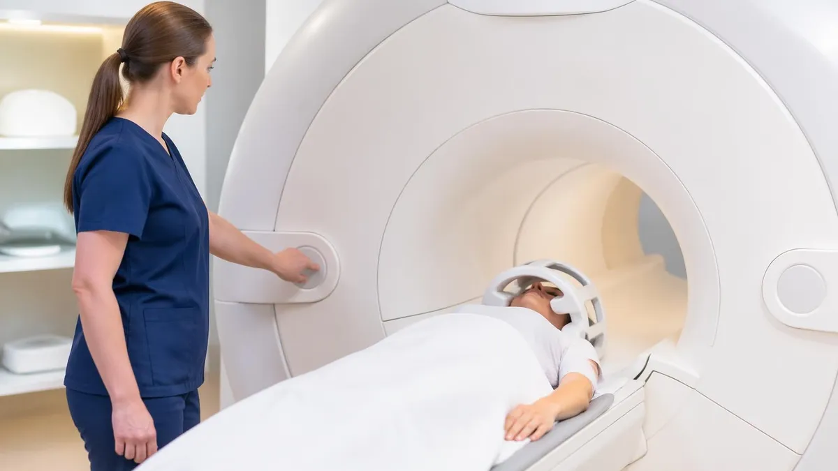
Claustrophobia affects a significant portion of patients and is one of the most common reasons MRI scans are incomplete or rescheduled. The bore of a standard MRI machine is a narrow cylindrical tube roughly 60-70 cm in diameter, and the scan can take 15-90 minutes. Open MRI machines are available at some facilities and are wider and less enclosed, though they typically produce lower-quality images than closed bore machines.
If you have significant anxiety about enclosed spaces, discuss it with your referring physician before your appointment — mild sedation or anxiolytic medication can be prescribed for the scan, and this is common practice. Preparing for MRI-related careers and understanding the full scope of MRI technologist responsibilities is covered in the what does mri stand for career guide.
Patients who are pregnant — particularly in the first trimester — are generally advised to avoid MRI unless the clinical need is urgent. MRI is not thought to cause fetal harm, but the precautionary principle applies because there is limited safety data for first-trimester exposure. Gadolinium contrast agents are specifically contraindicated during pregnancy as they cross the placental barrier. If you are or might be pregnant, inform the MRI facility before your scan so appropriate protocols can be followed.
Preparation for outpatient MRI scans also includes practical logistics. Wear comfortable clothing without metal hardware — yoga pants, cotton shirts without underwire, socks — to avoid changing into a gown. Arrive early enough to complete the metal screening questionnaire fully without rushing. If you wear hearing aids, they'll need to be removed.
If you take medications, confirm whether any of them are affected by the scan (most aren't). If you have significant anxiety and weren't prescribed anything for it, mention this when you check in — some facilities can provide anxiolytics on the day if your appointment wasn't pre-planned for sedation.
MRI: Key Facts
What to Expect During and After an MRI
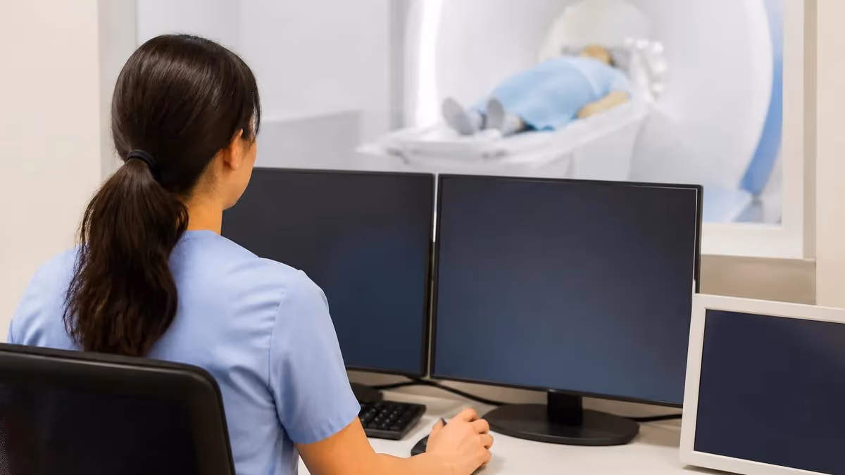
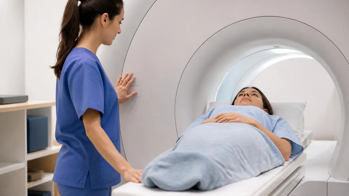
During an MRI scan, you'll lie still on a padded table that slides into the bore of the machine. Movement degrades image quality — even slight movement during a scan can blur the images enough to require repeating sequences. The technologist will communicate with you through an intercom and can hear you at all times. Most facilities provide earplugs or headphones because MRI machines are loud — the gradient coils that switch on and off during imaging create a series of loud knocking, banging, and buzzing sounds, typically 80-110 dB. Some facilities allow music through the headphones.
You'll be given a squeeze bulb or button to signal the technologist if you need to stop the scan. Using it will pause imaging, and the technologist will speak with you immediately. There's no penalty for using it — incomplete images from a stopped scan are simply repeated. If you feel unwell, anxious, or need to stop for any reason, using the call device is the right action. A completed, high-quality scan of a calm patient is better than a rushed or degraded scan.
After the scan, you can resume normal activities immediately unless you received sedation. If sedation was administered, you'll remain in a recovery area until the medication wears off and you'll need someone to drive you home. Results are typically interpreted by a radiologist and reported to your referring physician within 24-72 hours for routine scans, or faster if the scan was ordered urgently.
The radiologist's report describes findings in detail, and your physician will discuss the results with you in the context of your clinical situation. How MRI technologist certification works and what the registry exam covers is in the what does mri stand for exam guide.
Career satisfaction among MRI technologists tends to be high relative to other healthcare support roles. The work involves direct patient interaction, technical skill, and problem-solving — positioning challenging patients, troubleshooting image artifacts, selecting appropriate protocols for non-standard cases. Technologists who enjoy a combination of hands-on technical work and patient care find it a sustainable career that doesn't require the years of training and debt that physician-level roles involve. The physical demands are moderate — some lifting and positioning — and the work environment is generally stable and predictable compared to fields like emergency medicine.
MRI vs. Other Imaging and Safety
X-rays are the simplest and fastest imaging method. They use ionizing radiation to produce 2D images of dense structures — bones and calcifications appear clearly, while soft tissues don't. X-rays are useful for fractures, lung conditions, and quick screening, but limited for soft tissue detail. CT scans (Computed Tomography) use X-rays from multiple angles and a computer to reconstruct 3D cross-sectional images. CTs are faster than MRI, better for emergencies, and excellent for visualizing bones, blood vessels, and detecting hemorrhage. The tradeoff is significant radiation exposure compared to MRI.
MRI produces superior soft tissue contrast compared to both X-ray and CT. It can differentiate between healthy and abnormal tissue types that appear identical on CT, making it the preferred tool for brain, spinal cord, joint, and organ imaging. MRI takes longer than CT, costs more, and requires more patient cooperation (staying still). Patients with certain metal implants can't have MRI but can have CT. Emergency physicians often prefer CT because it's faster — a CT of the head takes minutes, while an MRI brain scan takes 30-45 minutes.
The choice between MRI and other imaging modalities is always made by the referring physician based on the clinical question, urgency, available contraindications, and what each modality can answer. MRI isn't inherently superior — it's the best choice for specific clinical questions, and CT or X-ray is the best choice for others. Understanding the strengths and limitations of each modality is fundamental knowledge for MRI technologists and for anyone working in medical imaging.
MRI as a Career: Becoming an MRI Technologist
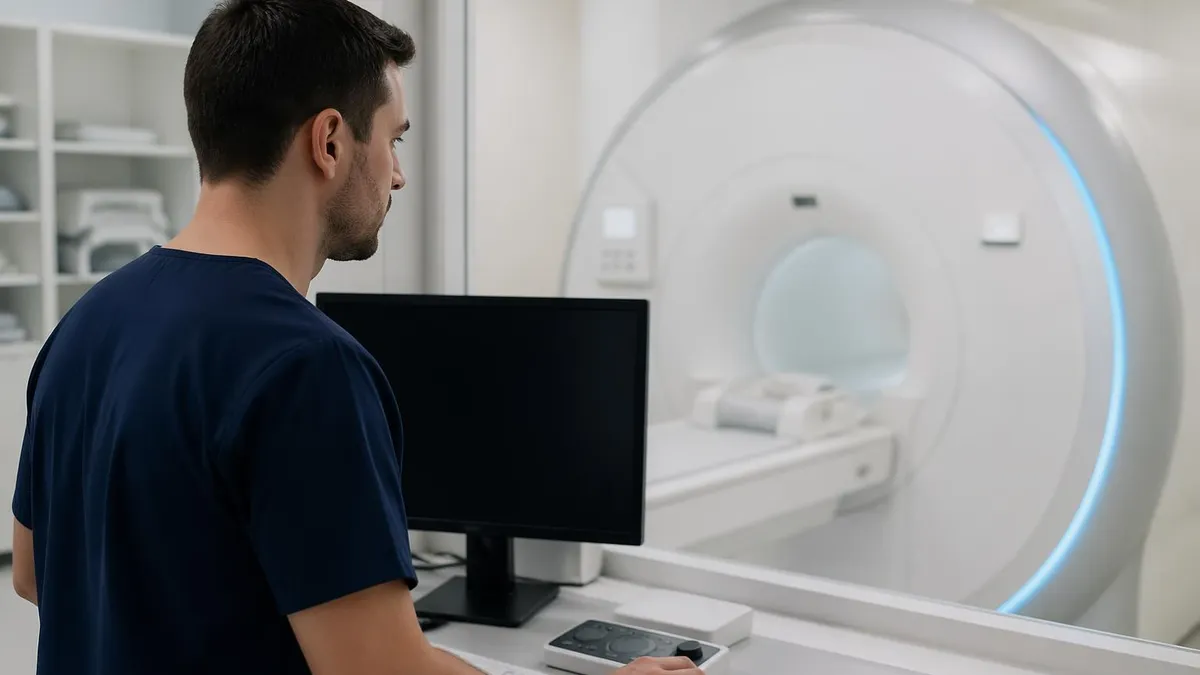
MRI technologists operate MRI equipment in hospitals, outpatient imaging centers, and specialty clinics. The career path to becoming an MRI technologist typically starts with a 2-year associate's degree in radiologic technology or a 4-year bachelor's program, followed by passing the ARRT board examination in radiography. After gaining experience as a radiologic technologist, many specialize in MRI by completing additional clinical hours and passing the ARRT MRI certification examination or the ARMRIT registry.
Some technologists enter MRI directly through dedicated MRI technology programs that are shorter than the full radiography pathway. These programs typically take 14-24 months and are available at community colleges and vocational schools. Direct MRI program graduates must still pass a registry examination to work as certified MRI technologists in most states.
Certification from ARRT or ARMRIT is the standard credential accepted by employers across the country. Information on preparation for the MRI registry examination — including what content domains are tested and how to study for them — is available in the what does mri stand for exam prep section.
Salaries for MRI technologists are strong relative to the education required. The median annual wage for MRI technologists in the US is approximately $77,000-$82,000, with experienced technologists at major medical centers or in high-cost-of-living markets earning $90,000-$110,000 or more. The demand for MRI technologists is driven by an aging population requiring more diagnostic imaging, expanding clinical applications for MRI in oncology, cardiology, and neurology, and the ongoing replacement of older equipment with newer high-field and specialty MRI systems. Job prospects for qualified MRI technologists are strong and expected to remain so over the next decade.
The MRI field is also one where continuing education and specialization create meaningful career differentiation. Technologists who develop expertise in specialized applications — cardiac MRI, neuroimaging, body MRI — or who move into quality management, equipment application specialist roles, or MRI safety officer positions can advance their careers without leaving the imaging field entirely. Large academic medical centers often have dedicated MRI research positions where technologists work alongside researchers on clinical trial imaging, protocol development, and new application research — a different kind of career path that appeals to technologists with strong technical curiosity and interest in advancing the field.
MRI Scans: Advantages and Limitations
- +No ionizing radiation — safer than CT or X-ray for repeated imaging, particularly in children and younger adults
- +Superior soft tissue contrast — can differentiate tissue types that appear identical on CT or X-ray
- +Multi-planar imaging — produces images in any orientation (axial, coronal, sagittal) without moving the patient
- +No known long-term health risks from the magnetic field or radio waves themselves
- +Functional and metabolic imaging possible — fMRI maps brain activity, spectroscopy measures tissue chemistry
- +Continuously improving technology — high-field and ultra-high-field systems provide increasing diagnostic detail
- −Long scan times — 15-90 minutes compared to seconds for X-ray or minutes for CT, requiring patient cooperation
- −Claustrophobia — the enclosed bore is uncomfortable or intolerable for a significant minority of patients
- −Metal contraindications — patients with certain implanted devices cannot have standard MRI
- −Cost — significantly more expensive than X-ray or CT, and not always available or covered in all settings
- −Sensitivity to motion — patient movement degrades image quality; sedation may be needed for children or anxious adults
- −Not suitable for all emergencies — CT is faster and more practical for acute emergencies like stroke or trauma triage
MRI: Questions and Answers
About the Author
Medical Laboratory Scientist & Clinical Certification Expert
Johns Hopkins UniversityDr. Sandra Kim holds a PhD in Clinical Laboratory Science from Johns Hopkins University and is certified as a Medical Technologist (MT) and Medical Laboratory Scientist (MLS) through ASCP. With 16 years of clinical laboratory experience spanning hematology, microbiology, and molecular diagnostics, she prepares candidates for ASCP board exams, MLT, MLS, and specialist certification tests.
Join the Discussion
Connect with other students preparing for this exam. Share tips, ask questions, and get advice from people who have been there.
View discussion (4 replies)