What Does a Back MRI Show — Complete Guide (2026)
What does a back MRI show? Disc herniation, stenosis, pinched nerves, fractures, tumors and infections — what radiologists actually see on lumbar and...
Back MRI by the Numbers
What a Back MRI Actually Reveals
Short answer: almost everything soft. A back MRI looks past bone and into the discs, nerves, spinal cord, and ligaments — the structures that cause most back pain but stay invisible on x-ray. If your doctor ordered one, they're hunting for something specific. Disc trouble. A pinched nerve. Maybe something rarer like an infection or a small tumor.
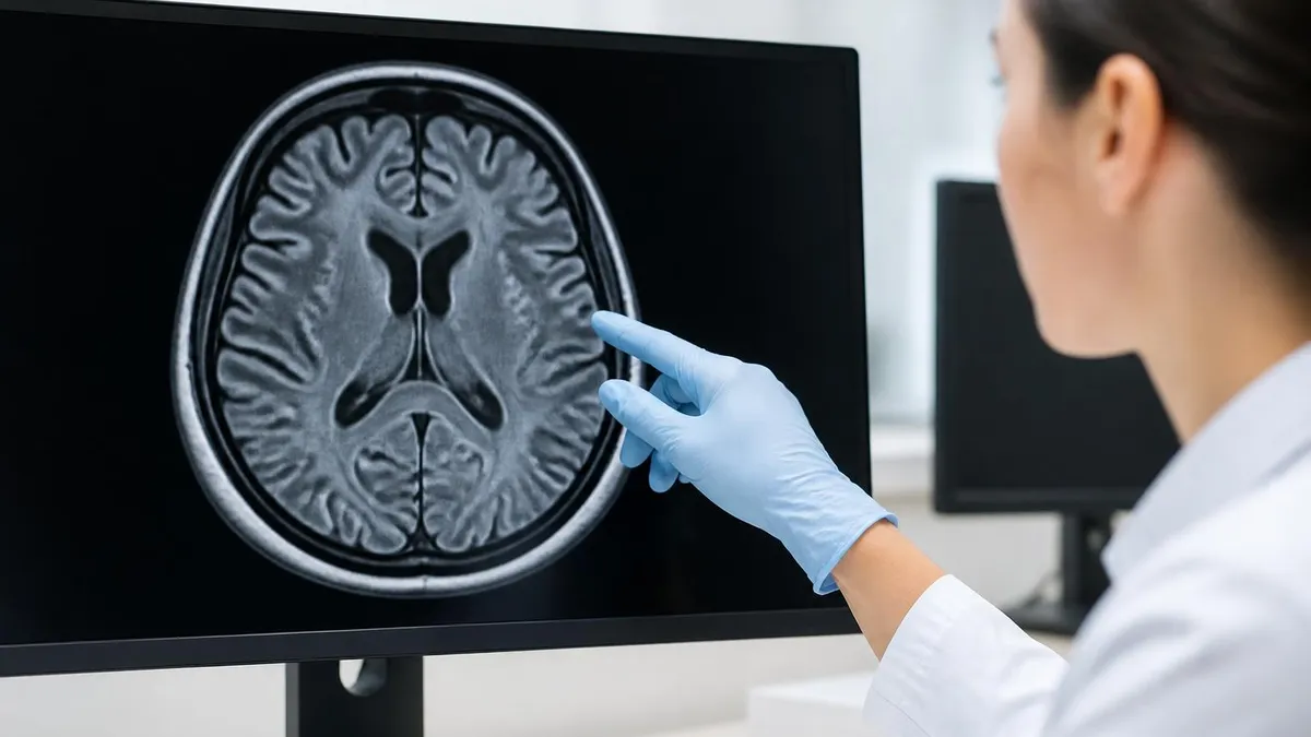
Your radiologist is reading slices of your spine roughly 3-5 millimeters thick, from the side, from the front, and from above. Each slice gets compared to the next. They're looking for asymmetry, bright signals where dark ones should be, and dark patches where the tissue should be lit up. That's the whole game.
An MRI of the back is split by region. A lumbar spine mri covers L1 through the sacrum — the lower five vertebrae that take the worst of standing, lifting, and sitting. A cervical spine MRI looks at the neck (C1-C7). Thoracic scans cover the middle, T1 through T12. Doctors sometimes order all three if your symptoms are vague or spread out.
Here's the thing: the spine isn't just bones. It's a stacked column of vertebrae, with rubbery discs between them, a tube of nerve tissue running through the middle, and dozens of ligaments holding it all together. MRI reads all four of those layers at once. That's why it's the gold-standard test for back pain when something serious is suspected.
What kicks off a back MRI order? Usually one of three things. Severe pain that won't quit after six weeks. Numbness or weakness in a leg or arm. Or red-flag symptoms — bladder problems, unexplained weight loss, fever. The scan answers a single question your doctor needs settled: is this structural, or is this muscle?
One myth worth busting: an MRI doesn't show pain. It shows tissue. Two patients can have nearly identical scans and feel completely different — one in agony, one fine. That's why the radiologist's report describes findings, not diagnoses. Your physician interprets those findings against your exam and history to decide what's actually wrong and what to do about it.
Why MRI Beats X-Ray for Back Pain
X-rays show bones. CT shows bones better. But back pain is almost never a bone problem — it's a disc, a nerve, or a soft-tissue issue. MRI is the only scan that maps these structures in detail without radiation. Roughly 80% of adults get back pain at some point, and when it doesn't resolve in six weeks, MRI is what answers why.
Three Regions of the Spine MRI
The workhorse scan. Covers L1-L5 plus the sacrum. Catches disc herniation, sciatica root cause, spondylolisthesis. About 70% of back MRIs order this region first.
- Vertebrae: L1-L5 + sacrum
- Top complaint: Low back pain, sciatica
- Scan time: 25-35 min
Top of the spine, C1-C7. Reads neck pain, arm tingling, and cord compression. The cord is closer to the bone here — small problems cause bigger symptoms.
- Vertebrae: C1-C7
- Top complaint: Neck pain, arm numbness
- Scan time: 20-30 min
Middle 12 vertebrae, T1-T12. Less commonly imaged because discs here are protected by the ribcage. Ordered for compression fractures, tumors, or suspected MS lesions.
- Vertebrae: T1-T12
- Top complaint: Mid-back pain, MS workup
- Scan time: 25-35 min
Disc and Nerve Findings — The Heart of the Report
Disc problems show up first. They're the most common reason for a back MRI, and the language radiologists use can sound scary — bulging, herniated, protruding, extruded, sequestered. These describe how far the gel-like center of the disc has pushed past its outer wall. A small bulge might mean nothing. A sequestered fragment pressing on a nerve root means something.
Healthy discs glow bright on T2 imaging. As they age, they lose water content — radiologists call this disc desiccation or dehydration. On the scan, a dehydrated disc looks dark instead of bright. It's normal after age 40. It's not the same as a problem, but combined with other findings, it tells a story about wear and tear that accumulated over decades.
Degenerative disc disease is the catch-all term. You'll see it written on most adult MRI reports, even for people without symptoms. The disc loses height, the outer ring cracks, the vertebrae above and below start to show wear changes called Modic changes. The MRI grades these by severity — Modic 1, 2, or 3, each meaning a different stage of inflammation or fat replacement in the bone next to the disc.
Spinal stenosis is the next big finding. The spinal canal — the tube that holds the cord and nerves — narrows. Sometimes from a thickened ligament called spine mri shows as ligamentum flavum hypertrophy. Sometimes from bone spurs growing into the canal. Sometimes from a disc bulging into the space. The scan measures the canal at each level and flags where it's too tight.
Nerve impingement — the technical reason for sciatica — happens when a herniated disc or stenotic canal pinches a nerve root as it leaves the spine. The radiologist names the specific nerve being squeezed. Left L5 nerve root impingement. Right S1 compression. That naming matters because the symptoms map back to the nerve. L5 = top of foot. S1 = bottom of foot and calf.
Spondylolisthesis is another finding that gets named explicitly. One vertebra slides forward over the one below it. Most common at L4-L5 or L5-S1. The radiologist grades it 1 through 4 based on how far the upper bone has shifted. Grade 1 is mild, often surgical only if symptoms are severe. Grade 3 or 4 usually means an operation. The MRI also reads the nearby pars defect — a tiny stress fracture in the bony arch — that often causes the slip in younger patients.
T1 vs T2 vs STIR — How Radiologists Read the Scan
What it shows — anatomy. Fat is bright. Water is dark. This is the structural map. Bone marrow, vertebral body shape, and the outline of discs all show clearly.
Used for — measuring vertebrae, spotting marrow replacement (tumor or infection signal), and post-contrast comparison. T1 is the reference scan everything else gets compared to.
Disc trouble on T1 — discs look dark or speckled. Not as diagnostic as T2 for disc water content but better for showing fat changes around damaged areas.
Fractures, Infections, Tumors and Cord Lesions
Compression fractures are the next big category. The vertebra collapses, often from osteoporosis or trauma. On MRI, a fresh fracture lights up on STIR — the bone is inflamed. An old healed fracture doesn't light up at all. That difference tells your surgeon whether the pain is from this fracture or one that healed years ago. The scan also catches spondylolisthesis — one vertebra sliding forward over the one below, usually L4 over L5 or L5 over S1.
Infections in the spine are rare but serious. Discitis is infection in a disc. Osteomyelitis is infection in the bone of a vertebra. Both glow brightly on T2 and STIR. The radiologist looks for fluid collections, called epidural abscesses, that can press on the cord. These usually need MRI with contrast to define the borders and rule out cord involvement. Patients with fever plus back pain almost always get this scan urgently, sometimes the same day they walk into the ER.
Tumors fall into two buckets. Primary tumors start in the spine itself — rare. Metastatic tumors traveled from somewhere else, usually breast, lung, prostate, or kidney cancer. On MRI they replace normal marrow signal, creating dark patches on T1 and bright ones on T2. Contrast makes them light up dramatically, which is why oncology patients with new back pain almost always get gadolinium scans. The report names the level, the size in millimeters, and whether it touches the cord.
The spinal cord itself runs through the cervical and thoracic spine. It ends around L1 — below that, you just have nerve roots in a bundle called the cauda equina. MRI catches cord problems too. multiple sclerosis plaques appear as small bright spots on T2 in the cord. Syrinx — a fluid-filled cavity inside the cord — shows as a dark tube on T1, bright on T2. Both findings change treatment dramatically.
Facet joint arthritis is the boring-but-common finding. The little joints behind each vertebra wear out, get arthritic, and cause back pain that mimics disc disease. MRI shows joint effusions, bone spurs, and synovial cysts pushing into the spinal canal. Most adult lumbar MRIs report some degree of facet arthropathy. It's not always the pain source — but sometimes it is. Pain specialists use the scan to plan facet injections targeting the right level.
How to Prep for Your Back MRI
- ✓Remove all metal — jewelry, piercings, watches, hairpins, belt, coins. Even hairpins with metal cores will pull toward the magnet.
- ✓Tell the tech about implants. Pacemakers, cochlear implants, surgical clips, and some older joint replacements may not be MRI-safe.
- ✓Wear loose comfortable clothes without metal fasteners. Most facilities will give you a gown anyway.
- ✓If claustrophobic, ask for a mild sedative ahead of time — your doctor can prescribe one. Or request an open or wide-bore scanner.
- ✓For contrast scans, fast 4 hours before. Drink water. Tell them about kidney problems, allergies, or pregnancy.
- ✓Allow 60-90 minutes total at the facility — check-in, gowning, scan time, then changing back.
With vs Without Contrast — When Gadolinium Matters
Most back MRIs run without contrast. The discs, bones, nerves, and ligaments all show up well on plain T1, T2, and STIR sequences. Adding contrast adds cost, time, and a small injection risk. Doctors only order it when they need it.
Three reasons trigger a contrast scan. First — looking for tumors. Cancer cells have leaky blood vessels and grab gadolinium fast, so they light up bright on post-contrast T1 images. Second — suspected infection. The inflammatory tissue around discitis or an epidural abscess enhances. Third — post-surgical imaging. After back surgery, scar tissue and recurrent disc herniation look similar on plain MRI. Scar takes up contrast. A new disc fragment doesn't. That difference changes whether you need another operation.
Gadolinium is given as a small IV injection mid-scan. The technologist runs the first sequences, pauses to inject, then runs the post-contrast set. The agent leaves your body through your kidneys over 24 hours. If your kidney function is poor (eGFR under 30), they'll either skip contrast or use a newer macrocyclic agent with a safer profile. The radiologist also compares pre and post-contrast T1 images side by side, looking for any new bright spots that weren't there before the injection.
Side effects are rare. Nausea or a warm flush happens in maybe 1% of patients. Severe allergic reactions are well under 0.01%. Long-term gadolinium retention in brain tissue has been observed but no clear health effect has been linked to it in patients with normal kidneys. mri without contrast is the default for straightforward back pain — and that covers the vast majority of cases.
One thing the contrast question doesn't change: nothing about your prep, the scanner, or the time in the bore. The injection takes 15 seconds. The whole scan still takes 30-45 minutes either way. You won't feel the contrast going in beyond a brief cold sensation in the arm. The IV comes out before you stand up. No driving restrictions, no post-scan recovery, no dietary changes for the rest of the day.
Worth knowing: contrast doesn't make the MRI better at every job. For straightforward back pain caused by a disc or stenosis, gadolinium adds nothing useful. The plain T2 sequence already shows everything needed. Adding contrast in those cases just costs more without changing what the radiologist sees on the report.
Closed Bore vs Open MRI for Back Imaging
- +Sharper images — 1.5T or 3T magnets produce higher resolution for subtle disc and nerve findings
- +Faster scans — stronger field means shorter sequence times, less time in the bore
- +Better for surgical planning — surgeons prefer high-field images for pre-op decisions
- +Standard of care for tumor and infection workups where small details matter
- +Covered by all insurance plans without preauthorization friction
- −Better for claustrophobic patients — wider opening or open sides eliminate the tunnel feel
- −Handles patients over 350 lbs — wide-bore and open scanners accept larger body sizes
- −Upright versions scan you standing — useful for positional back pain that disappears when lying flat
- −Less noise — open scanners are usually quieter than closed bores
- −Better for children or anxious patients who can't lie still for 30+ minutes in a tunnel
What a Back MRI Can't Show — And Post-Op Imaging
MRI is brilliant for soft tissue. It's mediocre for calcified bone. If your radiologist needs to measure how much bone spur is growing into a nerve foramen, CT is sharper. If you've broken a vertebra and need detailed fracture line mapping, CT wins. MRI tells you the spine is hurting; CT tells you exactly which bone fragment is where. That's why orthopedic surgeons often order both — MRI to see the disc and nerve, CT to see the bony fragments.
It also can't reliably grade pain. A herniated disc on the scan doesn't always match the patient's symptoms. Roughly 30-40% of adults without any back pain at all have disc bulges or protrusions visible on MRI. That's why your doctor pairs the scan with your exam — pain location, leg weakness, reflexes. The image isn't the diagnosis on its own. A 50-year-old with no symptoms can have an MRI that reads like a disaster on paper.
MRI doesn't show muscle spasms well either. The most common cause of back pain — sore, knotted paraspinal muscles — looks normal on every sequence. There's no MRI finding for a pulled muscle. That's why a normal scan can feel frustrating when you're in real pain. Normal doesn't mean nothing's wrong; it means nothing structural is wrong, which is the better answer. Muscle and ligament strains heal in weeks. Structural problems don't.
Post-surgical back MRI is a special category. After a discectomy or fusion, scar tissue forms and looks similar to recurrent disc herniation. The trick: scar takes up gadolinium contrast; disc fragments don't. So post-op patients with returning pain almost always get contrast scans. Metallic fusion hardware causes artifact — bright streaks that obscure nearby tissue — but newer MRI-compatible titanium hardware reduces this dramatically. mri results for post-op patients take longer to read because the radiologist compares the new scan to your pre-op films.
If you've had spinal fusion or hardware placed, mention it before the scan. Most modern implants are MRI-safe at 1.5T. Many are safe at 3T. Some older spinal stimulators and pumps are not. The tech will scan your chart and confirm compatibility before you go in. Bring the implant card if you have one — it lists the make, model, and MRI conditional rating.
Back MRI Cost Ranges (USA Cash Prices)
What the Scan Day Looks Like
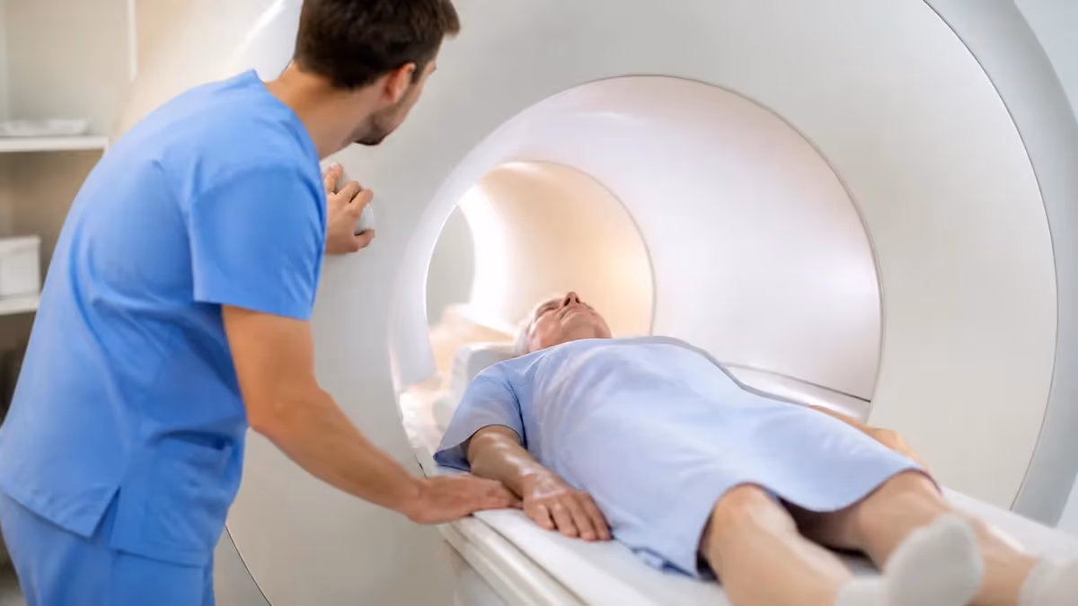
Plan on 60-90 minutes from arrival to walking out. Check-in takes about 15 minutes — paperwork, safety screening, and changing into a gown. The tech walks you through every metal-removal step and asks about implants, surgeries, and pregnancy.
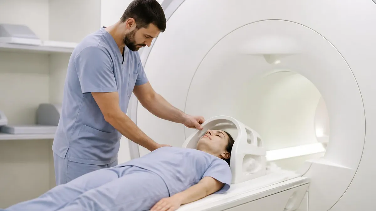
You lie on a sliding table. For a back MRI, the table moves you into the bore feet-first or head-first depending on which region they're scanning. Your back rests on a coil — a sensor pad that picks up the signal — and the tech positions you so the area of interest sits in the magnet's strongest field. Comfort matters here. You'll be still for 30-45 minutes, so they prop your knees with a bolster, give you a pillow, and hand you a panic button.
The noise is the surprising part. MRI scanners knock, buzz, and hum at 100+ decibels during scans. You'll get earplugs and headphones. Some facilities pipe in music. Once the scan starts, you stay still — every breath or twitch can blur a slice and force a repeat. Sequences run in 3-5 minute blocks with brief pauses between.
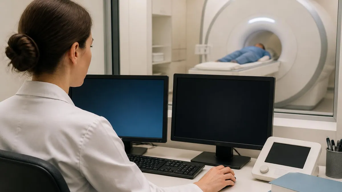
Results timing varies. The technologist sends images to a radiologist as soon as you're done. The radiologist reads, writes a report, and sends it to your ordering doctor — usually 24-72 hours later. Some imaging centers offer same-day reports for urgent cases. Your doctor's office calls or messages you with results once they've reviewed the report. Don't expect to hear directly from the imaging center.
If you want to see your own scan, ask for a copy on disc or through the patient portal. mri pictures are stored as DICOM files and viewable in free software like Horos or RadiAnt. But the report from the radiologist is what your doctor uses to make decisions. Self-reading is fine for curiosity — not for treatment planning.
One last note on results. The radiologist won't call you. Their report goes to the doctor who ordered the scan, and the conversation about next steps happens in that office. Don't expect a phone call from the imaging center unless something genuinely urgent shows up — a fresh fracture, a suspected cord compression, or a large mass. Those rare cases get fast-tracked back to your physician same day.
MRI Questions and Answers
More From the MRI Guide
About the Author
Medical Laboratory Scientist & Clinical Certification Expert
Johns Hopkins UniversityDr. Sandra Kim holds a PhD in Clinical Laboratory Science from Johns Hopkins University and is certified as a Medical Technologist (MT) and Medical Laboratory Scientist (MLS) through ASCP. With 16 years of clinical laboratory experience spanning hematology, microbiology, and molecular diagnostics, she prepares candidates for ASCP board exams, MLT, MLS, and specialist certification tests.