Lumbar Spine MRI: Complete Guide for Technologists and Patients
Lumbar spine MRI guide: protocols, sequences, anatomy, common pathologies, and patient prep. Boost your MRI exam prep with expert insight.
The lumbar spine MRI is one of the most frequently ordered musculoskeletal studies in modern radiology. Patients arrive with low back pain, sciatica, radiating leg numbness, or post-operative complaints, and the referring physician needs answers that only multi-planar, multi-sequence magnetic resonance imaging can deliver. For MRI technologists preparing for the ARRT (MRI) certification, the lumbar spine protocol is non-negotiable knowledge. It shows up on registry exams, in clinical practice, and in every conversation about workflow optimization.
Unlike a knee or shoulder study, the lumbar spine sits deep, surrounded by bowel motion, vascular pulsation, and patient body habitus that can wreck image quality. Coil selection matters. Sequence order matters. Slice angulation matters. So does breath-hold compliance, even though the lumbar region is technically below the diaphragm. Get one of those variables wrong and your radiologist is going to call you back. Worse, you will miss a pars defect, an annular fissure, or a small synovial cyst that could change the patient's surgical plan.
This guide walks through every element of the lumbar spine MRI exam: the anatomy you must recognize, the protocol sequences you must run, the pathologies you must flag, and the patient-care steps that keep your throughput steady. It is written for technologists at every level, from registry candidates to seasoned charge techs who train new staff. If you are a patient reading this before your appointment, you will also find clear answers about what to expect, how to prepare, and why the radiologist needs you to lie still for 25 minutes.
Lumbar Spine MRI by the Numbers
Why Radiologists Order a Lumbar Spine MRI
Most lumbar MRI referrals come from primary care, orthopedics, neurosurgery, and pain management. The clinical indications are remarkably consistent across specialties. Sciatica that fails six weeks of conservative therapy. New-onset back pain in a patient over 50 with red-flag symptoms. Suspected cauda equina syndrome, which is a true emergency. Post-laminectomy syndrome where the surgeon needs to differentiate scar from recurrent disc. Spinal stenosis workup before decompression. Tumor surveillance in oncology patients.
Each indication shapes the protocol. A cauda equina rule-out at 2 a.m. gets a fast screening exam: sag T2, ax T2 through the conus, and a quick STIR if metastatic disease is on the differential. A pre-op stenosis study for an elective case gets the full protocol with myelographic sequences, oblique cuts through the neural foramina, and sometimes a flexion-extension dynamic series. Knowing why the study was ordered helps you tailor the scan, saving table time and getting the radiologist the answer faster.
Cancer patients deserve special mention. A breast or prostate primary with new back pain needs marrow-sensitive imaging. STIR is your friend here. So is a post-contrast T1 with fat saturation if the radiologist suspects leptomeningeal disease or epidural extension. Always check the chart for prior imaging, prior surgery, and current renal function before injecting gadolinium.
Cauda equina syndrome, progressive neurological deficit, saddle anesthesia, bowel or bladder dysfunction, fever with back pain, and unexplained weight loss in a patient over 50 all warrant urgent or emergent lumbar spine MRI. These are not next-week studies. They are tonight or right now. If you are the on-call technologist and you see these indications on the requisition, prioritize the patient, notify the radiologist, and be prepared to add post-contrast sequences if infection or malignancy is suspected.
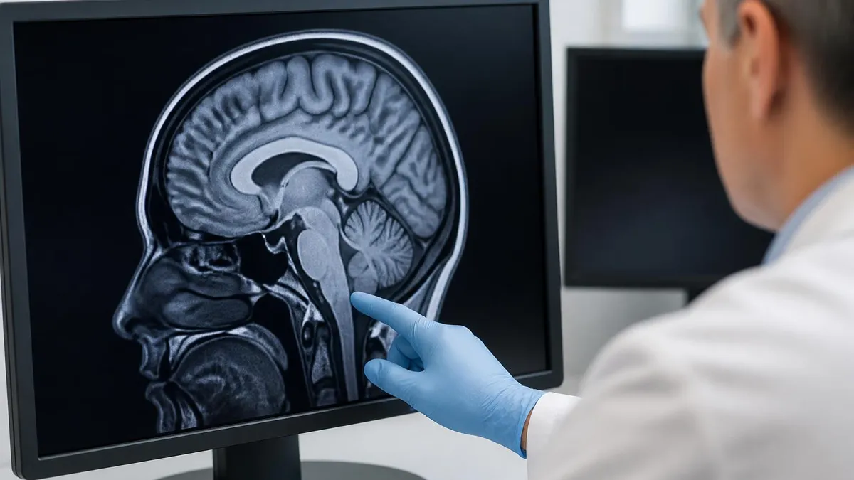
Lumbar Spine Anatomy You Must Recognize on MRI
Before you can run a great lumbar protocol, you need to know what you are looking at. The lumbar spine has five vertebral bodies (L1 through L5) plus the sacrum. Each vertebra has a body anteriorly, paired pedicles, transverse processes, lamina, a spinous process, and superior and inferior articular facets that form the facet joints. Between each pair of vertebrae sits an intervertebral disc with a tough outer annulus fibrosus and a softer nucleus pulposus that gives the disc its bright signal on T2.
The spinal canal contains the thecal sac, which houses cerebrospinal fluid (bright on T2, dark on T1) and the cauda equina nerve roots. The conus medullaris, where the cord tapers to its end, typically sits at the T12-L1 or L1-L2 level. Below the conus the canal contains only nerve roots, ligaments, and CSF. The neural foramina sit laterally at each level, transmitting the exiting nerve roots. Stenosis here causes radicular pain in the corresponding dermatome.
Ligamentous anatomy matters too. The anterior longitudinal ligament runs along the front of the vertebral bodies. The posterior longitudinal ligament runs behind them, inside the canal. The ligamentum flavum sits posteriorly between the lamina and frequently hypertrophies in degenerative disease, contributing to central canal stenosis. Recognizing these structures on every plane is what separates a confident scan from a guesswork scan.
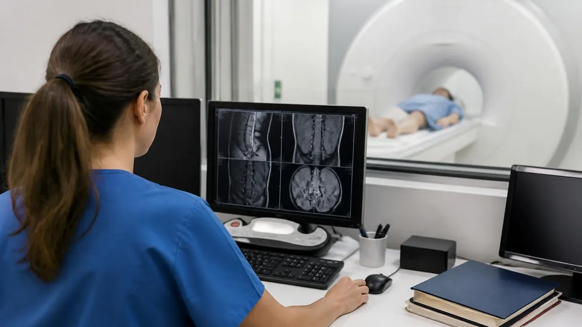
Standard Lumbar Spine MRI Sequences
Anatomic overview. Marrow signal, disc height, vertebral body morphology, and conus position. Pars defects show up here as dark interruptions in the pars interarticularis.
The workhorse. CSF is bright, discs are evaluated for hydration and herniation, ligamentum flavum thickness, and central canal caliber. Look for the disc-bulge pattern at each level.
Fat-suppressed inversion recovery. Detects marrow edema, fracture, infection, and metastasis. Mandatory in trauma and oncology workups.
Cross-sectional evaluation of each disc level. Quantifies central canal narrowing, lateral recess stenosis, and foraminal compromise.
Useful for evaluating fat planes around the thecal sac, post-operative scar, and confirming disc material composition.
Reserved for infection, tumor, or post-surgical workup. Enhancement differentiates active disease and scar from recurrent disc herniation.
Building the Perfect Lumbar Spine Protocol
Every department has its own protocol, but the core sequences are the same. Start with a three-plane localizer. Confirm the patient is centered, the coil is positioned over L1 through S1, and the spine is reasonably straight. If the patient has severe scoliosis, you may need to tilt your sagittal slab to follow the spinal axis rather than the magnet bore. Otherwise you will get partial-volume artifact through the discs and your radiologist will not be happy.
Next runs the sagittal T1. Use a fast spin echo with 3-4 mm slices, no gap or a small interleave, and a field of view that covers from T12 to the upper sacrum. The matrix should be at least 320 by 256 for diagnostic clarity. Phase-encode direction is usually superior-inferior to push respiratory artifact away from the spine. After the T1 comes the sagittal T2, often with identical geometry so the radiologist can flip between sequences without losing orientation.
The sagittal STIR or fat-saturated T2 follows. STIR gives more uniform fat suppression at the spine, where B0 inhomogeneity at the lung-spine interface can break a frequency-selective fat sat. Then come the axial sequences. Axial T2 is typically prescribed as angled stacks through each disc level (L1-L2, L2-L3, L3-L4, L4-L5, L5-S1), with the slices parallel to the disc rather than parallel to the magnet. This angulation is what makes a lumbar scan look professional. Straight-across axials miss the anatomy.
Some protocols add a coronal STIR for evaluation of the sacrum and SI joints, a 3D myelographic sequence for surgical planning, or oblique cuts through the neural foramina. Contrast is added when infection, tumor, or post-operative recurrent disc is in question. Inject 0.1 mmol/kg of a macrocyclic gadolinium agent, wait five minutes for tissue equilibration, then run sagittal and axial T1 with fat saturation.
Protocol Variations by Clinical Scenario
At 1.5 Tesla, SNR is lower than 3T, so expect slightly longer acquisition times for equivalent resolution. Typical parameters: sagittal T1 FSE TR/TE 500/12 ms, sagittal T2 FSE TR/TE 4000/100 ms, sagittal STIR TR/TE/TI 4500/40/150 ms, axial T2 FSE TR/TE 4500/120 ms. Slice thickness 4 mm sagittal, 4-5 mm axial. Total scan time around 22-25 minutes. Use a phased-array spine coil with at least eight elements.
Common Pathologies on Lumbar Spine MRI
Disc disease tops the list. Annular fissures appear as bright signal in the posterior annulus on T2, sometimes called a high-intensity zone. Disc bulges are broad-based extensions beyond the vertebral margin. Protrusions are focal extensions where the base is wider than the apex. Extrusions reverse that geometry, with the apex wider than the base, suggesting the disc material has herniated through a torn annulus. Sequestrations are free fragments that have separated from the parent disc, often migrating cranially or caudally in the canal.
Spinal stenosis comes in three flavors. Central canal stenosis narrows the thecal sac and compresses the cauda equina. Lateral recess stenosis pinches the descending nerve root before it exits. Foraminal stenosis compresses the exiting root within the neural foramen. Each pattern produces a different clinical syndrome, and grading severity (mild, moderate, severe) drives surgical decision-making.
Spondylolysis is a fracture of the pars interarticularis, classically at L5 in young athletes. It is often bilateral and can lead to spondylolisthesis, where one vertebral body slips forward on the one below. Grade the slip from I to IV based on percentage of slip. Modic changes describe vertebral endplate marrow signal alterations adjacent to degenerative discs: type 1 is edema (dark on T1, bright on T2), type 2 is fatty replacement (bright on both), and type 3 is sclerotic (dark on both).
Watch for the unexpected. Synovial cysts from facet arthropathy can cause severe radiculopathy. Tarlov cysts are perineural cysts that hollow out the sacrum. Schwannomas and ependymomas can grow inside the canal and present with vague back pain. Multiple myeloma riddles the marrow with focal lesions, especially in older patients. Discitis-osteomyelitis from staph or strep produces classic endplate destruction with paravertebral abscess on contrast imaging.
If sagittal T2 shows severe central canal stenosis with effacement of the thecal sac and the patient reports saddle anesthesia, new urinary retention, or bilateral leg weakness, stop and call the radiologist immediately. Do not let the patient leave the department. Cauda equina syndrome that goes more than 24-48 hours without decompression carries a high risk of permanent neurological deficit. Your speed and judgment matter.
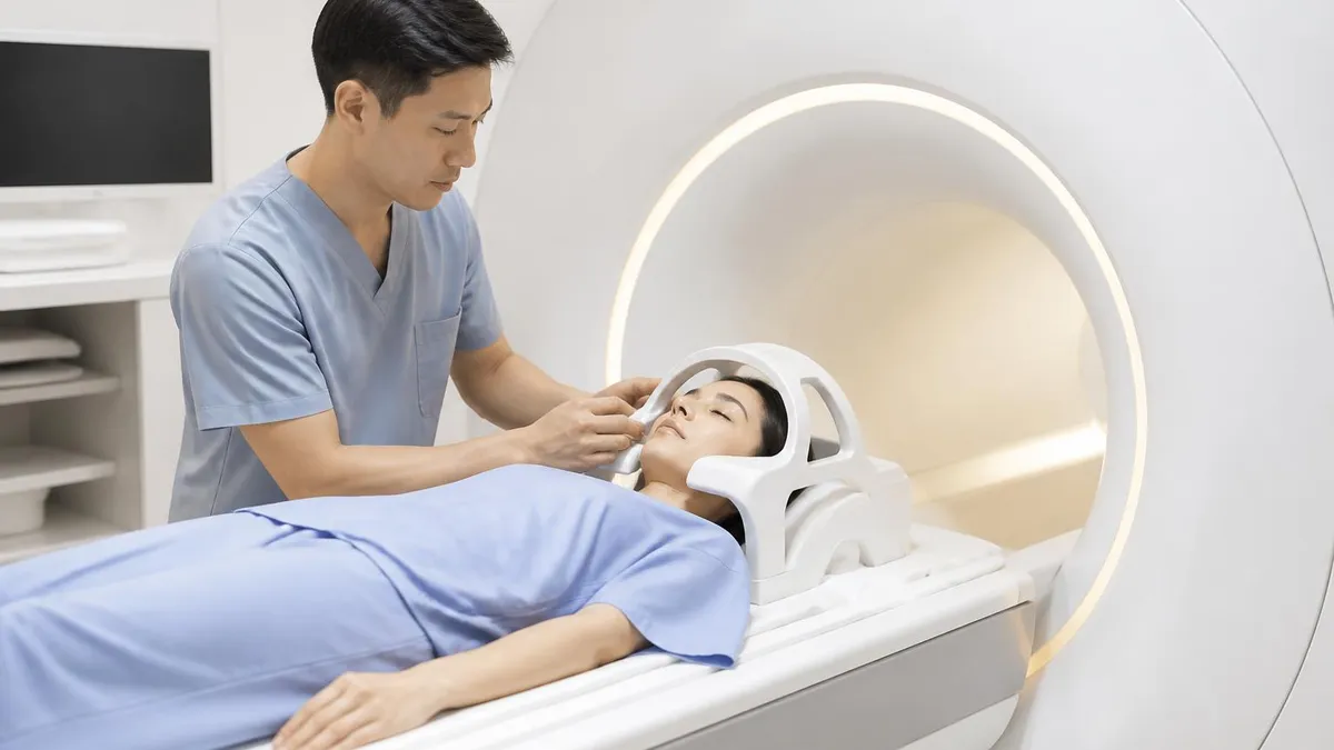
Patient Preparation and Safety
Screen every patient before they enter the magnet. Pacemakers, implanted cardioverter defibrillators, cochlear implants, certain aneurysm clips, and metallic foreign bodies in the eye are absolute or relative contraindications. Use a standardized MRI safety questionnaire and confirm device compatibility in MRIsafety.com or the manufacturer labeling. A patient with a spinal cord stimulator may need the device powered off or removed before imaging.
Ask about claustrophobia. The lumbar spine exam puts the patient's head near the bore opening, which helps, but anxious patients still struggle. Offer prone positioning if supine is intolerable, although prone reduces image quality slightly due to coil contact issues. Use mirrors, music, and frequent verbal reassurance. For severe claustrophobia, pre-medication with a benzodiazepine prescribed by the referring physician is appropriate.
Renal function matters when contrast is on the table. Check eGFR within the institutional window (often 30 days for outpatients, 48 hours for inpatients). Macrocyclic gadolinium agents have a much lower risk of nephrogenic systemic fibrosis than older linear agents, but you still document the dose, lot number, and administration time. Patients on dialysis can receive gadolinium with a hemodialysis session timed shortly after the scan.
Pre-Scan Checklist for Lumbar Spine MRI
- ✓Verify patient identity with two identifiers and confirm the body part ordered
- ✓Complete MRI safety screening including all metallic implants and prior surgeries
- ✓Document current weight in kg for any contrast calculation
- ✓Confirm renal function if gadolinium is anticipated
- ✓Remove all external metal: jewelry, hairpins, hearing aids, dentures with metal
- ✓Provide ear protection and a panic ball or squeeze bulb
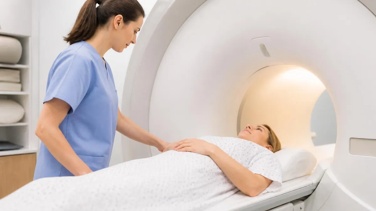
- ✓Position the patient supine with knees slightly flexed on a bolster for comfort
- ✓Center the spine coil over L1 through the upper sacrum and apply straps
- ✓Run a three-plane localizer and verify coverage before launching the protocol
- ✓Document patient cooperation level and any technical limitations in the worklist
Image Quality and Troubleshooting
Even on a perfectly running magnet, image artifacts happen. Motion is the most common offender. The patient shifts, breathes deeply, or coughs, and the sagittal images blur. Train your patients to take slow, shallow breaths and to keep their hands resting on their abdomen, not on their chest where breathing motion is most visible. For very long sequences, consider respiratory triggering or shorter scan times with parallel imaging.
Aliasing artifact, also called wrap, happens when anatomy outside the field of view folds back into the image. Increase the field of view, swap the phase-encode direction, or apply no-phase-wrap (oversampling) options. Chemical shift artifact appears as bright and dark bands at fat-water interfaces, most visible at vertebral endplates. Increase receiver bandwidth or shorten the echo train length to reduce it.
Susceptibility artifact from surgical hardware can obliterate the area of interest. Switch from gradient echo to fast spin echo. Increase bandwidth. Decrease echo time. Use view-angle tilting if your scanner supports it. If the patient has rods and screws, accept that you will get some signal void around the metal and focus the radiologist's attention on the levels above and below.
Truncation artifact creates parallel bands across the spinal cord on sagittal T2 and can be mistaken for a syrinx. Recognize it by its periodicity and confirm by switching the matrix or phase direction. Cerebrospinal fluid flow artifact can also create signal voids in the thecal sac that mimic vascular flow. Compensation techniques like flow compensation gradients or sampling at quieter phases of the cardiac cycle help reduce it.
Lumbar Spine MRI Pros and Cons
- +Superior soft tissue contrast for discs, nerves, and ligaments
- +No ionizing radiation, safe for repeat imaging
- +Multi-planar capability without repositioning the patient
- +Detects marrow pathology that CT cannot show
- +Gold standard for spinal cord and cauda equina evaluation
- +Gadolinium contrast distinguishes scar from recurrent disc
- −Long scan times compared to CT, around 25 minutes
- −Patient cooperation is essential; motion ruins the study
- −Claustrophobia can prevent completion of the exam
- −Metallic implants may be contraindicated or cause artifact
- −Cost is higher than CT or plain radiographs
- −Limited availability in rural settings and after-hours
Reporting and Communication With the Radiologist
Once the images are on the workstation, the radiologist takes over, but your job is not done. A short, well-written technologist note saves the radiologist time and improves patient care. Document the protocol run, any deviations (sequences skipped or added), the patient's body habitus, and any technical limitations such as motion or hardware artifact. If you noticed something concerning on the images, like a possible fracture or cord lesion, flag it.
For technologists preparing for the ARRT (MRI) registry, knowing how to communicate findings without overstepping your scope is a tested competency. You do not diagnose. You describe. Saying "sagittal T2 shows increased signal at L4-L5 disc" is fine. Saying "the patient has a herniated disc" is not. The radiologist makes the diagnosis. You make the images and the report-worthy observations.
Workflow efficiency is also a tested skill. Knowing how to schedule a lumbar MRI behind a brain MRI to share coil time, when to add a sequence on the fly, and how to handle a patient who needs to come off the table mid-scan are all real-world competencies that show up on the registry in scenario-based questions. The best technologists are calm, organized, and curious about every case that comes through the door.
MRI Questions and Answers
Putting It All Together
The lumbar spine MRI is more than a routine study. It is a diagnostic workhorse that asks the technologist to balance anatomy, physics, patient care, and clinical reasoning in real time. The best technologists do not just run the protocol. They tailor it. They watch the images as they come up. They notice when a sequence needs a repeat. They communicate clearly with the radiologist and with the patient.
If you are preparing for the ARRT (MRI) registry, study the lumbar spine protocol until you can build it from scratch on a piece of scratch paper. Know the sequence parameters, the slice angulations, the common pathologies, and the safety screening process inside and out. Practice questions are essential. The registry blueprint allocates a substantial portion of the exam to spine imaging, so this is high-yield material.
If you are a patient, remember that the technologist running your scan has trained for years to give you the best possible images. Ask questions if you have them. Tell the tech if you are uncomfortable. Hold still when the scan is running. Twenty-five minutes of cooperation can give your doctor the information needed to guide a treatment plan that could resolve your pain for good.
Bottom line: a lumbar spine MRI rewards careful preparation, patient communication, and steady protocol discipline. Master the basics, practice the angulations, and stay curious about every case. The next study that rolls onto your table could be straightforward degenerative disease or a hidden cord tumor — and either way, your work matters more than you think.
About the Author
Attorney & Bar Exam Preparation Specialist
Yale Law SchoolJames R. Hargrove is a practicing attorney and legal educator with a Juris Doctor from Yale Law School and an LLM in Constitutional Law. With over a decade of experience coaching bar exam candidates across multiple jurisdictions, he specializes in MBE strategy, state-specific essay preparation, and multistate performance test techniques.