Thoracic Spine MRI: Complete Guide to Imaging, Preparation, and Results
🎓 Learn about thoracic spine MRI scans including preparation, what to expect, common findings, and how to interpret results for back pain and spinal
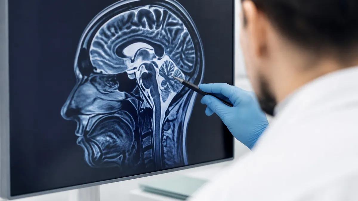
A thoracic spine MRI is one of the most valuable diagnostic imaging tools available for evaluating the middle portion of the vertebral column. This non-invasive procedure uses powerful magnetic fields and radiofrequency pulses to produce detailed cross-sectional images of the twelve thoracic vertebrae, intervertebral discs, spinal cord, and surrounding soft tissues. Physicians rely on thoracic spine MRI to diagnose conditions ranging from herniated discs and compression fractures to tumors and infections that affect the mid-back region of the body.
The thoracic spine is unique among spinal regions because it connects to the rib cage, providing structural stability that the cervical and lumbar spine do not possess. This anatomical characteristic means the thoracic vertebrae experience different mechanical stresses than other spinal segments. When patients present with mid-back pain, neurological symptoms, or unexplained sensory changes in the trunk, an MRI of the thoracic spine is typically the gold standard imaging modality that physicians order to identify the underlying pathology with precision.
Unlike X-rays or CT scans, MRI does not expose patients to ionizing radiation, making it a safer option for repeated imaging when monitoring chronic conditions or tracking treatment progress over time. The superior soft tissue contrast provided by magnetic resonance imaging allows radiologists to differentiate between muscles, ligaments, nerves, and fluid collections with exceptional clarity. This level of detail is critical when evaluating thoracic disc herniations, which can compress the spinal cord and potentially cause myelopathy if left untreated.
Patients referred for a thoracic spine MRI often experience symptoms such as persistent mid-back pain, radiating pain along the rib cage known as intercostal neuralgia, numbness or tingling in the torso, or progressive weakness in the lower extremities. These symptoms can indicate serious conditions including spinal cord compression, vertebral body fractures from osteoporosis, metastatic disease, or inflammatory processes like transverse myelitis that require prompt medical intervention and accurate diagnostic confirmation.
The examination typically takes between thirty and sixty minutes depending on whether contrast enhancement is required and how many sequences the radiologist prescribes for the specific clinical question. Standard protocols include T1-weighted, T2-weighted, and STIR sequences that each highlight different tissue characteristics. When infection or tumor is suspected, gadolinium-based contrast agents are administered intravenously to improve visualization of abnormal tissue enhancement patterns and help distinguish benign from malignant processes.
Understanding what a thoracic spine MRI involves, how to prepare for the examination, and what the results may reveal empowers patients to approach their appointment with confidence and engage meaningfully in discussions with their healthcare providers. This comprehensive guide covers every aspect of thoracic spine magnetic resonance imaging from the initial referral through to understanding your radiology report and the potential next steps in your medical care journey.
Whether you are a radiology technologist preparing for certification examinations, a student learning MRI physics and protocols, or a patient seeking reliable information about an upcoming scan, this article provides the detailed knowledge you need. We will explore imaging protocols, clinical indications, common pathological findings, and practical preparation tips that ensure the highest quality diagnostic images are obtained during your thoracic spine MRI examination.
Thoracic Spine MRI by the Numbers
The Thoracic Spine MRI Process
Referral and Scheduling
Patient Screening and Preparation
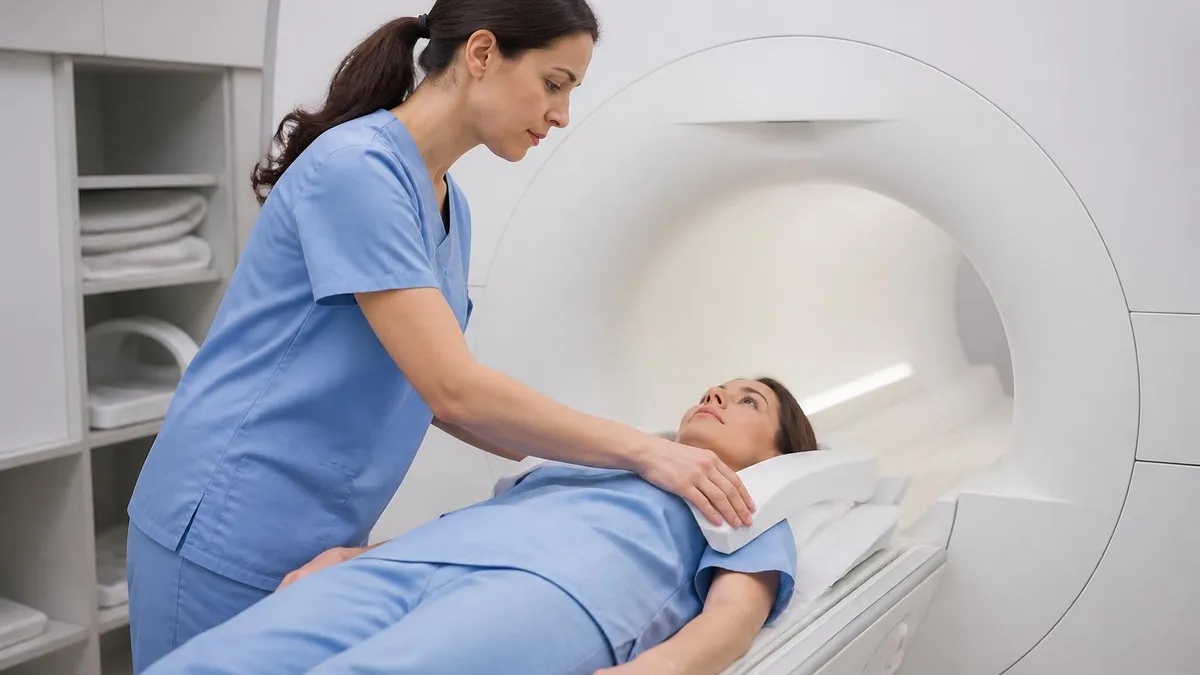
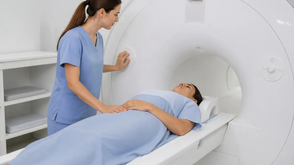
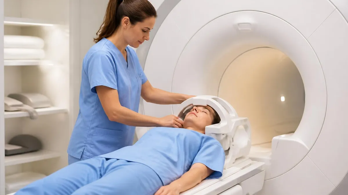
Positioning and Coil Placement
Image Acquisition
Contrast Administration if Needed
Image Review and Reporting
The thoracic spine encompasses twelve vertebrae labeled T1 through T12, forming the longest segment of the spinal column and providing attachment points for the twelve pairs of ribs. Each thoracic vertebra has a body anteriorly, pedicles and laminae forming the neural arch, spinous processes projecting posteriorly, and transverse processes extending laterally where rib articulations occur. The intervertebral discs between adjacent vertebral bodies serve as shock absorbers and allow limited flexibility in this relatively rigid portion of the spine.
Within the spinal canal, the thoracic spinal cord transmits motor and sensory signals between the brain and the lower body. The cord is narrower in the thoracic region compared to the cervical enlargement, making it particularly vulnerable to compression from disc herniations, bone spurs, or mass lesions. Any reduction in the available space within the thoracic spinal canal can produce myelopathic symptoms including gait disturbance, lower extremity weakness, and bowel or bladder dysfunction that may become permanent without intervention.
Common clinical indications for ordering a thoracic spine MRI include persistent mid-back pain that fails to respond to conservative treatment lasting six weeks or more. Physicians also order this examination when patients develop neurological deficits suggesting spinal cord involvement, including progressive lower extremity weakness, sensory level changes, or signs of upper motor neuron pathology. Traumatic injuries with suspected fractures or ligamentous damage also warrant thoracic spine MRI to fully characterize the extent of structural damage.
Oncologic indications represent a significant portion of thoracic spine MRI examinations. The thoracic vertebral bodies are common sites for metastatic disease because of their rich blood supply from the Batson venous plexus. Breast cancer, lung cancer, prostate cancer, and renal cell carcinoma frequently metastasize to thoracic vertebrae, causing pathologic fractures and spinal cord compression that require urgent imaging evaluation and treatment planning to preserve neurological function.
Infectious conditions including vertebral osteomyelitis, discitis, and epidural abscess also require thoracic spine MRI for accurate diagnosis and treatment monitoring. These infections can spread rapidly within the spinal canal and produce devastating neurological consequences if not identified promptly. Contrast-enhanced MRI is particularly valuable for infectious indications because it demonstrates the extent of tissue involvement, identifies abscess collections amenable to drainage, and helps monitor treatment response during antibiotic therapy.
Demyelinating diseases like multiple sclerosis frequently involve the thoracic spinal cord, producing lesions that cause sensory disturbances, weakness, and autonomic dysfunction. MRI can detect these plaques as high-signal areas on T2-weighted images and plays a crucial role in establishing the diagnosis by demonstrating dissemination in space and time. Serial imaging monitors disease activity and treatment response in patients receiving disease-modifying therapies for inflammatory conditions affecting the thoracic cord.
Congenital abnormalities and developmental conditions also benefit from thoracic spine MRI evaluation. Syringomyelia, tethered cord syndrome, diastematomyelia, and vascular malformations can all be identified and characterized using appropriate MRI sequences. These conditions may present at any age and often require longitudinal imaging surveillance to detect progression that might warrant surgical intervention to prevent irreversible neurological deterioration over time.
MRI Practice Test Questions
Prepare for the MRI - Magnetic Resonance Imaging exam with our free practice test modules. Each quiz covers key topics to help you pass on your first try.
MRI Knowledge
MRI Exam Questions covering Knowledge. Master MRI Test concepts for certification prep.
MRI Physics
Free MRI Practice Test featuring Physics. Improve your MRI Exam score with mock test prep.
MRI Anatomy and Pathology
MRI Test Prep for MRI Anatomy and Pathology. Practice MRI Quiz questions and boost your score.
MRI Anatomy and Positioning
MRI Questions and Answers on MRI Anatomy and Positioning. Free MRI practice for exam readiness.
MRI Contrast Agents
Free MRI Quiz on MRI Contrast Agents. MRI Exam prep questions with detailed explanations.
MRI Patient Care and Positioning
MRI Practice Questions for MRI Patient Care and Positioning. Build confidence for your MRI certification exam.
Thoracic Spine MRI Imaging Protocols
T1-weighted sequences provide excellent anatomical detail of the thoracic spine by highlighting fat-containing structures as bright signal and fluid as dark signal. These sequences clearly delineate vertebral body marrow, epidural fat planes, and the overall structural anatomy. Normal bone marrow appears bright on T1 images because of its fat content, making pathological processes that replace marrow such as metastatic disease or infection readily apparent as areas of abnormally decreased signal intensity compared to adjacent healthy vertebrae.
Standard T1 protocols for thoracic spine include sagittal spin-echo or turbo spin-echo sequences with repetition times around 400 to 600 milliseconds and echo times of 10 to 20 milliseconds. Axial T1 images are acquired through regions of interest identified on sagittal images. Post-contrast T1 sequences with fat saturation are essential when evaluating enhancing lesions because they suppress the normal high signal from fat, allowing gadolinium enhancement to stand out clearly against the suppressed background tissue signal.
Thoracic Spine MRI: Advantages and Limitations
- +Superior soft tissue contrast reveals spinal cord pathology invisible on CT or X-ray
- +No ionizing radiation exposure makes it safe for repeated imaging and pediatric patients
- +Multiplanar imaging capability provides sagittal, axial, and coronal views without repositioning
- +Contrast enhancement characterizes tumors, infections, and inflammatory lesions precisely
- +Detects bone marrow pathology earlier than other imaging modalities in many conditions
- +Non-invasive alternative to myelography for evaluating spinal canal contents
- −Longer examination time of 30-60 minutes requires patient cooperation and stillness
- −Claustrophobia affects approximately 5-10% of patients requiring sedation or open MRI
- −Contraindicated in patients with certain pacemakers, cochlear implants, or metallic foreign bodies
- −Motion artifacts from breathing and cardiac pulsation can degrade thoracic image quality
- −Higher cost compared to CT or X-ray imaging may limit accessibility for some patients
- −Gadolinium contrast carries rare risks including allergic reactions and nephrogenic systemic fibrosis
Thoracic Spine MRI Patient Preparation Checklist
- ✓Remove all jewelry, piercings, watches, and metallic accessories before entering the scan room
- ✓Inform technologists about any implanted medical devices including pacemakers, stents, or joint replacements
- ✓Disclose any history of metal fragments in eyes from grinding, welding, or industrial work
- ✓Wear comfortable clothing without metallic zippers, buttons, snaps, or underwire bras
- ✓Arrive fifteen to thirty minutes early to complete safety screening forms and registration
- ✓Inform staff if you are pregnant or suspect pregnancy before the examination begins
- ✓Discuss claustrophobia concerns with your ordering physician who may prescribe anti-anxiety medication
- ✓Fast for four to six hours before the scan if intravenous contrast administration is planned
- ✓Bring a list of current medications including dosages for the screening questionnaire
- ✓Arrange transportation home if you receive sedation medication for anxiety management
Thoracic Cord Compression Requires Urgent Imaging
Acute thoracic spinal cord compression from tumor, abscess, or disc herniation is a medical emergency. If you develop rapidly progressive leg weakness, loss of bladder or bowel control, or a sensory level across your trunk, seek immediate emergency care. MRI within hours of symptom onset guides urgent surgical decompression that can prevent permanent paralysis.
The most common findings on thoracic spine MRI include degenerative disc disease, Schmorl nodes, vertebral compression fractures, and disc herniations that may or may not produce clinical symptoms. Degenerative changes in the thoracic spine are extremely common in adults over forty years of age and frequently represent incidental findings unrelated to the patient's presenting complaints. Radiologists must carefully correlate imaging findings with clinical symptoms to distinguish clinically significant pathology from age-appropriate degenerative changes that require no treatment.
Thoracic disc herniations are less common than cervical or lumbar herniations because the rib cage limits motion in this spinal segment, but they carry greater clinical significance when present. The thoracic spinal canal is relatively narrow compared to the lumbar region, meaning even small disc herniations can compress the spinal cord and produce myelopathy. Large central thoracic disc herniations may require surgical intervention through anterior or posterolateral approaches that are technically more demanding than typical lumbar discectomy procedures.
Vertebral compression fractures represent another frequent finding on thoracic spine MRI, particularly in elderly patients with osteoporosis and those with metastatic cancer. MRI distinguishes acute from chronic fractures by identifying bone marrow edema on STIR and T2 sequences, which appears as bright signal within the fractured vertebral body. This distinction is clinically important because acute painful fractures may benefit from vertebroplasty or kyphoplasty procedures, while chronic healed fractures typically require only conservative management and osteoporosis treatment.
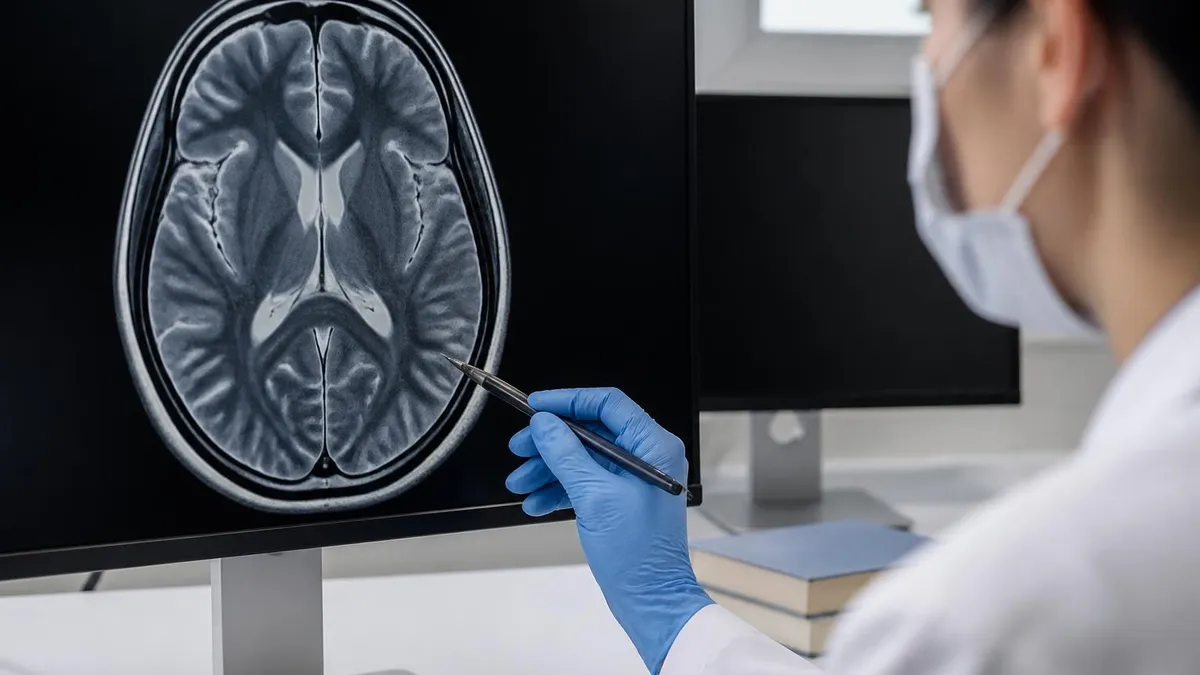
Spinal cord tumors visible on thoracic MRI include intramedullary tumors such as ependymomas and astrocytomas that arise within the cord substance, intradural extramedullary tumors like meningiomas and schwannomas that grow within the dural sac but outside the cord, and extradural tumors that compress the thecal sac from outside. Each category has characteristic imaging features on T1, T2, and post-contrast sequences that allow experienced radiologists to suggest the most likely diagnosis and guide the neurosurgical team in treatment planning.
Inflammatory and demyelinating conditions produce characteristic thoracic cord lesions on MRI. Multiple sclerosis plaques typically appear as short-segment lesions less than two vertebral bodies in length on sagittal images, often located peripherally within the cord. In contrast, neuromyelitis optica spectrum disorder tends to produce longitudinally extensive transverse myelitis spanning three or more vertebral segments. These imaging patterns help neurologists differentiate between autoimmune conditions that require different treatment approaches and carry different long-term prognoses.
Vascular pathology identified on thoracic spine MRI includes spinal cord infarction, which produces characteristic patterns of restricted diffusion and T2 signal abnormality corresponding to arterial territories. Dural arteriovenous fistulas cause progressive myelopathy from venous congestion and appear as flow voids along the dorsal cord surface on T2-weighted images. Early recognition of vascular pathology is essential because some conditions are treatable through endovascular embolization or surgical ligation when diagnosed before irreversible cord damage occurs.
Infectious processes on thoracic spine MRI typically demonstrate vertebral body endplate destruction, disc space involvement, and paravertebral or epidural abscess formation. Tuberculosis produces characteristic findings including subligamentous spread across multiple vertebral levels, large paraspinal abscesses, and relative preservation of disc spaces compared to pyogenic infections. Post-contrast imaging reveals rim-enhancing collections and helps define the extent of epidural disease that may require surgical drainage to prevent neurological deterioration from cord compression.
Your radiologist may recommend follow-up MRI at specific intervals to monitor indeterminate findings such as small hemangiomas, minor disc bulges, or equivocal signal changes. Always discuss follow-up recommendations with your referring physician who can determine if additional imaging is clinically warranted based on your symptoms and treatment response.
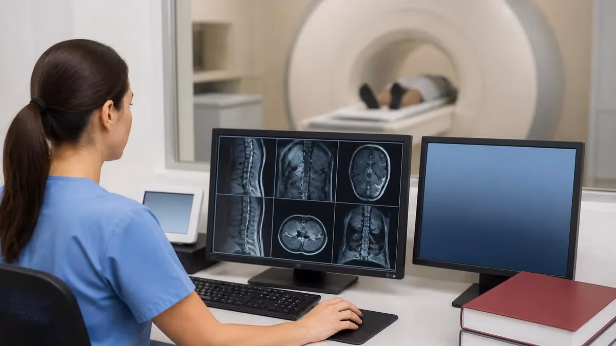
Understanding your thoracic spine MRI report requires familiarity with the standard format and terminology that radiologists use to communicate findings. Reports typically begin with a technique section describing which sequences were performed and whether contrast was administered. The findings section describes each anatomical structure systematically, usually proceeding from superior to inferior, noting alignment, vertebral body signal characteristics, disc morphology, spinal canal dimensions, cord signal, and any paravertebral soft tissue abnormalities identified during the examination.
The impression section at the end of the radiology report provides a concise summary of the most clinically relevant findings ranked by significance. This section is what your referring physician uses to make treatment decisions. If the impression mentions cord compression, significant stenosis, or suspicious lesions requiring further evaluation, your physician will likely discuss these findings with you and may refer you to a specialist such as a neurosurgeon, orthopedic spine surgeon, or neurologist for further management and treatment planning.
Normal thoracic spine MRI findings include uniform vertebral body height and signal, normal disc hydration appearing bright on T2 images, patent spinal canal with normal cord caliber and signal, and normal paravertebral soft tissues. Age-related changes that are generally considered normal include mild disc desiccation, small anterior osteophytes, and minor facet arthropathy. These findings are typically described in the report but noted as degenerative changes without clinical significance in the absence of neural compression or cord signal abnormality.
Several measurements help radiologists assess the thoracic spine quantitatively. The anteroposterior diameter of the spinal canal at thoracic levels normally measures at least nine to ten millimeters, with values below seven millimeters indicating significant canal stenosis. Vertebral body height loss greater than twenty-five percent suggests a compression fracture. Cord signal abnormality on T2 images at the level of maximum compression suggests myelomalacia or edema, which correlates with clinical myelopathic signs and often influences surgical decision-making.
When reviewing your MRI results with your physician, prepare questions about what specific findings mean for your condition, whether treatment is recommended, and what the expected timeline for improvement might be. Not all abnormal findings require treatment since many degenerative changes are asymptomatic and managed conservatively. However, findings suggesting cord compression, instability, infection, or malignancy typically require more urgent intervention and specialist referral for definitive management.
Follow-up imaging intervals depend on the specific diagnosis and clinical situation. Stable benign findings may never require repeat imaging, while tumors and infections typically need follow-up MRI at defined intervals to assess treatment response. Patients with multiple sclerosis undergo routine surveillance MRI to detect new lesions and monitor disease activity. Your physician will determine the appropriate follow-up schedule based on your specific diagnosis, treatment plan, and clinical response over time.
Comparing current MRI findings with prior examinations is one of the most valuable aspects of serial imaging. Radiologists document whether findings are stable, improved, or progressed compared to previous studies. This temporal information helps physicians adjust treatment plans, determine whether surgical intervention has succeeded, and identify new pathology that may have developed since the last examination. Always bring prior imaging studies or ensure they are available electronically for comparison during your thoracic spine MRI appointment.
Maximizing the diagnostic quality of your thoracic spine MRI begins with proper preparation and understanding what the examination entails. Patients who arrive informed and prepared produce better quality images because they cooperate more effectively with technologist instructions, remain still throughout the examination, and experience less anxiety that could lead to premature termination of the scan. Taking simple preparatory steps significantly improves your imaging experience and the resulting diagnostic information available to your medical team.
Breathing motion is the primary technical challenge during thoracic spine MRI because the chest wall moves with each respiratory cycle, potentially degrading image quality through motion artifacts. Technologists employ several strategies to minimize respiratory artifacts including navigator-triggered sequences, respiratory gating, and anterior saturation bands that suppress signal from moving anterior chest wall structures. Patients can help by breathing shallowly and regularly rather than taking deep breaths during sequence acquisitions.
Cardiac motion represents another source of artifacts in thoracic spine imaging, particularly affecting the upper thoracic region near the heart. Cardiac gating techniques synchronize image acquisition with the cardiac cycle to reduce pulsation artifacts. Spatial saturation bands placed over the heart and great vessels also help minimize flow-related artifacts that could obscure pathology in the upper thoracic vertebrae and spinal cord where diagnostic accuracy matters most for clinical decision-making.
For patients experiencing claustrophobia, several options exist to make the thoracic spine MRI more tolerable. Open MRI systems with wider bores reduce the confined feeling, though image quality may be slightly lower than closed-bore high-field systems. Mild sedation with oral benzodiazepines taken before the appointment helps many patients complete their examination successfully. Some facilities offer headphones with music, prism glasses for viewing outside the bore, or aromatherapy to reduce anxiety during the scanning procedure.
If your physician has ordered contrast-enhanced imaging, expect an intravenous line placement before or during your examination. Gadolinium-based contrast agents are generally well-tolerated with serious adverse reactions occurring in fewer than one in ten thousand administrations. Patients with significantly impaired kidney function require special consideration because of the rare risk of nephrogenic systemic fibrosis. Current guidelines recommend checking kidney function with a blood test before administering gadolinium to patients with known renal disease or risk factors for kidney impairment.
After your thoracic spine MRI is complete, you can typically resume all normal activities immediately unless you received sedation medication. There are no lasting effects from the magnetic field or radiofrequency energy used during the scan. Results processing time varies by facility, with many centers providing preliminary reports within twenty-four hours and final dictated reports within one to three business days. Your referring physician will contact you to discuss significant findings and next steps in your care plan.
For MRI technologists and students preparing for certification examinations, thoracic spine imaging protocols require understanding of coil selection, sequence optimization for the thoracic region, and artifact mitigation strategies specific to chest imaging. The ARRT MRI examination tests knowledge of thoracic spine anatomy, appropriate imaging planes, sequence selection rationale, and common pathological findings that technologists should recognize during quality assurance review of acquired images before the patient leaves the department.
MRI Questions and Answers

MRI Medical Abbreviation: What MRI Stands For and Why It Matters

Knee MRI Images: A Complete Guide to Reading, Understanding, and Interpreting Knee Scans

Noise of MRI Machine: Why MRI Scanners Are So Loud and What to Expect
Is Nickel Titanium MRI Compatible? A Complete Guide to MRI Safety Materials
What to Expect During MRI: Your Complete Patient Guide to the Scanning Process
About the Author
Medical Laboratory Scientist & Clinical Certification Expert
Johns Hopkins UniversityDr. Sandra Kim holds a PhD in Clinical Laboratory Science from Johns Hopkins University and is certified as a Medical Technologist (MT) and Medical Laboratory Scientist (MLS) through ASCP. With 16 years of clinical laboratory experience spanning hematology, microbiology, and molecular diagnostics, she prepares candidates for ASCP board exams, MLT, MLS, and specialist certification tests.
Join the Discussion
Connect with other students preparing for this exam. Share tips, ask questions, and get advice from people who have been there.
View discussion (6 replies)