
Thoracic MRI: Complete Guide to Imaging the Chest Spine and Mediastinum 2026
Learn everything about thoracic MRI — ✅ anatomy, protocols, pathology, and patient prep. Includes study tips for MRI registry exam success.
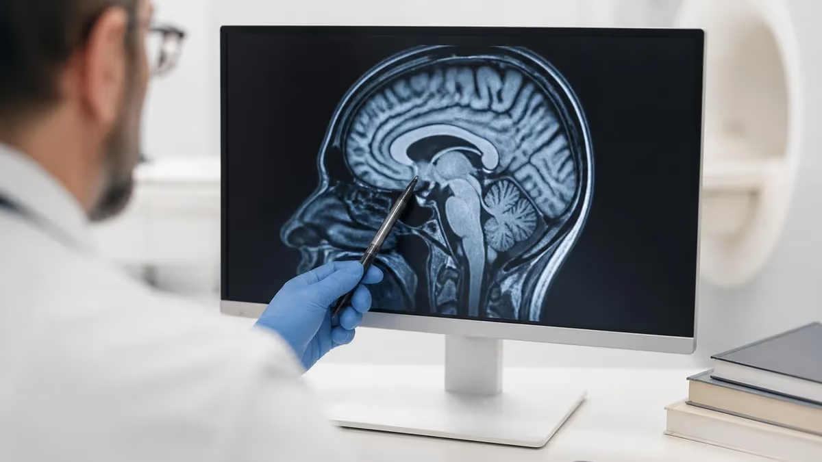
Thoracic MRI is one of the most technically demanding and clinically rewarding examinations in diagnostic radiology. The thoracic region encompasses the chest vertebrae (T1 through T12), the spinal cord and nerve roots, the mediastinum, the heart and great vessels, and the surrounding soft tissues — all of which present unique challenges during magnetic resonance imaging due to respiratory motion, cardiac pulsation, and the complex anatomical relationships packed into a relatively compact space. Practice with the free MRI - Magnetic Resonance Imaging practice test on PracticeTestGeeks (11 questions with answer explanations).
Understanding thoracic MRI begins with appreciating why this modality is often chosen over CT or X-ray. MRI provides superior soft-tissue contrast without ionizing radiation, making it the gold standard for evaluating spinal cord compression, disc herniations affecting the thoracic spine, paravertebral masses, brachial plexopathy, and cardiac or mediastinal pathology. For patients who require repeated imaging — such as those with multiple sclerosis or spinal tumors — this radiation-free approach is clinically significant over a lifetime of monitoring.
For MRI technologists preparing for registry examinations, the thoracic region demands mastery of both anatomy and pulse sequence selection. Knowing when to apply fat saturation, which plane best demonstrates a disc herniation, or how to compensate for cardiac artifact can determine whether a study is diagnostic or needs to be repeated. This guide covers everything from coil selection and patient positioning to pathology recognition and protocol optimization, giving you the comprehensive foundation needed to perform confidently on the board exam and in clinical practice.
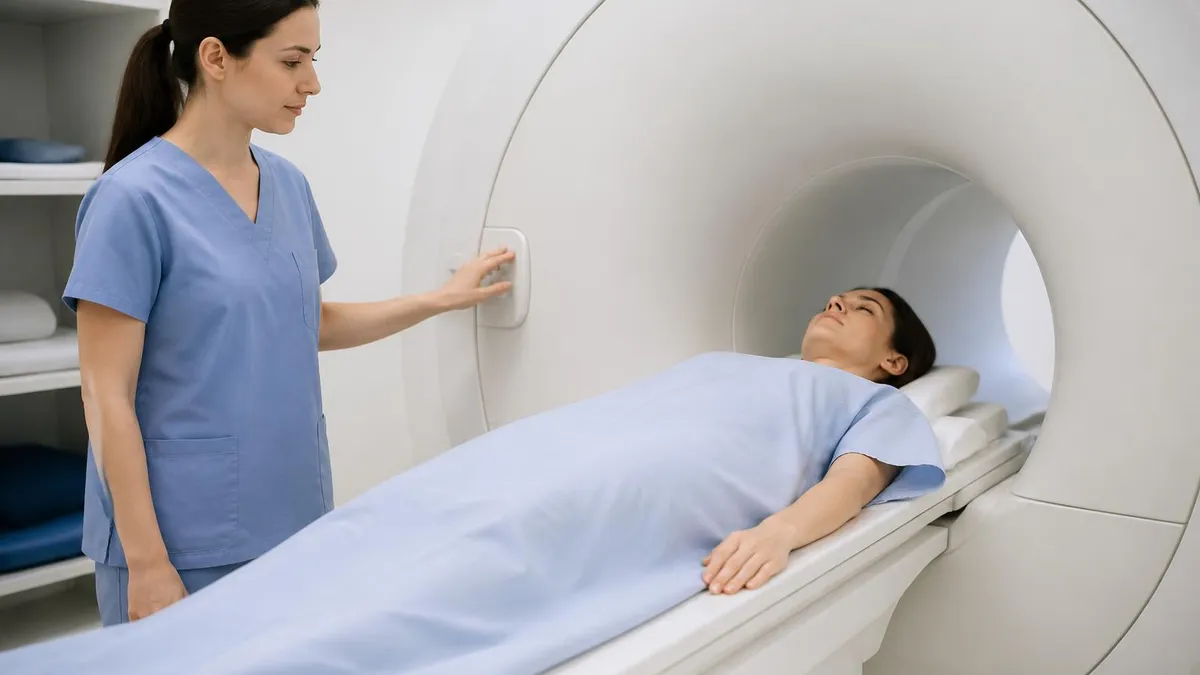
Patient preparation for thoracic MRI differs from other body regions primarily because of motion artifacts. Technologists must counsel patients on breath-holding instructions, explain the duration of the exam (typically 45 to 60 minutes for a comprehensive thoracic spine study), and screen carefully for contraindications including pacemakers, certain metallic implants, and claustrophobia. Sedation or anxiolytic medication may be coordinated with the ordering physician for patients who cannot tolerate the confined bore of the magnet.
The clinical indications driving a thoracic MRI order are broad. Neurologists request it for suspected myelopathy, transverse myelitis, or demyelinating disease. Orthopedic surgeons need it before operating on degenerative disc disease or spinal stenosis. Oncologists use it to stage spinal metastases or evaluate cord compression from epidural disease. Vascular surgeons may order a thoracic MRI with angiography sequences to evaluate the aorta. Each indication shapes the protocol, and the technologist plays a central role in customizing sequences to the clinical question.
From an MRI registry exam perspective, thoracic imaging questions frequently test knowledge of artifact identification and reduction. Ghosting from respiratory motion, CSF flow artifacts mimicking cord pathology, and chemical shift effects along vertebral endplates are among the most commonly tested concepts. Building a strong mental model of how physical principles translate into image artifacts — and how to correct them — is essential for both exam success and daily clinical performance.
Whether you are a student entering your first clinical rotation, a practicing technologist brushing up for recertification, or a radiologist seeking a quick protocol reference, this guide to thoracic mri will walk you through every layer of this complex but fascinating examination, from field strength considerations down to pathology recognition and reporting communication.
Free MRI - Magnetic Resonance Imaging Study Tools
Interactive MRI - Magnetic Resonance Imaging resources on PracticeTestGeeks, free to start:
- Free MRI Knowledge Questions and Answers — 11 questions with instant scoring and answer explanations
- MRI - Magnetic Resonance Imaging flashcards — review key terms and concepts card by card
- MRI - Magnetic Resonance Imaging study guide — topic-by-topic overview of what the exam covers
- MRI - Magnetic Resonance Imaging cheat sheet — one-page printable summary for last-minute review
MRI: upcoming dates and official changes
- Aug 17, 2026: ARRT launched a separate entry-level Imaging Assistant, Magnetic Resonance (I.A.) credential. A reduced $75 fee applied to applications made Aug. 17–Oct. 14, 2026, for testing Oct. 15–Nov. 30, 2026. Later applicants pay $225 and can test on or after Jan. 1, 2027.[3]
- The ARRT MRI exam for postprimary-pathway candidates has 220 questions: 200 scored and 20 unscored pilot items.[1]
- Postprimary-pathway candidates get 210 minutes for the ARRT MRI exam, inside a 230-minute (3 hours, 50 minutes) appointment.[1]
- To pass the ARRT MRI exam you need a total scaled score of 75 on ARRT's 1–99 scale. This is not a percentage.[2]
- The ARRT MRI exam through the postprimary pathway costs $225, or $450 for candidates whose supporting credential is from NMTCB or ARDMS.[3]
All verified MRI facts and sources
Numbers in brackets link to the official sources listed under Sources & last verified. Dates that have passed are removed automatically.
Thoracic MRI by the Numbers
Key Anatomical Regions in Thoracic MRI
The 12 thoracic vertebrae form the middle segment of the spine, each articulating with a pair of ribs. On MRI, the vertebral bodies, posterior elements, pedicles, and facet joints must all be systematically evaluated for fracture, infection, or tumor infiltration.
The spinal cord tapers from the cervicothoracic junction to the conus medullaris, typically ending at L1–L2. T2-weighted sequences make the bright CSF stand out sharply against the intermediate-signal cord, revealing intrinsic lesions such as contusions, syrinx, or demyelinating plaques.
Thoracic discs are smaller and more firmly supported by the rib cage than lumbar discs, making true herniations less common but more clinically significant when they occur. Degenerative endplate changes (Modic types) are well characterized on T1 and T2 sequences.
The anterior, middle, and posterior mediastinal compartments each harbor distinct pathologies. Paravertebral masses — including neurogenic tumors, lymphoma, and abscess — are optimally characterized with MRI contrast sequences that delineate extent and vascular involvement.
The artery of Adamkiewicz, the dominant supply to the lower thoracic cord, typically arises between T9 and T12 on the left. MR angiography can identify this vessel preoperatively to reduce risk during aortic or spinal surgery, a high-stakes clinical application of thoracic MRI.
MRI protocols for the thoracic spine are built around a core set of sequences that have been validated across decades of clinical practice and refined with advances in gradient hardware and parallel imaging. The foundation of any thoracic spine protocol begins with a sagittal T1-weighted sequence, which provides exquisite bone marrow signal for detecting infiltrative processes, fatty marrow replacement, and vertebral compression fractures. Normal marrow returns bright T1 signal; loss of this brightness in one or more vertebral bodies is a red flag that demands further evaluation with contrast or additional fat-saturated sequences.
Sagittal T2-weighted imaging is equally indispensable and forms the backbone of spinal cord evaluation. The long repetition time and long echo time create a natural myelographic effect, rendering the CSF bright and the cord intermediate in signal. Any focal T2 hyperintensity within the cord parenchyma — whether from demyelination, ischemia, tumor, or inflammation — stands out vividly against this background. A key technical consideration is suppressing respiratory ghosting, which can obscure cord signal on T2 sequences. Most modern scanners use respiratory triggering or navigator-based motion correction to address this problem.
Short tau inversion recovery (STIR) sequences are especially valuable in the thoracic region because they simultaneously suppress both fat signal and motion artifact, making them highly sensitive for marrow edema, disc infection, and cord contusion. When a vertebral compression fracture is identified, comparing T1 with STIR is the classic approach to differentiating acute (edematous) from chronic (fatty) fracture — a distinction that directly changes patient management, guiding decisions about vertebroplasty or kyphoplasty candidacy.
Axial sequences provide the complementary cross-sectional perspective needed to characterize disc herniations, foraminal stenosis, cord compression, and extramedullary pathology. Gradient echo axial images are particularly effective at demonstrating cord morphology at the level of a suspected herniation because they minimize CSF pulsation artifacts compared to spin echo techniques. The choice between T1, T2, and proton density weighting in the axial plane depends on the specific clinical question, and experienced technologists learn to anticipate which sequence will answer the radiologist's concern most directly.
Gadolinium contrast administration transforms the diagnostic yield of thoracic MRI when pathology is suspected in the cord itself, the meninges, or the epidural space. Leptomeningeal carcinomatosis, spinal cord arteriovenous malformations, intramedullary tumors like ependymoma and astrocytoma, and epidural abscesses all show characteristic enhancement patterns that are inaccessible without contrast. The standard adult dose is 0.1 mmol/kg of a macrocyclic gadolinium agent, and post-contrast T1 images with fat saturation are acquired in both sagittal and axial planes to maximize lesion conspicuity.
Field strength profoundly affects thoracic MRI quality. At 1.5 Tesla, signal-to-noise ratio is sufficient for routine clinical work, and the longer T1 relaxation times actually improve some contrast characteristics. At 3 Tesla, the doubled SNR can be traded for higher spatial resolution, thinner slices, or faster acquisition times — all clinically useful in the thoracic region where subtle cord pathology may be only a few millimeters in size. However, 3T also amplifies susceptibility artifacts near surgical hardware and intensifies chemical shift at vertebral endplates, requiring technologists to adjust bandwidth and echo time accordingly.
Specialized sequences expand the thoracic MRI toolkit for specific clinical scenarios. Diffusion-weighted imaging helps differentiate acute vertebral fracture from metastatic disease and can identify early cord ischemia. MR spectroscopy characterizes intramedullary lesion metabolism. Dynamic contrast-enhanced sequences evaluate tumor vascularity and treatment response. For patients with implanted devices that are conditionally MRI-safe, careful protocol modification — including reduced SAR sequences and field strength limitations — is mandatory, and the technologist's role in safe scanning is as important as their role in image quality optimization.
MRI Practice Test Questions
Prepare for the MRI - Magnetic Resonance Imaging exam with our free practice test modules. Each quiz covers key topics to help you pass on your first try.
MRI Knowledge
MRI Exam Questions covering Knowledge. Master MRI Test concepts for certification prep.
MRI Physics
Free MRI Practice Test featuring Physics. Improve your MRI Exam score with mock test prep.
MRI Anatomy and Pathology
MRI Test Prep for MRI Anatomy and Pathology. Practice MRI Quiz questions and boost your score.
MRI Anatomy and Positioning
MRI Questions and Answers on MRI Anatomy and Positioning. Free MRI practice for exam readiness.
MRI Contrast Agents
Free MRI Quiz on MRI Contrast Agents. MRI Exam prep questions with detailed explanations.
MRI Patient Care and Positioning
MRI Practice Questions for MRI Patient Care and Positioning. Build confidence for your MRI certification exam.
Thoracic MRI Pathology: Spine, Cord, and Mediastinum
Degenerative disease of the thoracic spine — including disc herniation, osteophyte formation, and facet arthropathy — is far less common than in the cervical or lumbar spine due to the stabilizing effect of the rib cage. However, when thoracic disc herniations do occur, they can cause severe myelopathy because the thoracic spinal canal is narrower relative to cord diameter. MRI is the definitive modality for assessing cord compression severity and planning surgical decompression. Calcified herniations, which are more common in the thoracic region, may require CT correlation.
Vertebral compression fractures from osteoporosis, trauma, or metastatic disease are among the most common indications for thoracic MRI. The classic MRI approach uses STIR to identify marrow edema in acute fractures and T1 to assess marrow fat replacement in chronic ones. Pathological fractures from metastases or myeloma show convex posterior cortex, soft tissue extension, and abnormal signal in the pedicles — features that reliably distinguish them from benign osteoporotic fractures. Identifying multilevel involvement or epidural extension changes surgical planning significantly and must be communicated promptly to the clinical team.
Thoracic MRI vs. CT: Advantages and Limitations
- +No ionizing radiation — safe for repeated imaging in young patients and pregnant women
- +Superior soft-tissue contrast for cord, disc, and mediastinal pathology characterization
- +Multiplanar capability without repositioning or additional radiation dose
- +Gadolinium contrast is less nephrotoxic than iodinated CT contrast at standard doses
- +Excellent sensitivity for bone marrow pathology including early metastases and infection
- +Functional sequences (DWI, spectroscopy, fMRI) add physiological information CT cannot provide
- −Longer scan times (45–60 min) increase motion artifact risk compared to CT (seconds)
- −Contraindicated or limited in patients with pacemakers, cochlear implants, or certain implants
- −Higher cost per study compared to CT, with less widespread availability in rural settings
- −Inferior depiction of cortical bone detail, calcifications, and acute hemorrhage vs. CT
- −Claustrophobia affects approximately 5–10% of patients, requiring sedation or open MRI
- −Respiratory and cardiac motion artifacts can degrade image quality without proper compensation
Pros and Cons at a Glance
| Pros | Cons |
|---|---|
| No ionizing radiation — safe for repeated imaging in young patients and pregnant women | Contraindicated or limited in patients with pacemakers, cochlear implants, or certain implants |
| Superior soft-tissue contrast for cord, disc, and mediastinal pathology characterization | Higher cost per study compared to CT, with less widespread availability in rural settings |
| Multiplanar capability without repositioning or additional radiation dose | Inferior depiction of cortical bone detail, calcifications, and acute hemorrhage vs. CT |
| Gadolinium contrast is less nephrotoxic than iodinated CT contrast at standard doses | Respiratory and cardiac motion artifacts can degrade image quality without proper compensation |
Thoracic MRI Patient Preparation Checklist
- ✓Complete the MRI safety screening questionnaire covering all metallic implants, devices, and prior surgeries.
- ✓Verify pacemaker, neurostimulator, or cochlear implant status — obtain device cards and contact manufacturer if conditionally MRI-safe.
- ✓Remove all ferromagnetic objects including jewelry, piercings, hairpins, and underwire bras before entering the scan room.
- ✓Assess and document renal function (eGFR) before administering gadolinium contrast if contrast is anticipated.
- ✓Identify and document any history of prior contrast reactions or gadolinium allergy.
- ✓Counsel the patient on expected exam duration (45–60 minutes) and the importance of remaining still.
- ✓Demonstrate breath-holding technique and provide practice repetitions before the exam begins.
- ✓Position the patient supine with spine coil elements both beneath and, for phased-array coils, applied anteriorly over the thoracic region.
- ✓Confirm the correct coil is selected and all coil elements register appropriate signal on the system console.
- ✓Document the clinical indication and communicate with the radiologist about any protocol modifications needed for the specific patient.
The Conus Level Matters More Than You Think
On thoracic MRI, always identify and document the level of the conus medullaris. A conus ending below L2 in adults suggests tethered cord syndrome, while a high conus or absent conus signal may indicate prior injury or developmental anomaly. This single anatomical landmark frequently determines whether findings represent clinically significant pathology or normal variant, and its misidentification is a common source of diagnostic error on registry examinations and in clinical practice.
MRI artifacts in the thoracic region represent some of the most challenging technical problems in all of body imaging, and understanding them thoroughly is essential both for producing high-quality diagnostic studies and for passing the MRI registry examination. The most pervasive artifact in thoracic MRI is respiratory ghosting, which appears as blurred or repeated copies of anterior chest wall structures propagated in the phase-encoding direction across the image.
On sagittal T2 sequences, these ghosts can overlie the spinal cord and simulate or obscure real pathology. The primary countermeasure is careful selection of phase-encoding direction — orienting it superoinferior rather than anteroposterior moves the ghost direction away from the cord — combined with respiratory triggering or navigator correction when available.
Cardiac pulsation artifact presents differently from respiratory ghosting but is equally problematic, particularly in the upper thoracic spine where proximity to the heart and great vessels is greatest. On spin echo T2 sequences, pulsatile CSF flow causes dark flow voids within the canal that can simulate cord pathology, most notably at the C7-T1 junction and the thoracolumbar junction where flow turbulence is greatest.
Cardiac gating — synchronizing sequence acquisition to the R-wave of the ECG — is the definitive solution, though it extends scan time. Gradient moment nulling (flow compensation) is a sequence-level approach that reduces flow-related phase shifts without requiring external triggering equipment.
Chemical shift artifact occurs at fat-water interfaces — prominently at vertebral endplates and around epidural fat — and appears as a dark band on one side of the interface and a bright band on the other. At 3T, chemical shift is twice as prominent as at 1.5T because the frequency separation between fat and water protons is proportional to field strength. Increasing the receiver bandwidth reduces chemical shift artifact at the cost of decreased signal-to-noise ratio, requiring the technologist to make a deliberate quality tradeoff based on the clinical priorities of the examination.
Susceptibility artifacts from metallic hardware — pedicle screws, rods, artificial disc replacements, and vertebroplasty cement — create signal voids and geometric distortion that can render adjacent tissue unevaluable.
The standard approach to reduce metal artifact is to use fast spin echo rather than gradient echo sequences (which are far more sensitive to susceptibility effects), increase the echo train length, apply metal artifact reduction sequences (MARS or MAVRIC at 1.5T; SEMAC or MAVRIC-SL at 3T), and reduce slice thickness to minimize partial volume averaging at the hardware-tissue interface. These techniques do not eliminate artifact but can dramatically improve diagnostic quality in post-operative studies.
Motion artifacts from uncooperative patients — whether from pain, anxiety, or altered mental status — require a fundamentally different approach than physiological motion correction. Practical strategies include explaining the importance of stillness before scanning, using foam padding and comfortable positioning to minimize musculoskeletal discomfort, shortening individual sequence acquisition times through parallel imaging acceleration, and, when clinically appropriate, coordinating with the ordering provider for anxiolytic or analgesic premedication. In emergency settings, accepting some degradation in image quality to obtain a timely diagnostic study is often the correct clinical decision.
Truncation artifact, also called Gibbs ringing, appears as parallel lines adjacent to sharp signal boundaries — most notably at the cord-CSF interface on high-resolution T2 sequences. These rings can simulate syringomyelia or central cord lesions, leading to false-positive diagnoses if not recognized. Truncation artifact is reduced by increasing the image matrix size (more phase-encoding steps), which improves spatial resolution but lengthens scan time.
Radiologists familiar with this artifact pattern will look for the characteristic regularly spaced nature of the rings, which differs from the rounded margins of a true syrinx, but technologists who understand the physics can prevent the artifact from being generated in the first place by choosing appropriate matrix parameters.
Aliasing (wrap) artifact occurs when the field of view is smaller than the anatomy being imaged, causing structures outside the FOV to fold back into the image. In thoracic MRI, sagittal sequences with a superior-inferior FOV that doesn't extend to the top of the skull or bottom of the sacrum may show aliased brain or pelvic structures superimposed on the thoracic spine. Solutions include increasing the FOV, applying no-phase-wrap (oversampling), or using saturation bands outside the FOV to suppress signal from aliased anatomy without lengthening the scan time significantly.
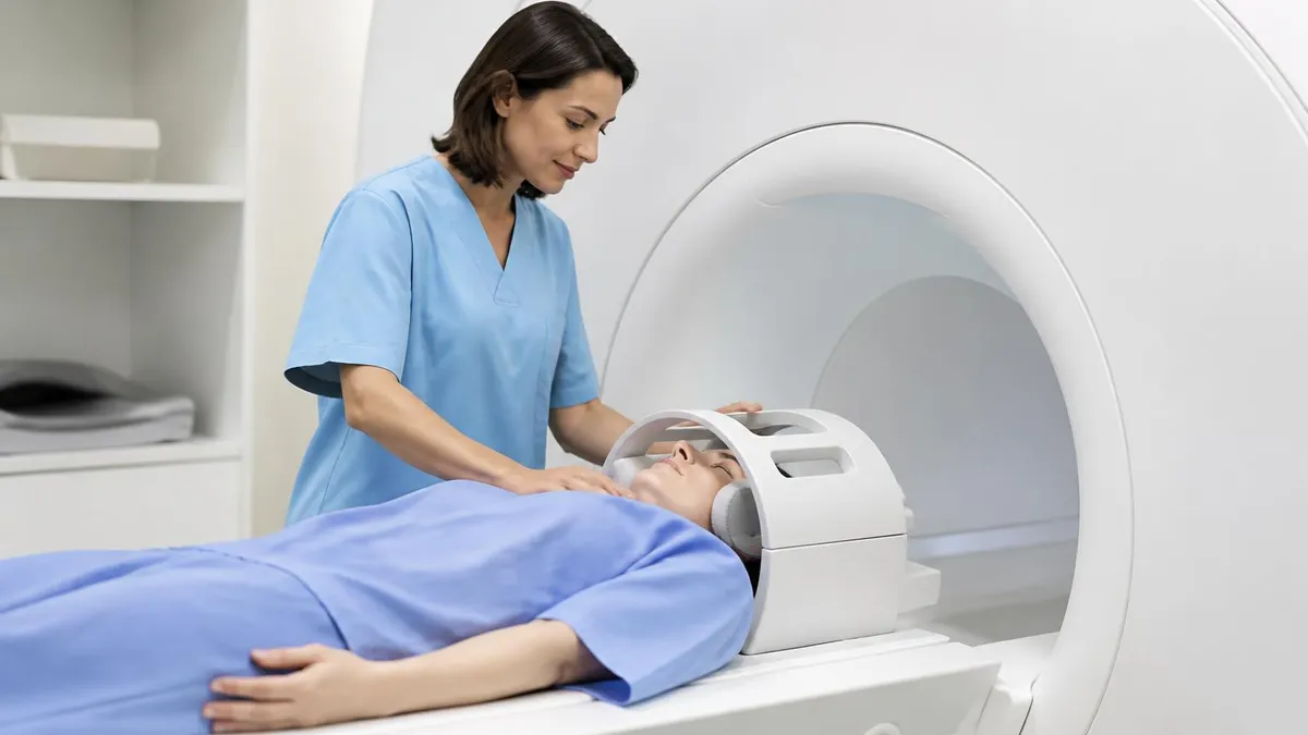
Nephrogenic systemic fibrosis (NSF) is a rare but serious complication of gadolinium administration in patients with severe renal impairment (eGFR below 30 mL/min/1.73 m²). Always verify eGFR before contrast administration for thoracic MRI, use macrocyclic agents (gadobutrol, gadoteridol, gadoterate meglumine) which carry the lowest NSF risk, and document informed consent. Patients on dialysis require coordination with their dialysis team for prompt post-contrast clearance.
Preparing for the MRI registry examination requires more than memorizing anatomy — it demands a systematic approach to understanding how clinical problems translate into imaging protocols, and how physical principles manifest as image quality characteristics. The ARRT MRI registry examination tests candidates across six major content categories: patient care, safety, image production, procedures, and physics principles. Thoracic MRI knowledge touches all of these domains, making it a rich area for concentrated study that yields broad examination benefits rather than narrow topic-specific returns.
The procedures section of the registry exam, which accounts for approximately 38% of the total question pool, includes substantial content on body MRI protocols including the thoracic spine and chest. High-yield topics in this category include coil selection rationale, sequence prescription for specific clinical indications, contrast administration protocols, and the ability to recognize and correct suboptimal image quality. Practicing with realistic case-based questions that present an imaging scenario and ask you to identify the best corrective action is one of the most effective study strategies for this content area.
Physics questions related to thoracic MRI frequently focus on artifact recognition and reduction — a topic that connects abstract physical principles to concrete clinical consequences. Understanding the phase-encoding direction, how to manipulate it to redirect ghosts, why gradient echo sequences are susceptibility-sensitive, and how k-space sampling affects image resolution are all recurring themes. Rather than memorizing isolated facts, build a mental model of how each variable (TE, TR, flip angle, bandwidth, matrix, FOV, NEX) affects the final image, and you will be equipped to reason through novel questions that present scenarios you haven't seen before.
Safety content related to thoracic MRI on the registry exam includes ferromagnetic screening, implant safety evaluation, specific absorption rate (SAR) monitoring, and quench protocols. Thoracic MRI presents specific safety challenges because patients with cardiac devices — pacemakers, ICDs, cardiac monitors — are increasingly being scanned under supervised conditional MRI protocols. The 2011 AHA/ACC guidance and subsequent manufacturer-specific conditional protocols have expanded MRI access for this population, but the technologist's role in verifying device model, programming changes, and post-scan device checks is critical and examinable content.
Time management during registry examination preparation is as important as content coverage. A structured study schedule that allocates time proportional to content weighting — more time on procedures and image production, less on topics that appear infrequently — maximizes your score per hour of study time. Practice examinations under timed conditions help build the mental stamina needed to maintain concentration across 230 questions, identify knowledge gaps early enough to address them, and calibrate your pacing so you complete all questions without rushing through the final section.
Clinical correlation is a study strategy that distinguishes high scorers from average performers. For every imaging concept you study — whether it's the MRI appearance of a Modic Type II endplate change, the direction of chemical shift artifact at a fat-water interface, or the SAR implications of a long echo train length — ask yourself what it would look like on an actual image, how it would affect patient care, and what a technologist's correct response would be. This three-dimensional thinking transforms passive reading into active preparation that translates directly to examination performance and professional competence.
Practice tests are the single most effective preparation tool available to MRI registry candidates. Research consistently shows that retrieval practice — actively recalling information from memory rather than passively re-reading it — produces superior long-term retention and better performance on high-stakes examinations. Taking multiple practice tests under realistic conditions, reviewing every incorrect answer in depth, and re-testing on missed concepts one week later creates a spaced-repetition cycle that drives knowledge into long-term memory. The resources available through PracticeTestGeeks, including the thoracic mri supplementary content and multiple MRI-specific practice test banks, are designed specifically to support this retrieval-practice approach.
Advanced thoracic MRI techniques are expanding rapidly, driven by hardware improvements, artificial intelligence-assisted reconstruction, and growing clinical demand for functional information beyond anatomical detail. Compressed sensing MRI, which uses mathematical undersampling of k-space combined with iterative reconstruction algorithms, can reduce thoracic spine scan times by 40 to 60 percent without meaningful loss of diagnostic quality — a breakthrough for patients who cannot tolerate prolonged immobility due to pain or anxiety, and for busy imaging departments managing high patient volumes.
Diffusion tensor imaging (DTI) of the thoracic spinal cord is an emerging technique that maps white matter tract integrity by tracking the directional movement of water molecules along axons. In patients with cord compression from disc herniation or epidural metastases, DTI can quantify microstructural injury before conventional T2 sequences show obvious signal change — potentially identifying patients who need urgent decompression before irreversible neurological damage occurs.
While DTI of the spinal cord remains more technically challenging than brain DTI due to the cord's small diameter and susceptibility to cardiac and respiratory motion, dedicated cardiac-gated DTI protocols at 3T are producing increasingly reliable results in research and selected clinical settings.
MR neurography of the thoracic region uses heavily T2-weighted sequences with fat saturation and 3D reformatting to trace individual nerve roots from the spinal cord through the neural foramina into the paravertebral and intercostal spaces. This technique is particularly valuable for evaluating thoracic outlet syndrome, intercostal neuralgia from herpes zoster or post-surgical injury, and nerve root involvement by paravertebral or Pancoast tumors. When combined with contrast enhancement, MR neurography can demonstrate inflammatory or neoplastic nerve involvement with a level of detail that conventional axial sequences cannot match.
Cardiac MRI, while a distinct subspecialty, overlaps significantly with thoracic imaging when evaluating mediastinal masses adjacent to the heart, pericardial disease, or great vessel pathology including aortic aneurysm and dissection. MRI-based functional cardiac assessment provides stroke volume, ejection fraction, and wall motion data in the same examination that evaluates mediastinal anatomy — a level of comprehensive information that CT, despite its speed advantage, cannot match without separate studies and additional radiation. For imaging departments building thoracic MRI programs, integrating cardiac MRI expertise into the workflow allows for truly comprehensive chest evaluation in complex patients.
Artificial intelligence applications in thoracic MRI are at an inflection point between research and routine clinical deployment. AI-assisted image reconstruction allows diagnostic quality images to be acquired with fewer signal averages, reducing scan time while preserving or improving SNR. Automated vertebral body labeling — reliably identifying T1 through T12 on scout images — reduces technologist setup time and prevents the labeling errors that occasionally affect surgical planning. AI-based detection algorithms for vertebral fracture, cord signal change, and disc herniation are in active clinical validation, with early results suggesting sensitivity comparable to experienced radiologists for high-prevalence pathologies.
For MRI technologists, staying current with these technological advances is both a professional obligation and a competitive advantage. ARRT continuing education requirements for MRI certification renewal include keeping pace with evolving imaging techniques, safety developments, and clinical applications. Reading peer-reviewed literature, attending SMRT and ISMRM educational conferences, and engaging with vendor educational resources are all pathways to maintaining the currency that registry recertification demands. The technologists who thrive in this evolving landscape are those who view each new technique not as a threat to existing knowledge but as an opportunity to expand their clinical value.
Ultimately, excellence in thoracic MRI is built on the same foundation as excellence in any complex technical-clinical field: deep anatomical knowledge, rigorous physical understanding, systematic protocol execution, careful attention to patient safety, and continuous learning. The interaction between these domains — where physics knowledge prevents artifacts, anatomical knowledge guides sequence planning, and clinical knowledge drives protocol customization — is what makes MRI technology a genuinely intellectually demanding profession.
Students and practitioners who master thoracic MRI develop transferable skills in image quality optimization, artifact analysis, and clinical-technical communication that improve their competence across every body region and imaging application they encounter throughout their careers.
You can continue preparing with the open bore mri, which includes answer explanations for every question.
You can continue preparing with the 72195 cpt code, which includes answer explanations for every question.
Sample MRI - Magnetic Resonance Imaging Practice Questions
Try these questions from our free MRI - Magnetic Resonance Imaging practice tests. The correct answer and an explanation follow each question.
When imaging the lumbar spine, which position best opens the intervertebral foramina and reduces lumbar lordosis for improved visualization?
- A. Prone with legs extended
- B. Supine with legs elevated on a cushion (knees and hips flexed)
- C. Lateral decubitus
- D. Prone with pillow under abdomen
Answer: B. Supine with legs elevated on a cushion (knees and hips flexed)
Flexing the hips and knees while supine flattens lumbar lordosis, opens intervertebral foramina, and improves disc and nerve root visualization.
Most superconducting magnets have a (horizontal or vertical) magnetiic field and are of the design type __.
- A. Selenoid magnet
- B. Permanent magnet
- C. Superconducting
- D. Resistive magnet
Answer: A. Selenoid magnet
Most high-field superconducting MRI magnets are designed as solenoid magnets. In this configuration, the superconducting wire coils are arranged to create a strong, uniform magnetic field that runs horizontally through the bore of the scanner. This solenoid design is highly efficient for generating the powerful and homogeneous fields required for advanced MRI imaging.
Cross-excitation (cross-talk) artifact in multislice MRI occurs because of:
- A. Imperfect slice profiles causing RF excitation of adjacent slices
- B. Gradient switching between adjacent slices
- C. Patient breathing between slice acquisitions
- D. Coil sensitivity overlap between two adjacent coil elements
Answer: A. Imperfect slice profiles causing RF excitation of adjacent slices
Real RF pulses have imperfect rectangular profiles; the tails of adjacent slice profiles overlap, causing partial saturation of neighboring slices — reduced by interleaved acquisition or adding gaps.
The gradient subsystem's primary goal is to
- A. Everything mentioned
- B. Choose the plane of slices.
- C. Encode the MR signal spatially
- D. Choose the imaging plane.
Answer: A. Everything mentioned
The gradient subsystem is crucial for multiple functions in MRI. It generates varying magnetic fields that enable precise slice selection by altering the Larmor frequency across the patient. Additionally, these gradients spatially encode the MR signal by creating frequency and phase shifts, which are essential for reconstructing a detailed image. Therefore, all listed options are primary goals of the gradient subsystem.
Take the full MRI - Magnetic Resonance Imaging practice test
MRI Questions and Answers
Sources & last verified
- assets-us-01.kc-usercontent.com — number of questions, time limit (official source). “MRI Scored Items 200 Pilot Items 20 Total Items 220 Test Time 210 min” Verified Oct 15, 2026.
- The American Registry of Radiologic Technologists — passing score (official source). “Our exam scoring scale ranges from 1 to 99, and you’ll need a total score of 75 to pass the exam.” Verified Oct 15, 2026.
- The American Registry of Radiologic Technologists — exam fee, other (official source). “Postprimary (M, CT, MR, BD, CI, VI, VS, BS, R.R.A.): $225 Postprimary (CT, MR, BD, or VS—using NMTCB as supporting category): $450 Postprimary (MR, VS,…” Verified Oct 15, 2026.
How we verify: editorial & fact-checking policy. Report an error: contact us.

MRI of Cervical Spine: Complete Guide to Imaging, Anatomy, Pathology, and Patient Preparation

Knee MRI Images: A Complete Guide to Reading, Understanding, and Interpreting Knee Scans
Ace Your Exam with MRI Practice Test 2026 Prep

MRI Tech School 2026 — Programs, Requirements, and What to Expect
MRI Procedure: What to Expect Before, During and After Your Scan
How this page was made: written with AI tools and published by Nathan Beja, founder of Practice Test Geeks, who is responsible for its content. Exam facts are checked against the official sources listed below. Not reviewed by a subject-matter expert. Editorial policy
Sources & references
Exam facts on this page (fees, format, dates, passing scores) are checked against these official sources; the date is our last check. Editorial policy
- arrt.org — Passing score, score release, fees, exam information · verified Oct 2026
- assets-us-01.kc-usercontent.com — Renewal, score validity, test delivery, eligibility, exam format, test dates, time limit · verified Oct 2026