Transverse Myelitis MRI: Imaging Findings, Diagnosis, and What to Expect
Learn how transverse myelitis MRI works, what imaging findings look like, how radiologists diagnose it, and what patients and techs need to know.
Transverse myelitis MRI is the cornerstone diagnostic tool for identifying and characterizing this rare but serious inflammatory condition of the spinal cord. When a patient presents with rapidly progressive limb weakness, sensory deficits, or bladder dysfunction, neurologists turn immediately to MRI because it provides unmatched soft-tissue contrast and multiplanar imaging capability. The spinal cord lesions associated with transverse myelitis have recognizable patterns on MRI — patterns that help clinicians distinguish this condition from spinal cord tumors, vascular events, and other demyelinating diseases. Understanding these imaging findings is critical for anyone working in diagnostic imaging or preparing for MRI certification examinations.
Transverse myelitis is defined as an inflammatory process that affects one or more spinal cord segments, disrupting both sensory and motor pathways as well as autonomic function. It can arise as a standalone condition — called idiopathic transverse myelitis — or as part of a broader systemic disease such as multiple sclerosis, neuromyelitis optica spectrum disorder (NMOSD), systemic lupus erythematosus, or Sjögren syndrome.
In some cases it follows a viral infection or vaccination. The precise incidence in the United States is estimated between 1,400 and 2,000 new cases per year, with two age peaks: children between 10 and 19 years and adults between 30 and 39 years. Both sexes are affected equally.
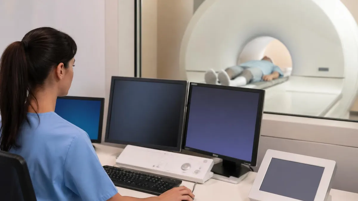
The role of the MRI technologist extends well beyond positioning and protocol selection. A tech who understands the pathophysiology of transverse myelitis can better anticipate which sequences the radiologist will rely on, ensure adequate coverage from the foramen magnum through the conus medullaris, and recognize when motion artifact or inadequate contrast timing may compromise a study. The referring neurologist depends on a clean, well-labeled examination, and the tech is often the last line of quality control before images reach the reading room. Familiarity with this disease directly improves patient outcomes.
MRI protocols for suspected transverse myelitis typically include sagittal T2-weighted and STIR sequences, sagittal and axial T1-weighted pre- and post-contrast sequences, and axial T2-weighted images through any identified lesion. At 3 Tesla, diffusion-weighted imaging (DWI) of the cord adds sensitivity for acute ischemic or highly inflammatory lesions. Brain MRI is almost always performed in the same session because demyelinating plaques in the brain change the diagnostic category — and the treatment plan — significantly. A comprehensive examination in a cooperative patient can be completed in under 60 minutes on a modern 1.5T or 3T scanner.
On T2-weighted sequences, transverse myelitis classically appears as a hyperintense signal abnormality within the cord parenchyma. The lesion typically spans two or more vertebral segments in length; lesions extending three or more vertebral body lengths are termed longitudinally extensive transverse myelitis (LETM) and are strongly associated with NMOSD and aquaporin-4 antibody positivity. In contrast, multiple sclerosis typically produces shorter lesions, often less than two segments in length and peripherally located in the cord. This length distinction is one of the most clinically important observations a radiologist can make from a single sagittal T2 image.
Gadolinium-based contrast administration adds another layer of diagnostic precision. Enhancement in transverse myelitis indicates active blood-brain barrier breakdown and ongoing inflammation. The enhancement pattern can be nodular, patchy, ring-like, or homogeneous, and it generally resolves over weeks as the acute inflammatory phase subsides. Persistent or recurring enhancement suggests ongoing disease activity and may prompt escalation of immunotherapy. Patients who are pregnant or have severe renal impairment require special consideration before contrast administration, and it is the technologist's responsibility to screen for these contraindications before the examination begins.
For MRI students and practicing technologists preparing for board examinations, understanding transverse myelitis imaging is essential because it appears regularly on the ARRT and ARMRIT registry exams. Questions frequently test your knowledge of sequence selection, lesion characterization, and protocol modifications for spinal cord pathology. Reviewing real imaging cases alongside transverse myelitis mri comparisons helps reinforce the abnormal patterns you need to recognize quickly under exam conditions. The following sections walk through the complete MRI picture of this condition in detail.
Transverse Myelitis MRI by the Numbers

Key MRI Sequences and Protocol Steps
Sagittal T2 / STIR — First Look
Axial T2 Through the Lesion
Pre-Contrast T1 — Baseline Signal
Gadolinium Contrast Administration
Post-Contrast T1 — Enhancement Pattern
Brain MRI — Complete the Picture
Understanding the MRI findings in transverse myelitis requires separating the condition into its major etiological subtypes, because each one produces a recognizable imaging signature. The three most clinically important categories are multiple sclerosis–associated transverse myelitis, neuromyelitis optica spectrum disorder (NMOSD), and idiopathic acute transverse myelitis. A fourth, rarer category includes myelitis caused by systemic autoimmune diseases such as lupus or antiphospholipid antibody syndrome. Each subtype has characteristic lesion length, location within the cord cross-section, enhancement pattern, and brain MRI appearance that collectively point toward the underlying etiology.
In multiple sclerosis, spinal cord lesions are typically short, involving fewer than two vertebral body segments. They are eccentrically located — meaning they favor the posterior or lateral columns rather than the central cord — and they rarely occupy more than half the cord cross-sectional area on axial imaging.
The cervical cord is the most commonly affected region in MS, and multiple discrete lesions at different cord levels can be present simultaneously. Enhancement, when present, is usually nodular or ring-like and resolves within four to six weeks. Brain FLAIR sequences almost always reveal accompanying periventricular, juxtacortical, or infratentorial plaques that confirm the dissemination in space required for an MS diagnosis.
NMOSD produces a dramatically different MRI picture. The aquaporin-4 water channel is expressed most densely in the central gray matter of the cord — particularly in the cervical and thoracic regions — and this distribution explains why NMOSD lesions are longitudinally extensive, spanning three or more vertebral segments, and centrally located within the cord.
On axial T2, the lesion often involves more than two-thirds of the cord cross-section, creating what radiologists call a "bright spotty lesion" pattern. Cord swelling can be pronounced in the acute phase, and gadolinium enhancement is common. Follow-up MRI often reveals cord atrophy and permanent T1 hypointensity ("black holes") in segments where the initial inflammation was most severe.
Idiopathic transverse myelitis occupies an imaging middle ground. Lesions are typically one to five segments long, involve the central cord, and show variable enhancement. Unlike MS, idiopathic TM tends to affect a younger demographic and presents monophasically — meaning it does not recur.
When a young patient presents with a first episode of TM and has a clean brain MRI, negative aquaporin-4 and MOG antibodies, and a lesion of intermediate length, the diagnosis of idiopathic TM is made by exclusion. MRI follow-up at three to six months typically shows partial or complete resolution of the T2 signal abnormality, with residual cord atrophy correlating with clinical recovery.
MOG antibody-associated disease (MOGAD) is a recently defined condition that produces yet another distinct imaging pattern. MOGAD lesions are often conus-predominant, involving the lower thoracic cord and conus medullaris — a distribution that is unusual for both MS and NMOSD. The lesions may be longitudinally extensive but tend to resolve more completely on follow-up MRI compared to AQP4-positive NMOSD. Leptomeningeal enhancement — a thin, linear enhancement along the cord surface — has been reported in MOGAD and is a useful differentiating clue when present. Familiarity with this emerging entity is increasingly tested on imaging board examinations.
Infectious myelitis deserves mention because it can closely mimic the imaging findings of autoimmune transverse myelitis. Viral causes include herpes simplex virus, varicella-zoster virus, cytomegalovirus, enteroviruses (particularly in children), and, more recently, SARS-CoV-2–associated myelitis. Bacterial and parasitic myelitis is rarer but occurs in immunocompromised patients.
The key differentiating imaging feature of infectious myelitis is often the distribution: viral myelitis caused by enteroviruses typically affects the anterior horn cells, producing a pattern of T2 signal abnormality restricted to the ventral gray matter. This "snake eyes" or "owl eyes" sign on axial T2 is highly characteristic and differs from the central or lateral column involvement seen in autoimmune TM.
Vascular myelopathy — specifically spinal cord infarction — is the most important acute mimic of transverse myelitis on MRI. Both conditions produce rapid-onset myelopathy with T2 cord hyperintensity. However, spinal cord infarction in the anterior spinal artery territory produces a very specific axial pattern: bilateral anterior horn involvement with sparing of the posterior columns, creating the so-called "owl eyes" pattern restricted to the anterior two-thirds of the cord.
DWI — now feasible at 3T with dedicated spinal cord coils — shows restricted diffusion in acute infarction but not in inflammatory TM. Recognizing this distinction is life-saving because the two conditions require completely different treatments.
Differential Diagnosis, Mimics, and Diagnostic Pitfalls
Distinguishing multiple sclerosis from neuromyelitis optica spectrum disorder on MRI is one of the most consequential decisions a neuroradiologist makes, because the two conditions require different disease-modifying therapies — and some MS drugs can actually worsen NMOSD. The key MRI discriminators are lesion length (short and peripheral in MS versus longitudinally extensive and central in NMOSD), brain lesion pattern (periventricular and juxtacortical in MS versus area postrema or hypothalamic in NMOSD), and the presence of cord atrophy on follow-up.
Aquaporin-4 IgG seropositivity confirms NMOSD regardless of lesion length, and MOG-IgG positivity now defines a third distinct entity, MOGAD, which has its own characteristic conus-predominant distribution. When serology is negative but imaging is suggestive of NMOSD, repeating the antibody test with a cell-based assay rather than ELISA is recommended, as sensitivity is significantly higher. Radiologists should always note lesion length, axial distribution, and brain involvement in their reports to facilitate the correct serological workup.
MRI for Transverse Myelitis: Strengths and Limitations
- +Superior soft-tissue contrast allows direct visualization of cord parenchyma that CT cannot provide
- +Gadolinium enhancement accurately identifies active inflammation and blood-brain barrier disruption
- +Multiplanar capability enables precise lesion localization in both axial and sagittal planes
- +No ionizing radiation, making serial follow-up examinations safe for long-term monitoring
- +Brain MRI in the same session enables immediate assessment of dissemination in space for MS diagnosis
- +DWI at 3T can differentiate acute infarction from inflammatory myelitis, preventing treatment errors
- −Early acute TM (within 24 hours) may appear normal on MRI before edema and inflammation fully develop
- −Motion artifacts from breathing and CSF pulsation degrade image quality, particularly in the thoracic cord
- −Gadolinium is contraindicated in patients with severe renal impairment (eGFR below 30 mL/min)
- −Implanted devices (pacemakers, certain spinal hardware) may preclude MRI or reduce diagnostic quality
- −Lesion appearance alone cannot definitively distinguish all TM subtypes — serology and CSF are also required
- −Access in acute settings can be limited; patients with severe autonomic instability may not tolerate prolonged scanning
MRI Tech Checklist for Suspected Transverse Myelitis
- ✓Screen the patient for MRI contraindications including implants, renal function (for gadolinium), and pregnancy.
- ✓Confirm field strength and select a dedicated spine coil or phased-array coil for optimal cord signal-to-noise ratio.
- ✓Acquire a full-spine sagittal localizer to identify the level of maximum T2 signal abnormality before tailoring coverage.
- ✓Include sagittal T2 and STIR sequences spanning the entire cord from foramen magnum to conus medullaris.
- ✓Add axial T2 slices (≤3 mm) through any identified lesion to assess cord cross-sectional involvement.
- ✓Obtain pre-contrast sagittal and axial T1 sequences as baseline before gadolinium injection.
- ✓Administer 0.1 mmol/kg macrocyclic gadolinium contrast agent; wait at least 5 minutes before post-contrast acquisition.
- ✓Acquire post-contrast sagittal and axial T1 sequences and document enhancement length and pattern in scan notes.
- ✓Extend the examination to include a complete brain MRI (FLAIR, T2, DWI, post-contrast T1) in the same session.
- ✓Label all series with anatomical level, orientation, and sequence type before sending to PACS for radiologist review.
Lesion Length Is the Single Most Important MRI Observation
A lesion spanning three or more vertebral body segments on sagittal T2 — longitudinally extensive transverse myelitis (LETM) — dramatically shifts the differential toward NMOSD and should prompt immediate aquaporin-4 IgG antibody testing. Missing this measurement can delay life-changing treatment. Always report the exact number of vertebral segments involved, not just "long segment" or "extensive."
Treatment response monitoring is one of the most valuable ongoing uses of MRI in transverse myelitis, and understanding what to expect on follow-up imaging is essential for both radiologists and technologists. The acute phase of transverse myelitis typically peaks within days to three weeks of symptom onset, and imaging performed during this window usually shows the maximum degree of cord edema, T2 signal abnormality, and gadolinium enhancement. As the acute inflammation subsides — typically over four to twelve weeks — the enhancement fades first, followed by gradual reduction in cord swelling and T2 signal intensity.
The degree of MRI resolution on follow-up does not correlate perfectly with clinical recovery, but it does provide important prognostic information. Patients who show near-complete resolution of their T2 signal abnormality at three months tend to have better functional outcomes than those who develop persistent T2 hyperintensity or progressive cord atrophy.
T1 hypointensity within the cord — so-called T1 black holes — indicates severe tissue destruction with axonal loss and is associated with persistent neurological deficits. Quantitative MRI techniques such as magnetic transfer ratio and diffusion tensor imaging (DTI) can detect microstructural damage even in cord segments that appear normal on conventional sequences.
Acute treatment for transverse myelitis typically consists of high-dose intravenous methylprednisolone (1 gram daily for three to five days), sometimes followed by oral taper. In severe or steroid-refractory cases, plasmapheresis (plasma exchange) is used and is associated with meaningful recovery in a subset of patients. NMOSD patients who are aquaporin-4 antibody positive are now treated with monoclonal antibodies targeting interleukin-6 receptors (satralizumab, tocilizumab) or CD19-positive B cells (inebilizumab), as well as the complement inhibitor eculizumab. Follow-up MRI is used to assess whether these therapies are suppressing new lesion formation and preventing cord atrophy.
For NMOSD patients, the stakes of inadequate imaging surveillance are particularly high. Unlike MS, NMOSD relapses tend to produce cumulative, severe, and often irreversible disability with each attack. Serial spinal cord MRI — typically every six to twelve months or sooner after any new symptom — is used to detect subclinical lesion activity and adjust immunotherapy accordingly.
A new T2 lesion or new enhancement in a patient on maintenance therapy signals treatment failure and warrants a change in regimen. Radiologists should always compare the current examination against all prior studies and report new lesions with their vertebral segment level and enhancement status.
Pediatric transverse myelitis presents unique imaging challenges. Children are more likely than adults to have infectious or post-infectious myelitis, and their cord lesions may span a greater proportion of the spinal cord relative to body size.
ADEM (acute disseminated encephalomyelitis) is an important consideration in children after viral infection or vaccination; it produces multifocal brain and cord lesions simultaneously, and the cord lesions in ADEM tend to be large, patchy, and asymmetric. MOG antibody positivity is more common in pediatric inflammatory myelitis than in adults and often predicts a monophasic course with good recovery, though recurrent MOG antibody disease does occur.
Pregnancy and the postpartum period are associated with an increased risk of transverse myelitis onset or relapse, particularly in women with underlying NMOSD. MRI without gadolinium is the preferred approach during pregnancy because the safety of gadolinium contrast for the developing fetus has not been definitively established, and the FDA has issued guidance advising gadolinium use in pregnancy only when absolutely necessary. In practice, a well-performed T2 and STIR examination can characterize the lesion sufficiently for initial clinical decision-making without contrast. Post-contrast images can be deferred to the postpartum period unless the clinical situation demands immediate enhancement data.
Long-term MRI follow-up of transverse myelitis also serves a medicolegal and prognostic documentation function. Disability determinations for social security, workers' compensation, and insurance purposes frequently require objective imaging evidence of cord damage. The correlation between MRI cord atrophy measurements and clinical outcome scores such as the Expanded Disability Status Scale (EDSS) and the American Spinal Injury Association (ASIA) impairment scale has been validated in research studies, providing objective imaging biomarkers that support disability assessments. Technologists who understand this downstream use of their examinations appreciate the importance of consistent, reproducible positioning and protocol adherence across follow-up studies.
Transverse myelitis is a neurological emergency. Imaging should be performed within hours of symptom onset, not days. A normal MRI in the first 24 hours does not rule out TM — the T2 signal abnormality may not yet be visible — so repeat imaging at 48–72 hours is warranted if clinical suspicion remains high. Early treatment with IV steroids significantly impacts functional recovery outcomes.
For MRI students and registered technologists preparing for certification or registry examinations, transverse myelitis occupies a predictable place in the question bank because it tests several overlapping competencies simultaneously: sequence selection and rationale, lesion characterization, anatomy of the spinal cord, contrast administration protocol, and differential diagnosis. The ARRT and ARMRIT examinations both include anatomy and pathology sections that cover demyelinating and inflammatory spinal cord diseases, and questions about transverse myelitis consistently appear in those sections. Knowing the characteristic imaging appearance — T2 hyperintensity, cord swelling, gadolinium enhancement, and the lesion length criteria — is the foundation of a correct answer.
A commonly tested scenario on registry examinations involves a patient with acute weakness and sensory deficits below the thoracic level. The question presents sagittal T2 images and asks the candidate to identify the most likely diagnosis, the optimal next sequence, or the most appropriate contrast protocol modification. Candidates who have studied real imaging examples of transverse myelitis will immediately recognize the longitudinally extensive T2 signal abnormality in the central cord and correctly select post-contrast T1 as the next sequence. Candidates who have only memorized definitions without visual pattern recognition often miss these questions.
The physics underlying why transverse myelitis lesions appear bright on T2 is also testable. Inflammation causes increased free water content within the cord parenchyma due to vasogenic and cytotoxic edema. Free water has a longer T2 relaxation time than normal brain or cord tissue, so inflamed areas retain transverse magnetization longer and appear hyperintense on T2-weighted sequences. STIR sequences eliminate the short-T1 signal from fat (which would otherwise contribute to background signal in the spine) without affecting the long-T2 lesion signal, making STIR particularly sensitive for cord pathology at any field strength.
Another high-yield exam topic involves the modification of standard protocols for patients with contraindications. A patient with an eGFR of 25 mL/min cannot safely receive standard-dose gadolinium due to the risk of nephrogenic systemic fibrosis (NSF). The correct approach is to perform the examination without contrast and rely on T2, STIR, and DWI sequences.
At 3 Tesla, DWI of the spinal cord has sufficient quality to detect restricted diffusion in acute lesions, providing a partial substitute for the information that gadolinium enhancement would otherwise yield. Knowing the eGFR thresholds (below 30 for group II agents, below 40 for group I agents according to ACR guidance) and the available alternatives is essential knowledge for any practicing MRI technologist.
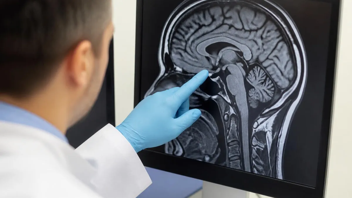
Preparing for the anatomy and pathology section of MRI registry exams requires more than memorizing lists of findings. The most effective study approach combines reading-based review with active image interpretation practice. When you look at a spinal MRI image and ask yourself "what level is this, what sequence is this, and what is abnormal," you are building the pattern recognition circuits that registry questions are designed to test. Practice tests that include labeled imaging exhibits — not just text questions — are particularly valuable for this type of learning.
The internal architecture of the spinal cord visible on high-resolution MRI is also testable. The central H-shaped gray matter contains anterior horns (motor neurons), posterior horns (sensory relay), and lateral horns in the thoracic cord (autonomic neurons). The surrounding white matter consists of ascending sensory tracts (dorsal columns for proprioception and fine touch, spinothalamic for pain and temperature) and descending motor tracts (corticospinal). Understanding which tract occupies which quadrant of the cord cross-section helps you predict the clinical deficits from a given lesion location — and helps you answer questions that map imaging findings to patient symptoms.
Students who want to deepen their understanding of cord anatomy in the context of normal baseline imaging should review reference materials comparing normal and abnormal cord signal. Comparing a healthy cord MRI to one affected by transverse myelitis side by side is one of the fastest ways to internalize the normal appearance and recognize subtle early abnormalities.
Boards questions often present borderline or early-stage pathology precisely to test whether candidates know the normal appearance well enough to detect deviation from it. Consistent practice with annotated image sets, combined with focused quiz sessions, will solidify both the diagnostic knowledge and the physics understanding you need to perform well on examination day.
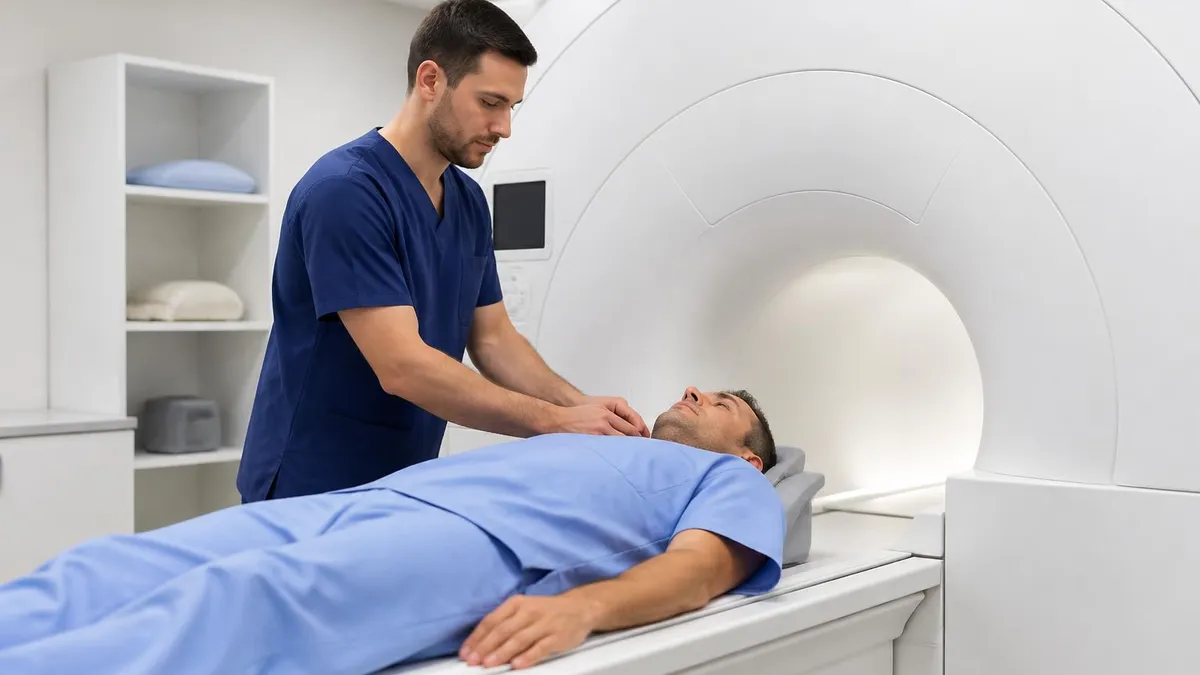
Practical preparation for working with transverse myelitis patients in the MRI suite begins well before the patient arrives in the room. Reviewing the clinical order, the referring neurologist's notes, and any prior imaging studies gives the technologist the context needed to make protocol decisions efficiently. If the ordering provider suspects NMOSD, the tech should ensure the brain protocol is loaded and ready, since the radiologist will almost certainly request brain images regardless of whether they were explicitly ordered. Proactive communication with the reading radiologist before the scan — especially for complex or urgent cases — prevents delays and repeat examinations.
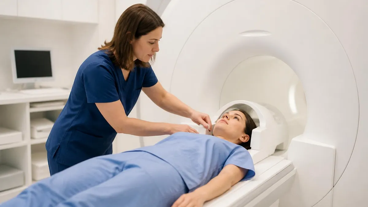
Patient communication is particularly important in transverse myelitis cases because many patients present with significant anxiety. A sudden onset of weakness or sensory loss is terrifying, and many patients have not yet received a diagnosis when they arrive in the MRI suite.
The technologist should explain the examination calmly and clearly, describe what the patient will hear and feel (including the warm sensation that some patients experience with gadolinium injection), and reassure them that the examination is non-invasive. Patients with residual motor deficits may need additional padding and positioning support to remain still throughout the examination, which can last 45 to 90 minutes for a combined spine and brain protocol.
Monitoring patients during gadolinium administration is a safety responsibility that falls directly on the MRI technologist. Acute hypersensitivity reactions to gadolinium contrast agents, while rare (estimated at 0.01–0.1% of all administrations), can occur within seconds to minutes of injection. The technologist must be present, observe the patient, and have a practiced response plan in place. Delayed reactions can also occur up to an hour after administration. All MRI suites should have emergency equipment within reach and staff trained in contrast reaction protocols according to ACR and institution-specific guidelines.
Post-processing and image labeling are the final quality gates before images reach the radiologist. Ensure that all series are correctly labeled with anatomical level and orientation. Confirm that reformatted images — sagittal, axial, and coronal reconstructions from 3D acquisitions — are complete and free of aliasing artifacts. If a dedicated spinal cord coil was used, verify that coil sensitivity correction maps were applied to prevent artificial signal intensity gradients. For urgent cases, call the reading radiologist directly after sending images to PACS rather than waiting for the standard read queue — in neurological emergencies, minutes matter.
Building a personal reference library of transverse myelitis cases over your career as a technologist or radiologist creates an invaluable resource. Document cases that taught you something new — an unusual enhancement pattern, an unexpected differential, a protocol modification that improved image quality in a challenging patient. Case-based learning of this type accelerates pattern recognition far more effectively than textbook reading alone because it anchors imaging features to real clinical contexts. Many professional societies including the ASNR, ACR, and SMRT maintain online case repositories that can supplement your personal collection.
Continuing education requirements for MRI certification explicitly include pathology and anatomy, making knowledge of conditions like transverse myelitis directly relevant to maintaining your credentials. The ARRT requires 24 continuing education credits per two-year renewal cycle for registered MRI technologists. Structured case review sessions, departmental teaching files, and online modules focused on spinal cord pathology all qualify as CE credits. Investing time in this specific topic area pays double dividends: it keeps your certification current and makes you a more confident and competent practitioner in the scanning room.
Finally, staying current with the evolving literature on transverse myelitis is professionally valuable because the field is moving quickly. The discovery of MOG antibodies, the approval of several new NMOSD therapies, and the increasing use of quantitative MRI biomarkers in clinical trials have all occurred within the past decade.
Emerging techniques such as spinal cord fMRI, quantitative susceptibility mapping, and automated lesion segmentation using machine learning are entering clinical practice and will change how follow-up studies are interpreted. The MRI technologist who understands the clinical and imaging landscape of this condition is positioned not just as a button-pusher, but as a genuinely skilled clinical team member whose expertise contributes directly to patient care.
MRI Questions and Answers
About the Author
Medical Laboratory Scientist & Clinical Certification Expert
Johns Hopkins UniversityDr. Sandra Kim holds a PhD in Clinical Laboratory Science from Johns Hopkins University and is certified as a Medical Technologist (MT) and Medical Laboratory Scientist (MLS) through ASCP. With 16 years of clinical laboratory experience spanning hematology, microbiology, and molecular diagnostics, she prepares candidates for ASCP board exams, MLT, MLS, and specialist certification tests.
Join the Discussion
Connect with other students preparing for this exam. Share tips, ask questions, and get advice from people who have been there.
View discussion (4 replies)