MRI Scanners: A Complete Guide to How MRI Machines Work, Types, and Clinical Applications
✅ MRI scanners explained: 1.5T vs 3T vs 7T, open vs closed designs, costs, safety, and clinical applications. Complete 2026 July guide for techs and patients.
MRI scanners are among the most sophisticated diagnostic machines in modern medicine, using powerful magnetic fields and radiofrequency pulses to generate detailed cross-sectional images of soft tissue, bone marrow, blood vessels, and the central nervous system. Unlike CT or X-ray, MRI scanners produce no ionizing radiation, which makes them the preferred imaging modality for repeated examinations, pediatric studies, and detailed neurological assessment. Understanding how these machines actually work is essential for technologists, radiologists, and patients preparing for their first scan.
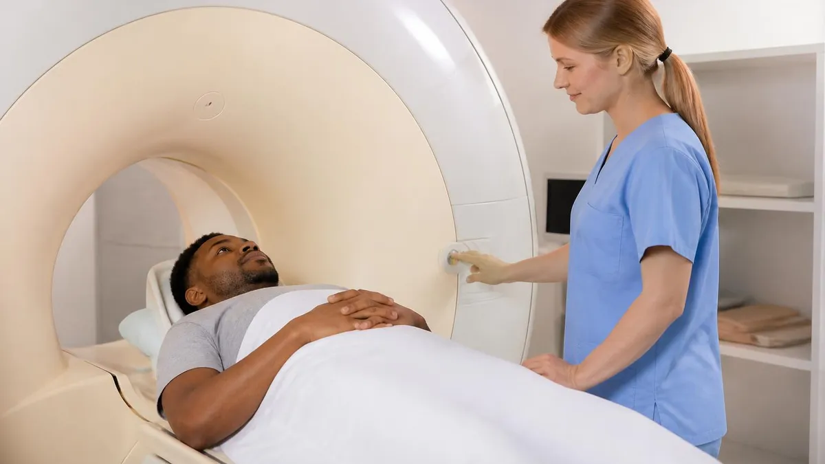
A modern MRI scanner is built around a superconducting magnet cooled by liquid helium to roughly 4 Kelvin, surrounded by gradient coils, radiofrequency transmit and receive coils, and a patient table that slides into a cylindrical bore. The combined system can resolve anatomical structures smaller than one millimeter, depending on field strength and sequence selection. Most US hospitals operate scanners between 1.5 Tesla and 3 Tesla, with research and specialty centers increasingly adopting 7 Tesla units for high-resolution neuroimaging and musculoskeletal work.
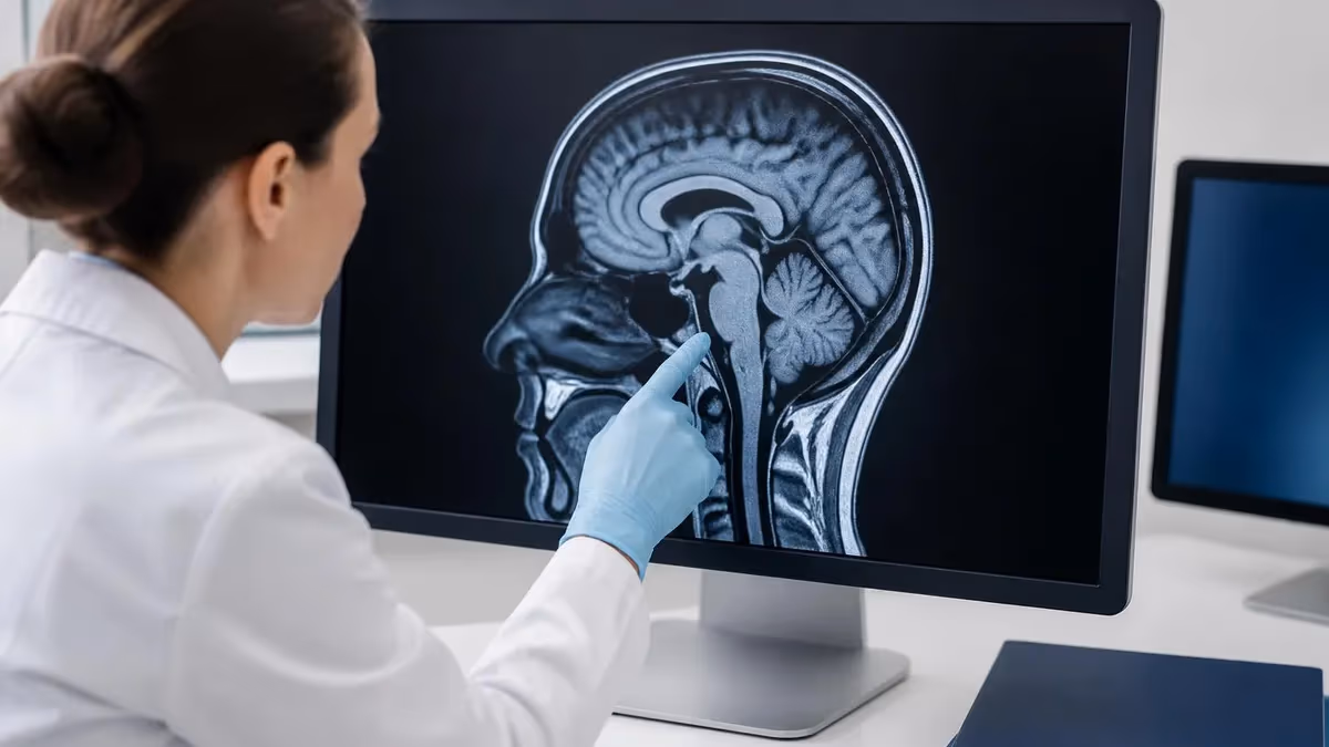
The clinical applications of MRI scanners have expanded dramatically since the first whole-body image was produced in 1977. Today, MRI is the gold standard for diagnosing brain tumors, spinal cord pathology, ligament tears, multiple sclerosis lesions, prostate cancer mri, and a host of cardiac conditions. Functional MRI maps brain activity in real time, while diffusion-weighted imaging detects acute stroke within minutes of symptom onset. These capabilities have transformed neurology, orthopedics, oncology, and cardiology over the past three decades.
Despite the technology's power, MRI scanners come with real limitations that every clinician and patient must understand. Strong magnetic fields create absolute contraindications for certain implants, the bore is confining for claustrophobic patients, scan times can stretch beyond an hour, and acoustic noise during gradient switching routinely exceeds 100 decibels. Cost is another factor — a single 3T scanner installation often exceeds three million dollars when site preparation, shielding, and cryogen infrastructure are included.
This guide walks through every major aspect of MRI scanners: the physics that makes them work, the major scanner types and field strengths, how a typical exam unfolds from check-in to image review, the safety protocols that protect patients and staff, and the practical economics that shape access in 2026. Whether you are studying for the ARRT MRI registry, preparing for your own scan, or simply curious about the technology, you will find concrete details rather than vague generalities throughout.
We also address common patient questions about scan duration, contrast agents, mri preparation and procedure claustrophobia management, and the difference between an open-bore and a wide-bore design. By the end, you should understand why certain protocols are chosen, what the technologist is doing during the exam, and how to interpret the basic reasoning behind sequence selection. The goal is practical literacy, not specialist mastery — though the technical depth here will serve registry candidates well.
Finally, a note on terminology: throughout this article, we use "MRI scanner," "MRI machine," and "MRI unit" interchangeably to refer to the complete imaging system. The terms "magnet" or "bore" refer specifically to the superconducting magnet assembly at the center of the scanner. Field strength is given in Tesla (T), where 1.5T and 3T are the dominant clinical workhorses in American hospitals and outpatient imaging centers as of 2026.
MRI Scanners by the Numbers
How an MRI Scanner Produces an Image
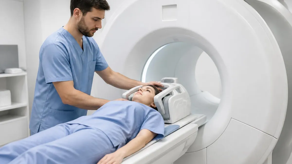
Patient Alignment in Static Field
Radiofrequency Excitation
Gradient Spatial Encoding
Signal Reception and Decay
Fourier Reconstruction
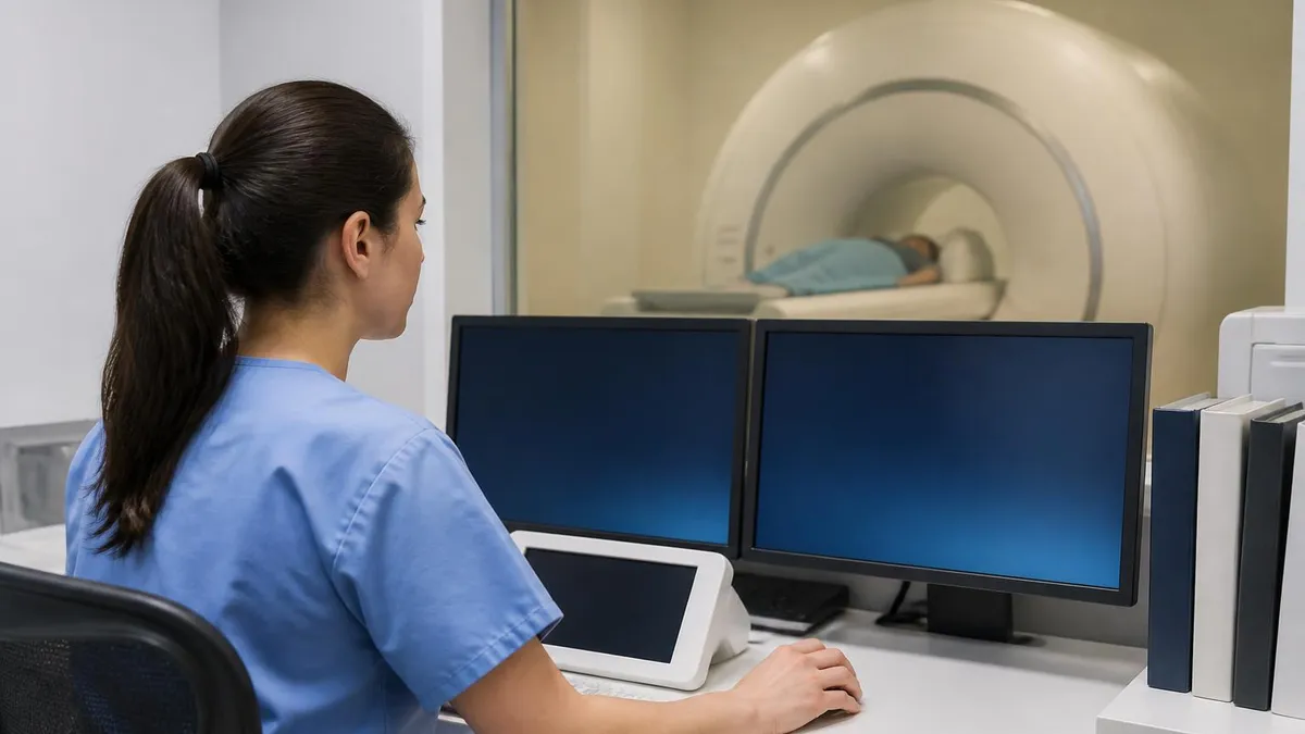
Image Review and Reporting
Field strength is the single most defining specification of any MRI scanner, measured in Tesla units after physicist Nikola Tesla. The dominant clinical strengths in 2026 remain 1.5T and 3T, with 0.55T systems making a quiet comeback for accessibility, 7T units expanding in research and select clinical neuro programs, and ultra-low-field point-of-care scanners under 0.1T appearing in stroke units. Each strength carries trade-offs in image quality, scan time, artifact behavior, safety, and cost.
A 1.5T scanner remains the workhorse of American radiology departments. Its lower field strength produces fewer susceptibility artifacts around metal implants, handles dental hardware more forgivingly, and tolerates a broader range of patient body habitus. Spatial resolution is excellent for most clinical questions — knee, cpt codes for mri abdominal, and pelvic imaging are routinely diagnostic. The lower specific absorption rate (SAR) also makes 1.5T safer for patients with certain conditional implants, which is one reason many community hospitals stick with this field strength.
A 3T scanner doubles the signal-to-noise ratio in theory, allowing thinner slices, smaller voxels, faster acquisition, or some combination of all three. This translates to superior detail in mri for migraines prostate MRI, breast MRI, and high-resolution musculoskeletal work. The trade-off is more pronounced susceptibility artifacts near metal, increased chemical shift, higher RF deposition, and more challenging cardiac imaging due to standing-wave effects. Most academic medical centers operate a mix of 1.5T and 3T units to match the magnet to the clinical question.
Open and wide-bore designs address patient comfort rather than image quality. A traditional closed-bore scanner has a 60 cm aperture, while modern wide-bore systems offer 70 cm openings, accommodating larger patients and reducing claustrophobia complaints by roughly forty percent in real-world studies. True open MRI scanners use vertical or C-shaped magnets at lower field strengths, typically 0.3T to 1.2T, accepting reduced image quality in exchange for an open environment ideal for severely anxious or bariatric patients.
Specialty scanners have carved out important niches. Intraoperative MRI units sit adjacent to neurosurgical suites, letting surgeons confirm tumor resection completeness mid-procedure. Extremity-only scanners image hands, wrists, knees, and ankles in a compact footprint at lower cost, popular in orthopedic outpatient practices. PET-MRI hybrid systems combine metabolic and anatomic imaging in a single exam, particularly valuable in oncology staging and pediatric imaging where minimizing radiation matters.
The 7T frontier is no longer purely experimental. The FDA cleared 7T clinical use in 2017, and dozens of US centers now offer 7T neuroimaging for epilepsy localization, multiple sclerosis lesion characterization, and presurgical planning. Image quality at 7T can reveal cortical layers, individual hippocampal subfields, and microbleeds invisible at lower field strengths. Challenges include B1 field inhomogeneity, increased SAR, longer pre-scan calibration, and a smaller list of compatible implants.
Looking ahead, ultra-low-field portable MRI scanners represent a genuine paradigm shift. Systems operating at 0.064T can be wheeled to an ICU bedside, run on a standard wall outlet, and produce diagnostic-quality brain images for stroke, hemorrhage, and hydrocephalus screening. While they will never replace high-field scanners for complex imaging, they extend MRI access to environments and patient populations previously excluded entirely, including remote clinics and resource-limited settings worldwide.
MRI Practice Test Questions
Prepare for the MRI - Magnetic Resonance Imaging exam with our free practice test modules. Each quiz covers key topics to help you pass on your first try.
MRI Knowledge
MRI Exam Questions covering Knowledge. Master MRI Test concepts for certification prep.
MRI Physics
Free MRI Practice Test featuring Physics. Improve your MRI Exam score with mock test prep.
MRI Anatomy and Pathology
MRI Test Prep for MRI Anatomy and Pathology. Practice MRI Quiz questions and boost your score.
MRI Anatomy and Positioning
MRI Questions and Answers on MRI Anatomy and Positioning. Free MRI practice for exam readiness.
MRI Contrast Agents
Free MRI Quiz on MRI Contrast Agents. MRI Exam prep questions with detailed explanations.
MRI Patient Care and Positioning
MRI Practice Questions for MRI Patient Care and Positioning. Build confidence for your MRI certification exam.
MRI Scanner Components Explained
The superconducting magnet is the heart of every modern MRI scanner. Niobium-titanium coils submerged in liquid helium at approximately 4 Kelvin carry electric current with zero resistance, generating field strengths from 1.5T to 7T. The magnet is housed inside a vacuum-insulated cryostat with multiple thermal shields, and modern designs use "zero boil-off" technology to virtually eliminate helium loss during normal operation, dramatically reducing long-term operating costs for hospital imaging departments.
Surrounding the main magnet are shim coils that fine-tune field homogeneity to within a few parts per million across the imaging volume. Without precise shimming, image quality collapses — fat suppression fails, geometric distortion appears, and spectroscopy becomes impossible. Active shielding uses additional coils to contain the fringe field, allowing scanners to be installed in tighter spaces without the massive iron rooms required by earlier-generation magnets from the 1980s and early 1990s.
MRI Scanners vs Other Imaging Modalities
- +No ionizing radiation, making MRI safe for repeated exams and pediatric imaging
- +Superior soft tissue contrast unmatched by CT or ultrasound
- +Multiplanar imaging in any orientation without repositioning the patient
- +Functional, diffusion, and spectroscopy techniques reveal physiology, not just anatomy
- +Excellent for neurological, musculoskeletal, and cardiac diagnosis
- +Contrast agents are generally safer than iodinated CT contrast for kidney function
- −Strong magnetic field excludes patients with certain pacemakers, cochlear implants, and ferromagnetic foreign bodies
- −Scan times of 30 to 90 minutes are far longer than CT or X-ray
- −Acoustic noise routinely exceeds 100 decibels during gradient switching
- −Claustrophobia affects roughly one in twenty patients in closed-bore scanners
- −Equipment and operating costs are substantially higher than other modalities
- −Motion artifacts degrade image quality and may require repeat sequences
Pre-Scan MRI Safety Checklist
- ✓Complete a thorough MRI safety screening form listing all implants, surgeries, and metal exposure
- ✓Remove all jewelry, watches, hearing aids, removable dental work, and body piercings before entering Zone IV
- ✓Change into a hospital gown to eliminate any metallic threads, zippers, or hidden snaps in clothing
- ✓Verify any implant by serial number against MRI conditional labeling at the documented field strength
- ✓Screen for prior occupational metal exposure with orbital radiographs if there is any history of metal fragments in the eyes
- ✓Confirm pregnancy status and discuss benefits versus theoretical risks if applicable
- ✓Check serum creatinine and eGFR when gadolinium contrast is planned for at-risk patients
- ✓Ask about claustrophobia and discuss sedation, music, or open-bore alternatives in advance
- ✓Insert IV access for contrast studies and confirm patency before the patient enters the bore
- ✓Provide hearing protection — foam earplugs plus headphones are now standard for every MRI exam
Never assume an implant is safe — verify every time
Even patients who have had multiple prior MRIs without incident can be harmed if their implant status changes or if they move to a higher field strength scanner. The American College of Radiology recommends documented verification of every implant against manufacturer MRI labeling for the specific scanner, field strength, and coil being used. When in doubt, do not scan — call the manufacturer or refer to MRIsafety.com before proceeding.
The economics of MRI scanners shape access in ways that surprise most patients. A new 1.5T mri experience a major manufacturer typically lists between 1.2 and 1.8 million dollars, while a 3T system runs 1.8 to 3 million dollars before installation. Wide-bore versions add roughly ten percent. A 7T system can exceed seven million dollars when site preparation is included. Installation alone — RF shielding, magnetic shielding, cryogen plumbing, structural reinforcement, and chiller infrastructure — frequently doubles the headline equipment price.
Ongoing operating costs are substantial. A typical MRI scanner consumes 25 to 40 kilowatts when actively scanning, plus another 5 to 10 kilowatts for the chiller that keeps the magnet cold. Annual service contracts from the original equipment manufacturer run 100,000 to 300,000 dollars depending on field strength and contract terms. Helium refills, while less frequent on modern zero-boil-off magnets, can cost 30,000 to 80,000 dollars when needed. Staffing requires at least one MRI-certified technologist per active scanner shift.
From the patient side, an outpatient MRI in the United States costs anywhere from 400 dollars at a transparent self-pay imaging center to over 5,000 dollars at a hospital outpatient department, with insurance negotiated rates typically falling between those extremes. Medicare reimbursement for a brain MRI without contrast under the 2026 Hospital Outpatient Prospective Payment System sits near 350 dollars, while commercial insurance rates can be triple that figure. This pricing variability has fueled growth of independent imaging centers.
Geographic distribution of MRI scanners varies dramatically across the United States. Urban areas like Boston, Manhattan, and Los Angeles have scanner densities exceeding 15 per 100,000 residents, while rural counties may have none at all. The OECD reports the US operates roughly 38 MRI scanners per million population, well above the OECD average of 17, yet utilization patterns mean mri results wait times for non-urgent outpatient MRI can still stretch to three or four weeks in certain markets.
Workforce shortages have become as significant as equipment availability. The American Society of Radiologic Technologists estimates a national MRI technologist vacancy rate above eight percent in 2026, with some markets reporting fifteen percent or higher. This drives evening, weekend, and overnight scanning at many hospitals to make use of expensive scanner capacity. It has also pushed salaries for ARRT(MR) registered technologists into the 80,000 to 110,000 dollar range nationally, with travel positions exceeding 140,000 dollars annually.
Reimbursement pressures continue to reshape the MRI marketplace. The Centers for Medicare and Medicaid Services have shifted certain advanced imaging services toward bundled payments, prior authorization requirements have expanded, and clinical decision support is now mandatory for outpatient advanced imaging ordered by Medicare providers. These changes aim to reduce inappropriate utilization but have added administrative burden for ordering physicians and imaging facilities alike.
Looking ahead, several economic forces will reshape MRI access during the late 2020s. AI-enabled scan acceleration techniques are cutting acquisition times by 30 to 50 percent without quality loss, effectively expanding throughput without new capital. Helium-free magnet designs eliminate one of the largest operational expenses. Portable low-field units extend MRI to bedside use, emergency departments, and rural clinics. Together, these trends point toward broader, faster, and more affordable MRI access through the next decade.
A quench occurs when the superconducting magnet suddenly loses superconductivity, rapidly boiling off liquid helium into the scan room. Modern scanners vent helium safely outside the building, but failure of the vent system can displace oxygen and cause asphyxiation. Every MRI suite must have a working oxygen monitor, a clearly marked quench button protocol, and trained staff who never enter the magnet room during a suspected quench until oxygen levels are confirmed safe.
Clinical applications of MRI scanners span virtually every organ system, but the modality dominates in three domains: neurological imaging, musculoskeletal evaluation, and cancer staging. Brain MRI is the gold standard for assessing stroke, tumor, multiple sclerosis, dementia workup, epilepsy, and trauma. Diffusion-weighted imaging detects ischemic stroke within minutes of symptom onset, often before changes appear on CT. Functional MRI maps eloquent cortex before brain tumor surgery, while susceptibility-weighted imaging reveals microhemorrhages from diffuse axonal injury invisible to other modalities.
Spinal MRI evaluates disc herniation, spinal stenosis, cord compression, infection, and primary or metastatic tumors. The exquisite soft tissue contrast distinguishes nerve roots, ligaments, marrow, and the spinal cord with detail no other modality approaches. For patients with new neurological deficits, MRI is often the only imaging study capable of identifying the lesion responsible. Cervical, thoracic, and lumbar regions each require dedicated coils and sequence selections optimized for the anatomy being studied.
Musculoskeletal MRI has revolutionized orthopedic care. A knee mri images reliably identifies meniscal tears, anterior cruciate ligament injuries, articular cartilage defects, and bone bruises that change surgical decision-making. Shoulder MRI characterizes rotator cuff tears, labral injuries, and impingement anatomy. MRI arthrography, where dilute gadolinium is injected directly into the joint before scanning, improves sensitivity for subtle labral and ligamentous pathology in athletes and military populations.
Cardiac MRI provides functional, anatomic, and tissue characterization information in a single exam. Cine sequences measure ventricular volumes and ejection fraction with unmatched accuracy. Late gadolinium enhancement identifies myocardial fibrosis, scarring after infarction, and infiltrative cardiomyopathies. T1 and T2 mapping quantify edema, iron deposition, and amyloidosis. Cardiac MRI has become indispensable in evaluating unexplained cardiac mri clinical topics, suspected myocarditis, and complex congenital heart disease in both adults and children.
Abdominal and pelvic MRI excel in characterizing liver lesions, pancreatic masses, adrenal nodules, prostate cancer, and gynecologic pathology. multiparametric mri MRI using T2, diffusion, and dynamic contrast mri-enhanced sequences has transformed prostate cancer diagnosis, allowing targeted biopsies of suspicious lesions rather than blind systematic sampling. Hepatobiliary contrast agents like gadoxetic acid (Eovist) enable functional assessment of hepatocyte uptake, improving differentiation of benign focal nodular hyperplasia from hepatocellular adenoma or carcinoma in the liver.
Breast MRI plays a defined role in high-risk screening, evaluating extent of newly diagnosed breast cancer, and assessing implant integrity. Annual screening MRI is recommended alongside mammography for women with greater than 20 percent lifetime breast cancer risk, including BRCA carriers. The high sensitivity comes with moderate specificity, meaning callback rates and biopsy recommendations are higher than mammography alone — a trade-off most high-risk patients and their providers accept given the early detection benefit.
Pediatric MRI deserves special mention because the modality's lack of ionizing radiation makes it the imaging study of choice whenever possible. Pediatric protocols use faster sequences, child-friendly bore designs with murals and lighting, and frequently feed-and-swaddle techniques for infants to avoid sedation. For older children, distraction tools like in-bore video goggles dramatically reduce sedation rates. Specialized pediatric imaging centers report sedation-free completion rates above 90 percent for children as young as four years old.
Practical tips for getting the most from an MRI exam start before the patient arrives at the imaging center. Wearing two-piece clothing without metal allows changing only into a gown if needed. Eating a normal meal is fine for most exams except abdominal MRI, MRCP, and small bowel studies, which require four to six hours of fasting. Hydrate normally — well-hydrated patients tolerate contrast better and have improved vascular imaging. Bring a current list of all medications, prior imaging studies on disc, and any implant cards documenting MRI conditional status.
For patients with claustrophobia, request a wide-bore or open scanner in advance rather than discovering the issue at the moment of the scan. Most imaging centers can prescribe a single dose of oral lorazepam taken 30 to 60 minutes before the appointment, but you must arrange a driver for the trip home. Practicing slow nasal breathing, closing your eyes before entering the bore, and using the call button as a reassurance anchor rather than only an emergency tool all help meaningfully reduce panic during the exam.
Patients should understand that the loud knocking, beeping, and clicking sounds are normal and not signs of malfunction. The noise comes from gradient coils flexing as the scanner switches magnetic field gradients hundreds of times per second. Foam earplugs reduce peak sound pressure by approximately 30 decibels, and many scanners offer music playback through MRI-compatible headphones. Some advanced systems include silent scanning sequences that reduce noise to near-conversational levels at the cost of slightly longer acquisition times for select protocols.
Communication with the technologist is essential. During the exam you will hear instructions through the intercom — breath-hold cues for abdominal sequences, head-still reminders for brain scans, and check-ins between sequences. The squeeze ball or call button in your hand provides direct communication if you need to stop or report discomfort. Tell the technologist immediately about any warming sensation, tingling, or sudden anxiety. Mild warmth is common, but localized hot spots or muscle twitching require immediate attention from the operator.
If gadolinium contrast is part of your protocol, expect a small IV inserted before scanning and a flushing or cool sensation during injection. Modern macrocyclic agents have an excellent safety profile, with serious allergic reactions occurring in less than 0.01 percent of doses. Patients with severe kidney disease require special consideration due to the rare risk of nephrogenic systemic fibrosis. Most facilities check creatinine within 30 to 90 days for patients with risk factors and use Group II agents preferentially because of their superior safety record.
After the scan, you can typically resume normal activities immediately unless you received sedation. Contrast is cleared by the kidneys within 24 hours, and drinking extra fluids that day is reasonable but not strictly required. Images are usually available to the ordering physician within a few hours, with the formal radiology report typically completed within 24 hours for routine outpatient exams. Urgent and emergency studies are read in real time, often within minutes of acquisition by an on-call radiologist.
For students preparing for the ARRT MRI registry exam, working through structured practice questions is one of the most efficient ways to identify knowledge gaps. Focus areas should include patient care, mri with braces dental production, sequences, and procedures. Schedule study time around your clinical hours, prioritize the highest-weight content areas first, and use spaced repetition to retain physics concepts that don't appear in daily clinical workflow. Most successful candidates report 200 to 400 hours of focused study over three to six months before sitting for the exam.
MRI Questions and Answers
What Is an MRI Test? How Magnetic Resonance Imaging Scans Diagnose Disease in 2026

MRI Medical Abbreviation: What MRI Stands For and Why It Matters

The History of MRI: From Discovery to Modern Medicine

Knee MRI Images: A Complete Guide to Reading, Understanding, and Interpreting Knee Scans

Noise of MRI Machine: Why MRI Scanners Are So Loud and What to Expect
About the Author

Medical Laboratory Scientist & Clinical Certification Expert
Johns Hopkins UniversityDr. Sandra Kim holds a PhD in Clinical Laboratory Science from Johns Hopkins University and is certified as a Medical Technologist (MT) and Medical Laboratory Scientist (MLS) through ASCP. With 16 years of clinical laboratory experience spanning hematology, microbiology, and molecular diagnostics, she prepares candidates for ASCP board exams, MLT, MLS, and specialist certification tests.
Join the Discussion
Connect with other students preparing for this exam. Share tips, ask questions, and get advice from people who have been there.
View discussion (6 replies)