MRI Scan With Braces: Safety, Image Quality, and What to Expect in 2026 July
MRI scan with braces — learn safety rules, image artifacts, retainer policies, and what to expect during a dental brace MRI in 2026 July. 🗨️
An MRI scan with braces is one of the most common imaging concerns raised by orthodontic patients, parents, and even some referring physicians. The short answer is reassuring: in nearly all modern cases, having fixed dental braces, bonded retainers, or orthodontic wires does not prevent you from safely undergoing magnetic resonance imaging. However, the longer answer involves understanding metal compatibility, image artifact patterns, scanner field strength, and the exact location of the body being imaged, all of which influence whether your study will be diagnostic.
Most contemporary orthodontic brackets are manufactured from stainless steel, titanium, ceramic, or nickel-titanium alloys. These metals are either non-ferromagnetic or only weakly paramagnetic, meaning they do not experience strong attractive forces inside the magnet bore. Patients with full upper and lower fixed braces routinely complete MRI exams of the cpt codes for mri knees, shoulders, and abdomen without incident, although the picture changes when the radiologist needs to evaluate structures inside the mouth, jaw, or sinuses themselves.
Concerns about MRI with braces fall into three buckets: safety (will anything move, heat, or dislodge), image quality (will artifacts obscure the anatomy I need to see), and protocol modification (does the scan need to change because of the hardware). Radiology technologists who pass their MRI certification exams must be able to triage all three on the spot, which is why this topic appears regularly on registry questions and clinical safety screening checklists across the United States.
The Food and Drug Administration and the American College of Radiology both classify the vast majority of orthodontic appliances as MR Conditional, meaning they are safe to scan under specific stated conditions — typically up to 3 Tesla field strength, with normal-mode SAR limits, and with the patient screened for any retained loose metallic fragments. Your orthodontist or treating dentist can provide the manufacturer's MR compatibility documentation if your radiologist requests it.
This guide walks through what happens when you have a brain MRI with braces, why your face might feel a mild tugging or warming sensation, how technologists minimize susceptibility artifact, which appliances can be temporarily removed, and what to do when the MRI must image the very region where the brackets sit. We will also cover bonded lingual retainers, palatal expanders, clear aligners, and the difference between mri safety implants and orthodontic hardware, because patients often confuse the two during screening.
If you are scheduled for imaging in the next few weeks, share your appointment letter with your orthodontist now. They can confirm bracket material, document any recent adjustments, and, in cases where a removable retainer or aligner is being worn, simply have you bring it in a labeled case so it stays out of the bore. With clear communication between the dental office, the imaging center, and the radiologist, an MRI with braces is almost always a routine, uneventful exam.
One last framing note before we dive in: while the language sounds technical, the underlying reality is friendly. Millions of teenagers and adults wear braces in the United States at any given moment, and MRI machines see them every single day. The protocols are well established, the safety record is excellent, and the artifact, when it does occur, follows predictable physical rules that experienced technologists know how to work around.
MRI With Braces by the Numbers
Are Dental Braces Safe Inside an MRI Scanner?
Stainless steel and titanium brackets are MR Conditional up to 3 T. They do not migrate, but they cause localized image distortion in nearby tissues such as the tongue, palate, and lips.
Nickel-titanium archwires are weakly magnetic. They stay firmly bonded under normal scanning, though patients occasionally report a faint tugging or vibrating sensation during gradient-heavy sequences.
Thin retainer wires fixed behind the front teeth rarely cause problems beyond mild artifact. They are not removed for imaging and have an excellent long-term safety record at 1.5 T and 3 T.
Clear plastic aligners such as Invisalign contain no metal and pose zero MRI risk. Patients are still asked to remove them before the scan and store them in a labeled case in the dressing room.
Fixed expanders with stainless components are usually safe but cause significant artifact in sinus and brain base imaging. The radiologist may request expander removal before complex maxillofacial protocols.
To understand why an MRI scan with braces is overwhelmingly safe, you need a quick tour of the metals involved. Orthodontic appliances are not made from a single substance; they are an engineered combination of brackets, archwires, bands, ligatures, springs, and bonding adhesive. Each of these components has a different magnetic susceptibility, which is the property that determines how strongly a material responds to an external magnetic field. The lower the susceptibility, the smaller the safety risk and the smaller the image artifact.
Stainless steel brackets fall into two categories. Austenitic stainless steels, such as the 304 and 316L grades commonly used in modern orthodontics, are paramagnetic with susceptibility values close to zero. They do not experience meaningful translational force in the bore. Older martensitic stainless steels, which are now rare in clinical practice, are more ferromagnetic and can produce larger artifacts. If your appliance was placed within the last decade in the United States, it almost certainly belongs to the safer austenitic group.
Titanium and titanium alloys are the gold standard for MRI compatibility. They are nearly invisible to the magnetic field, produce minimal heating, and create only modest susceptibility artifact. Many premium self-ligating bracket systems and orthodontic mini-implants use titanium specifically to make patients more imaging-friendly. Nickel-titanium archwires, despite the nickel content, are also classified as MR Conditional in the standard clinical configurations they ship in.
Ceramic brackets, which are tooth-colored and increasingly popular with adult patients, are made from polycrystalline alumina. They are not magnetic at all. The wires that thread through them are still metallic, so a patient with ceramic brackets is not artifact-free, but the bracket bases themselves contribute nothing to the magnetic interaction. Self-ligating designs reduce the amount of ligature wire required, which can slightly reduce overall artifact volume.
Bonding adhesive, the resin cement that fixes the bracket to the enamel, is non-metallic and non-magnetic. It does not interact with the MRI field. Likewise, elastic ligatures, the small colored rubber bands children love to choose at adjustment appointments, are plastic and entirely MR safe. The combination of safe adhesive, safe ligatures, and MR Conditional brackets explains why orthodontic patients routinely complete imaging without protocol modification.
If you are studying for a registry exam or simply curious, remember the difference between MR Safe, MR Conditional, and MR Unsafe labels. MR Safe means a device poses no known hazards in any MRI environment. MR Conditional, which describes most braces, means the device is safe under stated conditions such as a maximum field strength, gradient slew rate, and SAR limit. MR Unsafe items, like older aneurysm clips or pacemakers without conditional labeling, must never enter the scan room.
For most patients, the practical takeaway is simple. Stainless steel braces, titanium brackets, ceramic brackets, nickel-titanium wires, and bonded retainers are all routinely scanned without removal at 1.5 T and 3 T. The technologist will document the appliance, screen for any history of broken or loose hardware, and proceed with the prescribed sequences. Major problems are rare enough that most centers see fewer than one safety event per several thousand orthodontic patients scanned.
MRI Practice Test Questions
Prepare for the MRI - Magnetic Resonance Imaging exam with our free practice test modules. Each quiz covers key topics to help you pass on your first try.
MRI Knowledge
MRI Exam Questions covering Knowledge. Master MRI Test concepts for certification prep.
MRI Physics
Free MRI Practice Test featuring Physics. Improve your MRI Exam score with mock test prep.
MRI Anatomy and Pathology
MRI Test Prep for MRI Anatomy and Pathology. Practice MRI Quiz questions and boost your score.
MRI Anatomy and Positioning
MRI Questions and Answers on MRI Anatomy and Positioning. Free MRI practice for exam readiness.
MRI Contrast Agents
Free MRI Quiz on MRI Contrast Agents. MRI Exam prep questions with detailed explanations.
MRI Patient Care and Positioning
MRI Practice Questions for MRI Patient Care and Positioning. Build confidence for your MRI certification exam.
Understanding Image Artifacts From Dental Braces
Susceptibility artifact is the dominant problem when scanning patients with braces. Because metals distort the local magnetic field, protons near the brackets precess at slightly different frequencies than expected. The result is signal loss, geometric distortion, and bright rim artifacts on gradient echo sequences. The pattern is most severe near brackets and least severe a few centimeters away from the appliance.
The good news is that susceptibility artifact obeys predictable physics. It is worse at 3 T than at 1.5 T, worse on gradient echo than on spin echo, and worse with long echo times. Radiologists working around braces simply choose sequences that minimize these effects. For brain imaging, this rarely meaningfully changes the diagnostic picture, since the brain itself sits well above the level of the dental arches.
Should You Keep Braces On for an MRI?
- +Avoids a costly emergency orthodontist visit to remove and rebond brackets
- +Preserves ongoing treatment progress and tooth alignment timeline
- +Most exam locations away from the head and neck show negligible artifact
- +Modern bracket materials are MR Conditional up to 3 T
- +No documented cases of bracket migration during routine clinical MRI
- +Allows the scan to proceed on schedule without rescheduling delays
- −Susceptibility artifact may obscure portions of the lower face and oral cavity
- −Some patients report a mild tugging or warming sensation near the appliance
- −Sinus and skull base imaging quality can be reduced near maxillary brackets
- −Specialized metal artifact reduction sequences add 10 to 20 minutes of scan time
- −TMJ-specific imaging may require partial bracket removal for optimal evaluation
- −Patients with non-documented older orthodontic hardware may need additional screening
Pre-Scan Checklist for an MRI With Braces
- ✓Tell the scheduler and technologist that you currently wear fixed or removable orthodontic appliances
- ✓Request your bracket and wire material documentation from your orthodontist
- ✓Bring a labeled case for any removable retainer, aligner, or expander
- ✓Remove all jewelry including tongue, lip, and cheek piercings before arrival
- ✓Confirm there are no loose or broken brackets that could detach during the scan
- ✓Disclose any history of metal fragments in the eyes, face, or jaw from injuries or surgery
- ✓Ask whether the scan targets the head, neck, jaw, or sinus region
- ✓Plan to keep the jaw relaxed and avoid clenching during long imaging sequences
- ✓Tell the technologist immediately if you feel heat, tugging, or unusual sensation
- ✓Schedule the MRI for a day when no orthodontic adjustment has occurred within 24 hours
Braces almost never stop a clinically necessary MRI
Across virtually every body region, MRI scans proceed safely and diagnostically with braces in place. The only common modification is added time for artifact reduction sequences when imaging the jaw, sinuses, or oral cavity. Never delay urgent imaging because of orthodontic hardware without first speaking to the radiologist.
Despite the strong safety record, several special situations deserve careful attention. The first is the patient with a history of trauma that may have left residual metallic fragments in the face, eye, or jaw. A bracket bonded to a tooth is a known quantity, but a sliver of metal from an old workshop accident is not. Imaging centers screen all patients for occupational and traumatic metal exposure, and orthodontic patients are not exempt. If there is any doubt, an orbital radiograph is performed before scanning.
A second situation is the patient who has recently had brackets repositioned or wires changed. Newly placed bonds reach full adhesive strength within twenty-four hours, but the gentle vibration of gradient switching during long sequences can occasionally dislodge a freshly placed bracket. Most centers ask patients to wait at least one day after an orthodontic adjustment, especially for cardiac or musculoskeletal MRIs that involve aggressive gradient performance.
A third concern involves heating. The radiofrequency pulses used during MRI deposit energy into tissue, and any metallic object can theoretically act as a small antenna. In practice, fixed orthodontic appliances are too small and too discontinuous to cause clinically meaningful heating, and decades of clinical experience confirm this. Larger appliances such as palatal expanders, lingual arches, or full-coverage bands warrant slightly more caution, but documented thermal injuries from braces are essentially absent from the published literature.
Patients with dental implants, surgical plates, or maxillofacial fixation hardware should not confuse those devices with orthodontic braces. Implants are typically titanium and MR Conditional, but their compatibility documentation lives separately from brace records. If you have both braces and an implant, share both sources of documentation with the imaging center. The combined artifact pattern can be more pronounced than either alone, particularly on facial and TMJ studies.
Pediatric considerations come up frequently because so many brace wearers are children and adolescents. Young patients may be more anxious about the scanner, more sensitive to noise, and more likely to fidget. Sedation policies vary by institution, but pediatric MRI protocols are well established and braces do not change them. Headphones, weighted blankets, and child life specialists help young patients tolerate the bore while their appliances remain in place.
Pregnant patients undergoing necessary MRI should know that braces add no additional risk to the pregnancy. MRI itself is generally avoided in the first trimester when not urgent, but the presence of orthodontic hardware is not a contraindication at any stage. The technologist will follow standard pregnancy protocols, including avoiding gadolinium contrast unless absolutely required, regardless of whether braces are in place.
Finally, the question of cochlear implants, neurostimulators, and other electronically active devices in combination with braces sometimes arises in adult patients who have multiple medical histories. In these cases, the active implanted device dictates the scanning conditions, not the braces. Always defer to the highest-risk device when planning the scan, and bring documentation for every implanted or attached item to the imaging appointment.
Although clinically significant heating of orthodontic appliances is extremely rare, any sustained warmth, burning, or tingling sensation during the scan must be reported at once. The technologist will pause the sequence, check on you through the intercom, and adjust the protocol or end the exam if needed. Your safety always takes priority over completing a sequence.
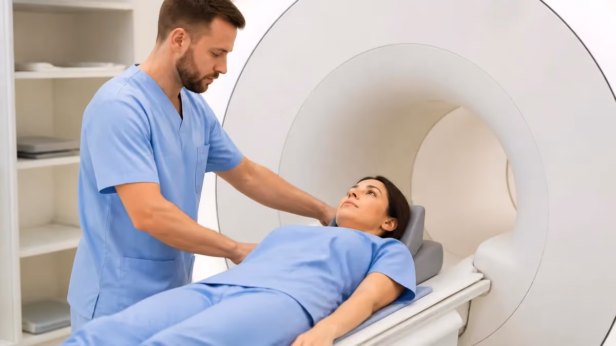
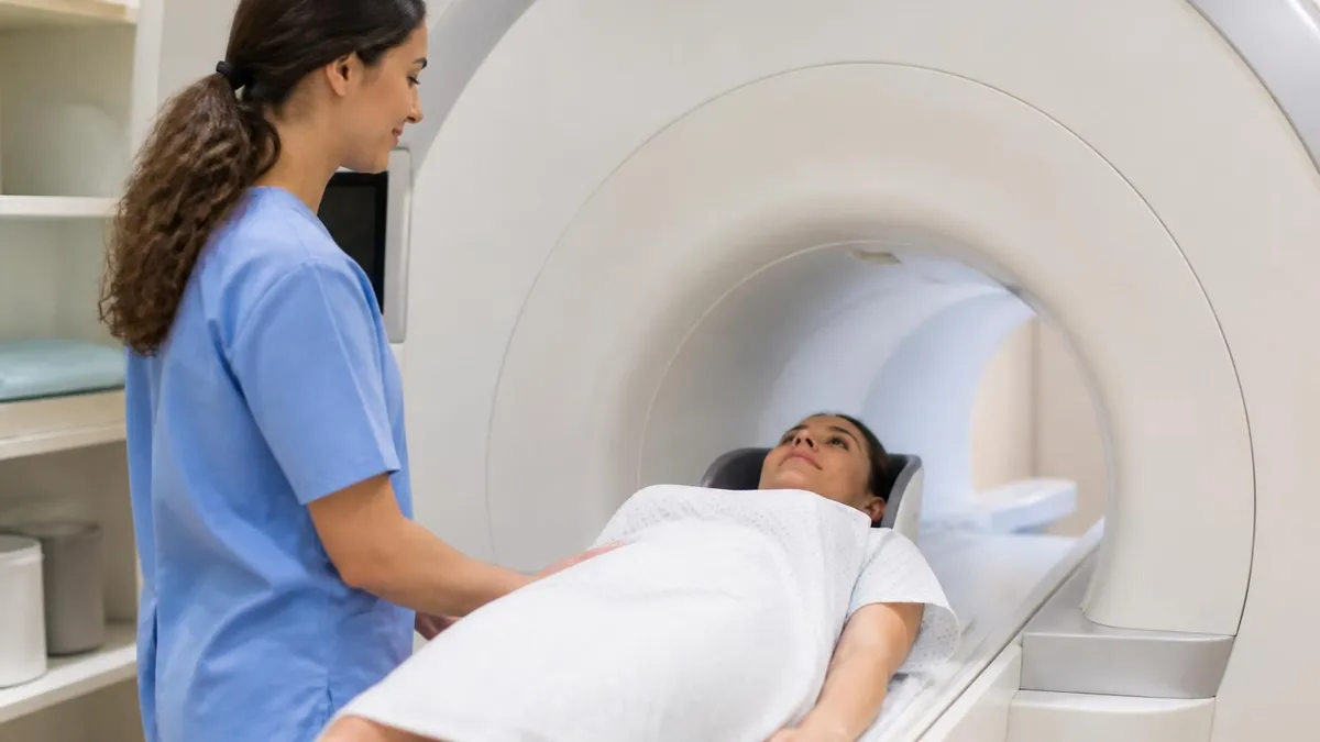
Knowing what to expect on the day of your MRI makes the experience easier, especially when you are wearing braces. From the moment you arrive at the imaging center, the workflow is designed to identify any safety risk and adjust the scan accordingly. You will check in, complete or update a safety screening questionnaire, and meet the technologist who will perform your study. Bring any orthodontic documentation in writing or as a photo on your phone; it streamlines the process considerably.
The screening questionnaire asks about implants, surgeries, occupational metal exposure, and prior imaging. There is almost always a question that specifically addresses dental work, braces, and retainers. Answer honestly and in detail. If you are unsure about the material in your brackets, the technologist can look it up by manufacturer or call your orthodontist for confirmation. This conversation usually takes less than five minutes.
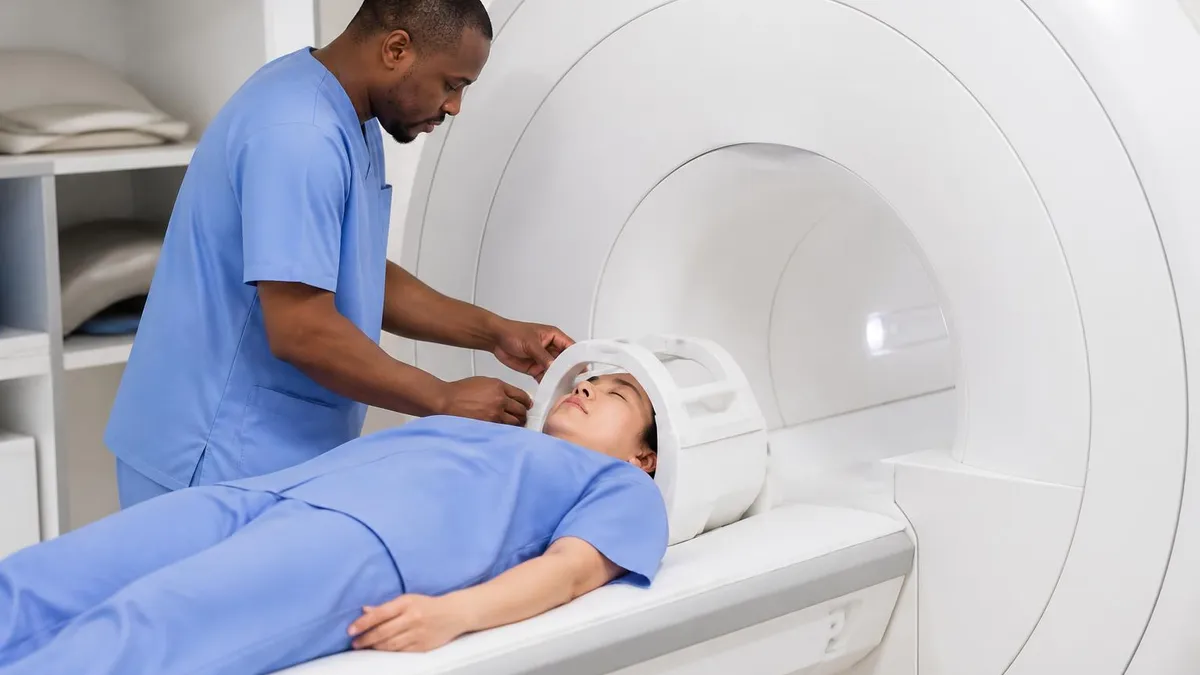
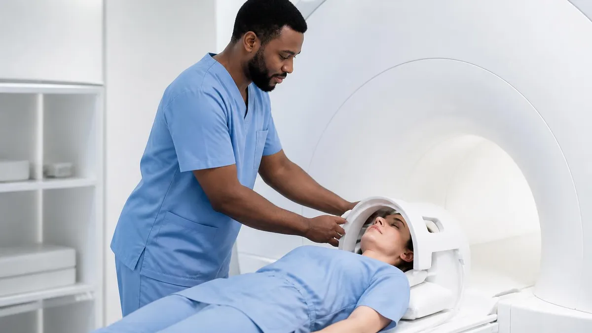
Next comes the dressing room, where you change into a gown and lock your belongings away. Remove any removable orthodontic appliances at this stage, label them, and store them with your other belongings. Wedding rings, watches, hearing aids, hairpins, and even some clothing with metallic threads must come off. Fixed braces stay in place. The technologist will perform a final magnet-side check with a handheld ferromagnetic detector before you enter the room.
Inside the scan room, you will lie on the table and be positioned for the body part being studied. For a brain MRI, your head goes into a head coil that resembles a helmet. For a knee or shoulder MRI, the coil wraps the joint while your head remains outside the bore. Even with full braces, none of these positions are uncomfortable, and the appliance has no effect on coil placement. Foam pads keep you still, and headphones reduce the scanner noise.
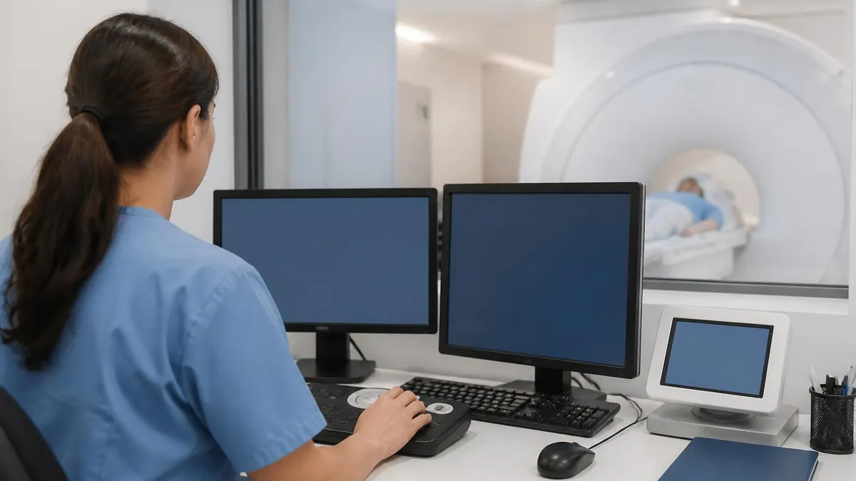
Once the scan begins, you will hear a series of loud clicks and hums that correspond to the gradient coils switching. Many patients with braces notice no sensation at all from their appliance. A few report a faint vibration or a sensation like a gentle tug at the lips during the loudest sequences. This is normal and not a sign of any problem. If you have a concern, press the squeeze ball and the technologist will respond immediately. For a deeper dive into the soundscape, see our explainer on the mri machine noise sequences.
Average total exam time depends on the body part. A brain MRI runs about thirty minutes, a knee about forty-five, and a complex spine study with contrast can exceed an hour. Braces do not significantly extend most protocols, although your radiologist may add one or two metal-friendly sequences if the area of interest is close to the dental arches. The added time is usually less than ten minutes.
After the exam, you change back into your clothes, retrieve your retainer or aligner if you brought one, and head home. There are no activity restrictions, no recovery period, and no follow-up care needed because of the braces themselves. Images are sent to the radiologist for interpretation, and your referring physician will share the results within a few business days. The entire experience is, for most patients, completely uneventful.
Beyond the procedural basics, a few practical tips help patients with braces get the most from their MRI experience. First, schedule the scan during a stable phase of your orthodontic treatment. Avoid booking imaging in the week following major adjustments such as wire upgrades, new bands, or expander activations, when your mouth is most sensitive. A well-settled appliance is more comfortable in the bore and less likely to feel any odd vibration during gradient-heavy sequences.
Second, communicate clearly with both teams. Ask your orthodontist for a brief letter that lists the bracket brand, wire material, and any recent modifications. Hand it to the MRI technologist when you arrive. This small piece of paper resolves most uncertainty about MR Conditional ratings on the spot and saves time that might otherwise be spent calling around for documentation. Many orthodontic offices in the United States now provide this letter automatically when patients mention an upcoming scan.
Third, manage your own comfort. Bring earplugs in addition to the headphones the center supplies. Wear something warm because scan rooms run cool, and dental brackets can make the lips and cheeks feel chilly. Stay well hydrated before the appointment, but avoid drinking large quantities of water in the thirty minutes before the scan so you do not need to interrupt sequences. A relaxed jaw produces the cleanest images, so practice unclenching while you wait.
Fourth, plan for follow-up. If the radiologist needs more imaging due to artifact obscuring a region of interest, ask whether a targeted repeat with different sequences can be scheduled before any consideration of bracket removal. In most cases, simple sequence adjustments resolve the problem. Removing brackets purely for imaging purposes is uncommon and usually reserved for situations where the area of interest is directly beneath the appliance and no alternative imaging is acceptable.
Fifth, learn the basics about your hardware. Knowing whether you have stainless steel or titanium brackets, traditional or self-ligating designs, and metal or nickel-titanium wires turns you from a passive patient into an informed partner. This knowledge also helps if you require imaging at multiple facilities or in an emergency, where staff may not have time to track down your orthodontist. A quick screenshot of your appliance list on your phone is enough.
Sixth, take advantage of the educational opportunity. MRI is a remarkable technology, and the protocols that allow scans with braces showcase years of physics, engineering, and clinical refinement. If you or your child are curious about how it works, ask the technologist. Most are happy to explain the role of the magnet, gradients, and radiofrequency coils. Understanding the why behind the scan often reduces anxiety significantly.
Finally, share your experience. If you complete an MRI with braces uneventfully, tell friends and classmates who may be scheduled for their own scans. Misinformation about dental hardware and imaging is common, and a calm firsthand account from a peer often does more to reassure a nervous teenager than any written brochure. The more accurately patients understand the safety profile, the smoother the entire imaging pipeline becomes.
MRI Questions and Answers
What Is an MRI Test? How Magnetic Resonance Imaging Scans Diagnose Disease in 2026

MRI Medical Abbreviation: What MRI Stands For and Why It Matters

The History of MRI: From Discovery to Modern Medicine

Knee MRI Images: A Complete Guide to Reading, Understanding, and Interpreting Knee Scans

Noise of MRI Machine: Why MRI Scanners Are So Loud and What to Expect
About the Author
Medical Laboratory Scientist & Clinical Certification Expert
Johns Hopkins UniversityDr. Sandra Kim holds a PhD in Clinical Laboratory Science from Johns Hopkins University and is certified as a Medical Technologist (MT) and Medical Laboratory Scientist (MLS) through ASCP. With 16 years of clinical laboratory experience spanning hematology, microbiology, and molecular diagnostics, she prepares candidates for ASCP board exams, MLT, MLS, and specialist certification tests.
Join the Discussion
Connect with other students preparing for this exam. Share tips, ask questions, and get advice from people who have been there.
View discussion (6 replies)