1.5 Tesla MRI Machine Types: Complete Guide to Field Strengths, Bore Sizes, and Clinical Uses
1.5 tesla MRI machine types explained — compare 0.3T, 1.5T, 3T, 7T scanners, open vs closed bore designs, and clinical applications. 🎯
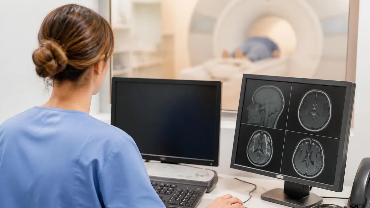
The 1.5 tesla MRI is the workhorse of modern radiology, and understanding the different MRI machine types available today helps patients, students, and technologists make sense of why one scanner is chosen over another. Field strength, bore design, magnet configuration, and gradient performance all influence image quality, scan time, and patient comfort. Hospitals frequently operate several scanner classes side by side because no single machine fits every clinical question or every body habitus.
MRI scanners are classified primarily by magnetic field strength, measured in tesla (T). Low-field systems range from 0.2T to 0.5T, mid-field systems sit between 0.5T and 1.0T, high-field scanners include the popular 1.5T platforms, and ultra-high-field machines reach 3T, 7T, and beyond in research settings. Each tier offers distinct trade-offs in signal-to-noise ratio, spatial resolution, artifact sensitivity, and cost per study.
Beyond field strength, magnet design separates scanners into closed-bore, wide-bore, open, and extremity configurations. Closed-bore cylindrical magnets dominate clinical practice because they deliver the most homogeneous field and the best diagnostic images. Open MRI systems, by contrast, prioritize claustrophobic and bariatric patient access, often at the cost of lower field strength. Extremity scanners target a single joint and reduce scan time dramatically for orthopedic indications.
The history of medical imaging shows that today's diverse scanner landscape did not happen overnight — you can trace the evolution from early prototypes through modern superconducting magnets in this history of MRI. Each generation of machines added gradient strength, parallel imaging coils, and software refinements that compressed exam times from over an hour down to fifteen minutes for many protocols.
Choosing the right scanner type matters clinically. A neuroradiologist evaluating multiple sclerosis lesions may insist on 3T for superior gray-white matter contrast, while an orthopedic surgeon mapping a meniscal tear may be perfectly satisfied with a 1.5T extremity unit. Pacemaker patients, large-bodied patients, pediatric patients, and patients with implanted hardware each create different scanner-selection pressures that radiology departments must balance every shift.
This guide walks through every major MRI scanner classification used in the United States, breaks down practical differences in image quality and patient experience, and explains how machine selection affects diagnostic confidence. Whether you are preparing for a registry exam, scheduling your first scan, or working as a new technologist learning the equipment, the sections ahead will clarify what each scanner does best and where its limits lie.
By the end, you will recognize the visual and technical signatures of each major MRI platform, understand why a 1.5T closed-bore remains the most common scanner in American hospitals, and appreciate the niche roles played by open, ultra-high-field, and intraoperative systems. The choice of machine is rarely arbitrary — it reflects clinical question, patient factors, and institutional resources working together.
MRI Machine Types by the Numbers
MRI Field Strength Categories Explained
Older or specialty systems often used for extremity imaging or open designs. Lower SNR means longer scans, but reduced susceptibility artifacts can help around metal implants and pacemakers in select cases.
A transitional category once common in community hospitals. Offers reasonable image quality at lower operating cost than 1.5T but is increasingly replaced by modern wide-bore high-field systems with better gradient performance.
The clinical gold standard for most body, brain, spine, and musculoskeletal exams. Balances image quality, scan time, safety, and cost. Compatible with the widest range of implants and accessories.
Doubles signal-to-noise ratio compared to 1.5T, enabling thinner slices, faster scans, and superior neuro, vascular, and functional imaging. Higher specific absorption rate and increased susceptibility artifacts require careful protocoling.
Reserved for academic and research centers. Produces extraordinary detail of small brain structures, cortical layers, and metabolic imaging via spectroscopy. Limited body applications and stricter safety constraints than clinical scanners.
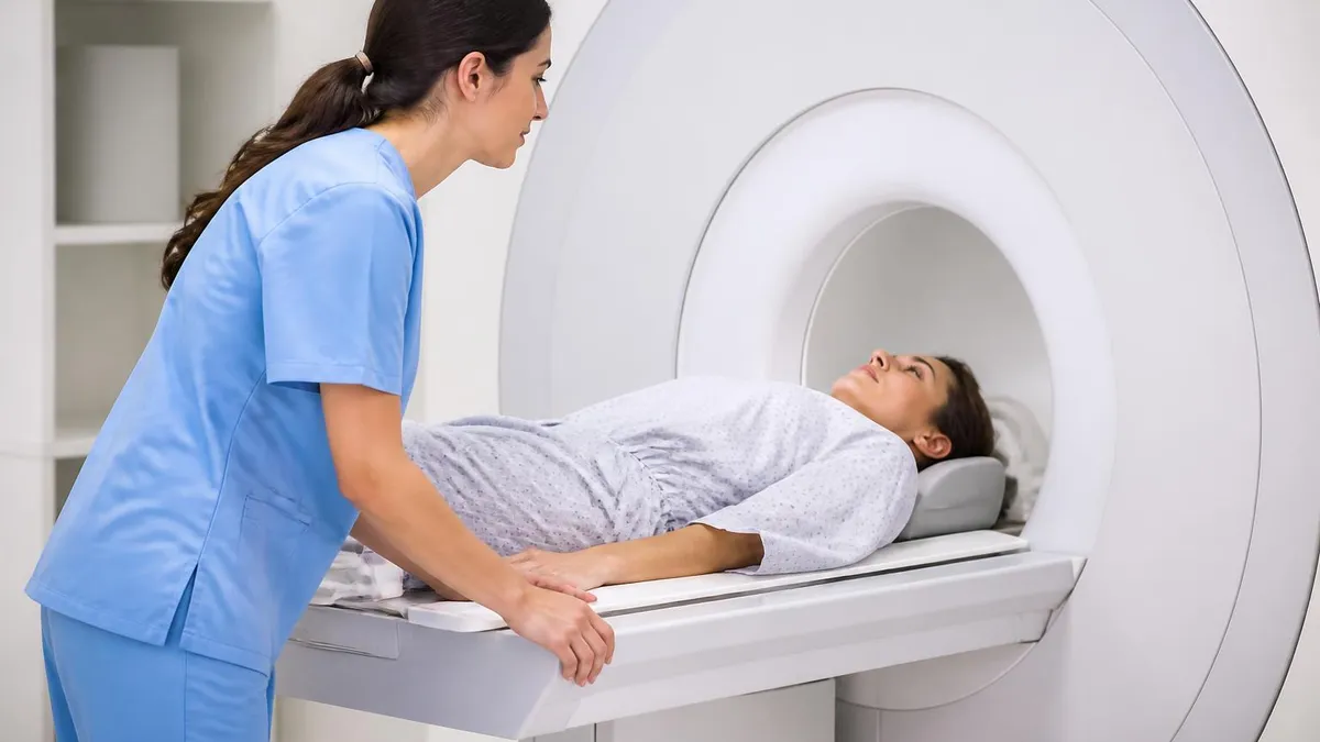
Closed-bore MRI scanners — the cylindrical tunnels most patients picture when they hear "MRI" — remain dominant because the cylindrical superconducting magnet geometry produces the most uniform magnetic field. Field homogeneity directly determines image quality, fat suppression reliability, and the success of advanced sequences like diffusion-weighted imaging and spectroscopy. Modern closed-bore systems typically operate at 1.5T or 3T with bore diameters of 60 cm standard or 70 cm wide-bore.
Wide-bore scanners deserve special mention. By widening the patient tunnel from 60 cm to 70 cm, manufacturers reduced claustrophobia complaints, accommodated patients up to roughly 550 pounds, and made positioning easier for shoulder and hip studies that require arms-at-sides geometry. Wide-bore 1.5T and 3T platforms now represent the majority of new installations in the United States. They retain full diagnostic image quality while improving access.
Open MRI systems use a fundamentally different magnet design — typically two parallel pole pieces above and below the patient — and they operate at lower field strengths, commonly 0.3T to 1.2T. Open scanners eliminate the tunnel sensation entirely. They are invaluable for severely claustrophobic patients, very large patients, pediatric patients who need a parent nearby, and patients requiring imaging in non-standard positions. The trade-off is reduced image quality and longer scan times.
Extremity MRI scanners are compact units that scan only a hand, wrist, elbow, foot, ankle, or knee. The patient sits or lies beside the unit while only the affected limb enters a small bore. These dedicated systems often operate at 1.0T to 1.5T, deliver excellent musculoskeletal images, and free up the main scanner for body and neuro work. Many orthopedic clinics own extremity units to control workflow.
Standing or upright MRI is a small niche where weight-bearing assessment of the spine matters — for example, evaluating disc herniation that only appears under load. These scanners operate at low field strength and produce images inferior to closed-bore systems, but their unique positioning capability is medically valuable in selected cases. Most patients will never encounter one outside specialized spine clinics.
Intraoperative MRI suites place a scanner inside or adjacent to a surgical theater, allowing neurosurgeons to image the brain mid-procedure and confirm complete tumor resection before closing. These hybrid environments combine MR-conditional surgical tools, ceiling-mounted scanner transport, and rigorous safety zones. Procedures involving mri with and without contrast cpt may both be performed during a single intraoperative session to maximize diagnostic confidence.
Finally, PET-MRI hybrid systems combine positron emission tomography with MR imaging in a single gantry. These rare and expensive scanners are used at major cancer and neurology centers to fuse metabolic and anatomical data in one acquisition. Although the global installed base is small, PET-MRI illustrates the direction of integration that the next decade of imaging is heading toward — multimodal scanners that answer multiple questions per visit.
MRI Practice Test Questions
Prepare for the MRI - Magnetic Resonance Imaging exam with our free practice test modules. Each quiz covers key topics to help you pass on your first try.
MRI Knowledge
MRI Exam Questions covering Knowledge. Master MRI Test concepts for certification prep.
MRI Physics
Free MRI Practice Test featuring Physics. Improve your MRI Exam score with mock test prep.
MRI Anatomy and Pathology
MRI Test Prep for MRI Anatomy and Pathology. Practice MRI Quiz questions and boost your score.
MRI Anatomy and Positioning
MRI Questions and Answers on MRI Anatomy and Positioning. Free MRI practice for exam readiness.
MRI Contrast Agents
Free MRI Quiz on MRI Contrast Agents. MRI Exam prep questions with detailed explanations.
MRI Patient Care and Positioning
MRI Practice Questions for MRI Patient Care and Positioning. Build confidence for your MRI certification exam.
1.5T, 3T, and 7T MRI Compared
The 1.5 tesla MRI scanner is the most widely deployed clinical platform in the United States and globally. It strikes the best balance between image quality, scan time, safety, implant compatibility, and operational cost. Almost every musculoskeletal, abdominal, pelvic, cardiac, and routine neurological exam can be performed at 1.5T with excellent diagnostic confidence.
Its broad implant compatibility list is a major reason 1.5T dominates. Many pacemakers, neurostimulators, cochlear implants, and aneurysm clips carry MR-conditional labeling specifically at 1.5T. Specific absorption rate is lower at 1.5T than at 3T, reducing tissue heating concerns during long sequences and making 1.5T the safer choice for many pregnant patients and patients with conductive implants.
Is a Higher Field Strength Always Better?
- +Greater signal-to-noise ratio enables thinner slices and finer anatomical detail
- +Faster scan times for many sequences improve patient throughput
- +Superior performance for functional MRI, spectroscopy, and diffusion tensor imaging
- +Better detection of small lesions in brain, pituitary, and inner ear
- +Enhanced vascular imaging without contrast at 3T and above
- +Improved fat suppression on advanced spectral sequences
- +Higher confidence for subtle musculoskeletal cartilage assessment
- −Increased susceptibility artifacts near metal implants and air interfaces
- −Higher specific absorption rate increases tissue heating risk
- −Smaller list of MR-conditional implants compared with 1.5T
- −More pronounced chemical shift artifacts in spine and abdomen
- −Higher purchase and maintenance costs for hospitals
- −Some patients experience dizziness, vertigo, or metallic taste
- −Pediatric and pregnant patient protocols require additional caution
How to Choose the Right MRI Machine Type
- ✓Confirm the clinical question and target anatomy before booking
- ✓Check implant compatibility cards against the scanner's MR-conditional label
- ✓Assess patient body habitus and choose wide-bore if over 60 cm shoulder width
- ✓Screen for claustrophobia and offer open MRI when severe
- ✓Match field strength to lesion size — small neuro lesions favor 3T
- ✓Consider sedation needs for pediatric or anxious patients
- ✓Verify coil availability for the body part being scanned
- ✓Account for scan duration and patient ability to remain still
- ✓Coordinate contrast administration timing with renal function screening
- ✓Choose extremity units for isolated orthopedic indications to free main scanners
Field strength is only one variable.
Gradient performance, coil technology, and software acceleration often matter as much as raw tesla. A modern 1.5T wide-bore with advanced multi-channel coils and compressed-sensing reconstruction frequently outperforms an older 3T system on routine clinical work. Always evaluate the whole platform — not just the magnet number.
Specialty MRI systems address clinical questions that general-purpose scanners cannot answer efficiently. Cardiac MRI suites combine 1.5T or 3T scanners with ECG gating, respiratory navigators, and dedicated cardiac coils to image a beating heart in three dimensions. These platforms quantify ejection fraction, detect myocardial scarring with late gadolinium enhancement, and evaluate cardiomyopathies that echocardiography may miss. Cardiac MRI typically requires technologists with additional training and longer appointment slots.
Breast MRI uses a dedicated breast coil and high-resolution dynamic contrast-enhanced protocols at 1.5T or 3T. It plays a central role in screening high-risk women, evaluating implant integrity, and staging known cancers. Some centers offer abbreviated breast MRI in under fifteen minutes as a screening adjunct to mammography. The patient lies prone with the breasts suspended into the coil, a position uniquely supported by breast MRI hardware.
Prostate MRI has rapidly grown as a tool for prostate cancer detection and staging. Multiparametric protocols combining T2-weighted, diffusion-weighted, and dynamic contrast sequences are now standard, and structured PI-RADS reporting has improved consistency across radiologists. Both 1.5T with an endorectal coil and 3T with a surface coil produce diagnostic-quality studies, with 3T trending as the preferred field strength when available.
Pediatric MRI presents unique challenges that drive scanner selection. Younger children may need sedation or general anesthesia to lie still. Wide-bore scanners with quieter gradients, faster sequences, and child-friendly suite designs — themed murals, audiovisual goggles, mock scanners — all reduce sedation needs. Some institutions invest in feed-and-wrap protocols for infants that obviate anesthesia entirely when scans can be completed quickly.
Interventional MRI extends imaging into procedural medicine. MR-guided biopsies, focused ultrasound treatments for tremor and prostate cancer, and laser interstitial thermal therapy for epilepsy and brain tumors all rely on real-time MRI guidance. These suites require MR-conditional instruments, advanced thermometry sequences, and tightly choreographed workflows between radiologists, surgeons, and technologists. The scanner essentially becomes a procedural tool, not just a diagnostic one.
Mobile MRI trailers bring scanners to hospitals that cannot afford permanent installations or that need temporary capacity during renovations. These trailers contain 1.5T scanners in a shielded environment, complete with control rooms and patient prep areas. Image quality matches fixed installations, although appointment volume is limited by trailer size. Rural hospitals frequently rely on mobile units rotating weekly to maintain MRI access for their communities.
Finally, neonatal MRI scanners are emerging in NICU settings. These small-bore 1.5T systems are designed for infants under 4.5 kg and are physically located inside or adjacent to the neonatal intensive care unit. They eliminate the dangerous transport of fragile premature infants to the main radiology department and enable bedside-level imaging of hypoxic-ischemic injury, periventricular leukomalacia, and congenital anomalies — a major advance for newborn neurology.
Every patient must be screened against the specific scanner's MR-conditional labeling before entering the magnet room. A device cleared for 1.5T may be unsafe at 3T. Always verify model numbers and labeling — assumptions cause serious harm. When in doubt, escalate to the MRI safety officer or radiologist before proceeding with the exam.
The next decade of MRI hardware development is being driven by three forces: artificial intelligence reconstruction, helium-free magnets, and access expansion through low-field renaissance. Each trend is already reshaping which scanner types you will see in clinical practice, and each has implications for technologists preparing for certification exams as well as for hospitals planning capital investments. Understanding the trajectory helps clinicians interpret manufacturer announcements with realistic expectations.
AI-based image reconstruction is the most disruptive immediate change. Vendors now sell software that reconstructs undersampled k-space data using deep learning networks, cutting acquisition times by 40 to 60 percent without sacrificing diagnostic quality. A knee MRI that once took 25 minutes can now complete in 10 minutes, dramatically increasing scanner throughput. These tools also denoise low-field images, partially closing the quality gap between 0.55T and 1.5T scanners.
Helium-free or low-helium magnet designs respond to global helium supply volatility and the operational cost of cryogen refills. New scanners use sealed superconducting magnets requiring far less helium, lowering installation and maintenance burdens. This trend benefits rural hospitals, ambulatory imaging centers, and developing-world deployments where helium logistics historically blocked MRI adoption. Expect future installations to advertise low-helium or zero-boil-off cryogenic systems prominently.
Low-field MRI is returning, but not in its old form. Modern 0.55T scanners combine wide bores, AI reconstruction, and advanced gradient design to produce surprisingly good clinical images at one-third the price of 1.5T units. These scanners excel for lung imaging — paradoxically aided by lower susceptibility at 0.55T — and for patients with metal implants where 1.5T or 3T would generate severe artifacts. Reading about MRI medical abbreviation conventions helps decode the alphabet soup of new product specs.
Portable point-of-care MRI is the most radical development. Compact 0.064T scanners are now FDA-cleared for bedside neuroimaging in stroke units, ICUs, and emergency departments. While image quality is far below conventional scanners, portable systems answer specific binary questions — Is there a large hemorrhage? Is there a midline shift? — at the patient's bedside without transport risk. They will not replace standard MRI, but they will expand its reach significantly.
7T clinical adoption will continue slowly. FDA clearances have expanded to include musculoskeletal indications alongside neuroimaging, and as RF and gradient technology mature, the technical hurdles that limit 7T body imaging will gradually fall. Whether 7T becomes routine outside academic centers within fifteen years remains uncertain, but the platform's research output justifies continued investment. Trainees should expect at least exposure to ultra-high-field work during fellowships.
Finally, integrated hybrid imaging — PET-MRI, MRI-LINAC for radiation therapy guidance, and MRI-guided focused ultrasound — will grow in tertiary care centers. Each hybrid system combines MRI with another modality to answer questions impossible to solve with either tool alone. The skill set required to operate these platforms is broader than traditional MRI training, and educational pathways are evolving to match. The diversity of scanner types in 2030 will far exceed the diversity available today.
Practical preparation for an MRI exam, whether you are the patient or the technologist running the scan, starts with knowing your scanner. If you are scheduled for an MRI, ask the imaging center which field strength they will use and whether the bore is standard or wide. This single question prevents the day-of surprise of arriving at a tight 60 cm bore when your shoulders measure 55 cm and your anxiety spikes. Centers should accommodate reasonable requests for wide-bore or open scanners when available.
Bring a complete and current implant list to your appointment. Modern MRI with braces and other dental hardware is usually safe at 1.5T, but the technologist still needs to know to anticipate artifacts and adjust sequences. Hardware compatibility cards from your surgeon — pacemakers, neurostimulators, cochlear implants, aneurysm clips, even retained shrapnel — must be available before screening begins. Photograph the cards and store them in your phone for instant access at any imaging facility.
Wear loose, metal-free clothing or accept the scrubs the facility provides. Underwire bras, snap closures, embedded metallic thread, and certain transdermal patches contain materials incompatible with MRI. Removing makeup is wise too — some cosmetics contain iron oxide pigments that can cause skin warming and image artifacts near the eyes. Hearing protection, given the loud gradients, will be provided, but you can bring your own foam earplugs if you have sensitivity.
Technologists preparing for board exams should understand the scanner-type questions that registries love to ask. Be able to identify a wide-bore from a standard bore by diameter, recognize the safety zones around a magnet room, list implant categories cleared at 1.5T versus 3T, and compare expected SAR values across field strengths. Registry questions often pair scanner specifications with clinical scenarios — knowing the equipment cold is essential.
If you are scheduling a follow-up MRI, ask whether it can be performed on the same scanner used previously. Direct comparison between scans is easier when the field strength, coil configuration, and protocol match. Multiple sclerosis monitoring, tumor surveillance, and post-surgical follow-up are all areas where consistency improves interpretation. Some institutions explicitly schedule follow-ups on identical platforms for this reason — ask when booking.
For complex or unusual clinical questions, advocate for the right scanner rather than accepting the first available slot. A pituitary microadenoma rule-out, a small acoustic neuroma, or a suspected MS plaque may benefit from 3T even if 1.5T is available sooner. Conversely, a patient with extensive spinal hardware will likely get better diagnostic information from 1.5T than 3T because of reduced susceptibility artifacts. Your radiologist or referring physician can guide these choices.
Finally, recognize that MRI machine types will keep evolving. The scanner you use this year may not exist in five years, replaced by faster, quieter, AI-enhanced versions. Stay curious about the technology, ask technologists questions during your scan when timing permits, and treat each MRI as both a medical procedure and a window into one of the most sophisticated imaging tools ever developed. The more you understand it, the better you can advocate for your own care or for your patients.
MRI Questions and Answers


About the Author
Medical Laboratory Scientist & Clinical Certification Expert
Johns Hopkins UniversityDr. Sandra Kim holds a PhD in Clinical Laboratory Science from Johns Hopkins University and is certified as a Medical Technologist (MT) and Medical Laboratory Scientist (MLS) through ASCP. With 16 years of clinical laboratory experience spanning hematology, microbiology, and molecular diagnostics, she prepares candidates for ASCP board exams, MLT, MLS, and specialist certification tests.
Join the Discussion
Connect with other students preparing for this exam. Share tips, ask questions, and get advice from people who have been there.
View discussion (6 replies)