Open Bore MRI: How Wide-Bore Scanners Work, What to Expect, and Who Should Choose One
📝 Open bore MRI explained: bore size, magnet strength, claustrophobia relief, image quality, costs, and how wide-bore scanners compare to traditional
An open bore MRI is a wide-bore magnetic resonance imaging scanner designed to give patients more breathing room, more headroom, and a far less confining experience than the narrow tunnels of older systems. While the term sometimes gets confused with truly open MRI machines that have no tunnel at all, an open bore MRI still uses a cylindrical magnet, but the opening is dramatically larger, typically 70 centimeters across instead of the traditional 60 centimeters. That extra ten centimeters changes everything about how a scan feels.
Patients who once refused MRI due to claustrophobia, body size, or anxiety often tolerate an open bore MRI without sedation, and radiologists still get the diagnostic image quality they need because most wide-bore systems operate at the same 1.5T or 3.0T field strength as conventional scanners. This combination of comfort and clinical performance has made the open bore MRI one of the most popular scanner designs in modern imaging centers across the United States.
The growth of wide-bore technology reflects a deeper shift in radiology priorities. Hospitals and outpatient imaging centers no longer compete on magnet strength alone. They compete on patient experience, throughput, and the ability to scan bariatric patients, pediatric patients, and people with implanted devices who require very specific positioning. An open bore MRI accomplishes all of these goals while preserving signal-to-noise ratio and gradient performance, which is why nearly every major manufacturer now sells one as a flagship model.
If you are scheduled for a scan and your provider mentioned a wide-bore machine, you are probably going to have a more comfortable visit than a friend who described a tight, noisy tube from a decade ago. Understanding what an open bore MRI is, how it differs from both closed and truly open systems, and what the trade-offs are will help you walk in prepared. It also helps to understand a little of the history of MRI so the modern designs make more sense in context.
This guide covers the engineering behind wide-bore magnets, the patient experience from check-in to discharge, image quality comparisons with closed systems, the role of 3T wide-bore scanners in advanced imaging, costs and insurance coverage in the US market, and a deep FAQ section addressing the questions patients and technologists ask most often. Whether you are a patient researching options, a student studying MR physics, or a clinician choosing a system for a new suite, you will find clear, practical information here.
By the end, you will understand exactly why an open bore MRI is rarely a compromise, when it is genuinely the wrong tool for the job, and how to advocate for one if claustrophobia or body habitus has stopped you from getting an important scan in the past. The technology has matured to the point where wide-bore is the new default for many indications, not the exception.
Let us begin with the numbers that define the modern open bore MRI landscape, then move into the physics, patient experience, and decision-making frameworks that matter most in real clinical practice today.
Open Bore MRI by the Numbers
Anatomy of a Wide-Bore MRI Scanner
A 1.5T or 3.0T magnet cooled by liquid helium provides the static field. Wide-bore designs use a larger warm-bore aperture but keep the same homogeneity standards required for diagnostic imaging.
Three orthogonal gradient coils encode spatial information. Wide-bore systems use re-engineered coils with higher peak amplitudes to compensate for the larger volume and maintain fast imaging performance.
A built-in body coil transmits radiofrequency pulses, while surface coils receive the signal. Wide-bore systems support dense multi-channel arrays for parallel imaging acceleration.
The motorized table on a wide-bore MRI typically supports up to 550 pounds and slides through the bore with sub-millimeter positioning accuracy for repeat exams and follow-ups.
Modern wide-bore scanners include dual-layer acoustic dampening and quiet pulse sequences that reduce noise by 70 to 80 percent compared to older closed-bore systems.
The defining specification of an open bore MRI is the diameter of the magnet opening, called the bore. Traditional closed MRI scanners have a bore of 60 centimeters, roughly 23.6 inches, which feels tight for anyone with broad shoulders, a larger torso, or even average build when the arms must be tucked. A wide-bore or open bore MRI increases that opening to 70 centimeters, about 27.6 inches, which sounds like a small change but represents a 36 percent increase in cross-sectional area. The added space dramatically reduces the sensation of being trapped.
Magnet strength on an open bore MRI is not compromised to achieve the wider opening. Most clinical wide-bore systems operate at 1.5 Tesla, the workhorse field strength of modern radiology, while premium models reach 3.0 Tesla for neurological, musculoskeletal, and oncologic imaging that benefits from higher signal-to-noise ratio. Engineers achieve this by using larger superconducting coils, more precise shimming, and advanced gradient designs. The result is a scanner that delivers the same diagnostic image quality as a closed system in nearly every clinical scenario.
The length of the bore matters just as much as the diameter. A short-bore wide-bore MRI, sometimes only 125 to 145 centimeters long, allows many head, neck, and extremity exams to be performed with the patient's head outside the magnet entirely. This feet-first positioning eliminates the visual trigger that causes most claustrophobic reactions. Combined with mirrors, prism glasses, and in-bore lighting, the open bore MRI experience can feel more like lying under a low ceiling than being inside a tunnel.
Gradient performance on open bore systems has improved substantially over the past decade. Early wide-bore scanners traded some gradient strength for the larger aperture, leading to slightly slower sequences for advanced applications like diffusion-weighted imaging and cardiac MRI. Modern designs have closed that gap. High-performance wide-bore systems now offer gradient amplitudes of 45 to 80 millitesla per meter and slew rates above 200 tesla per meter per second, matching or exceeding many older closed-bore scanners used for routine clinical work.
Patient weight capacity is another practical advantage. Conventional 60-centimeter bore systems often max out at 350 to 450 pounds, which excludes a significant portion of the US adult population. Wide-bore systems typically support 500 to 550 pounds, and several manufacturers offer bariatric configurations rated to 660 pounds. Combined with the larger aperture, this makes the open bore MRI the default choice for many imaging centers that serve diverse patient populations. The understanding of MRI fundamentals starts with the basic MRI medical abbreviation and expands into these engineering details.
One subtle but important point: an open bore MRI is not the same as an open MRI. True open MRI systems use a C-shaped or H-shaped magnet with no tunnel at all, and they typically operate at much lower field strengths between 0.2 and 1.2 Tesla. Image quality on those systems is acceptable for orthopedic work but generally inadequate for detailed brain, cardiac, or vascular imaging. The open bore MRI bridges the gap, offering the comfort patients want with the diagnostic performance clinicians require, which is why it has become the dominant new-installation choice in the United States.
Understanding these specifications helps patients ask the right questions when scheduling. If you have struggled with previous scans, ask whether the facility has a 70-centimeter bore system, whether it is 1.5T or 3.0T, and how long the bore is. Those three answers tell you almost everything you need to know about how comfortable and how diagnostic your scan will be.
MRI Practice Test Questions
Prepare for the MRI - Magnetic Resonance Imaging exam with our free practice test modules. Each quiz covers key topics to help you pass on your first try.
MRI Knowledge
MRI Exam Questions covering Knowledge. Master MRI Test concepts for certification prep.
MRI Physics
Free MRI Practice Test featuring Physics. Improve your MRI Exam score with mock test prep.
MRI Anatomy and Pathology
MRI Test Prep for MRI Anatomy and Pathology. Practice MRI Quiz questions and boost your score.
MRI Anatomy and Positioning
MRI Questions and Answers on MRI Anatomy and Positioning. Free MRI practice for exam readiness.
MRI Contrast Agents
Free MRI Quiz on MRI Contrast Agents. MRI Exam prep questions with detailed explanations.
MRI Patient Care and Positioning
MRI Practice Questions for MRI Patient Care and Positioning. Build confidence for your MRI certification exam.
Open Bore MRI vs Closed vs Truly Open Systems
An open bore MRI uses a cylindrical superconducting magnet with a 70-centimeter aperture and short bore length. It operates at full diagnostic strength of 1.5T or 3.0T, supports advanced sequences like diffusion, perfusion, and spectroscopy, and accommodates most bariatric patients up to 550 pounds. Image quality is essentially identical to a traditional closed system, making it the modern default.
Patients lying inside still feel partially enclosed, but the extra ten centimeters of diameter combined with mirrors and in-bore lighting reduces claustrophobic reactions dramatically. Scan times are equivalent to closed systems. The wide-bore approach is now the most common new installation in US outpatient imaging centers and hospital radiology departments due to its balance of comfort, capacity, and clinical performance.

Open Bore MRI: Pros and Cons
- +Significantly reduces claustrophobic reactions compared to traditional 60 cm bore systems
- +Accommodates bariatric patients up to 550 pounds without compromise
- +Maintains full diagnostic field strength of 1.5T or 3.0T
- +Shorter bore length keeps the head outside the magnet for many exams
- +Compatible with all standard MRI protocols including advanced sequences
- +Reduces need for sedation, lowering costs and recovery time
- +Improves throughput by reducing aborted scans and patient anxiety delays
- −Higher purchase and installation cost than traditional closed systems
- −Slightly reduced gradient performance on some older wide-bore models
- −Image quality at the bore periphery may be marginally lower than centerline
- −Still feels enclosed for severely claustrophobic patients who need truly open systems
- −Requires larger room footprint and stronger structural support
- −Helium refill costs and quench-pipe requirements remain unchanged from closed systems
Pre-Scan Preparation for an Open Bore MRI
- ✓Confirm with the facility that the scanner has a 70 cm wide bore, not a 60 cm closed bore
- ✓Disclose all implants, pacemakers, neurostimulators, and metal fragments to the technologist
- ✓Remove all jewelry, watches, hair pins, hearing aids, and removable dental work before entering
- ✓Wear clean, metal-free clothing or change into the gown the facility provides
- ✓Arrive 15 to 30 minutes early to complete the MRI safety screening form
- ✓Inform staff if you are pregnant, breastfeeding, or have kidney disease before any contrast
- ✓Eat normally unless your exam specifically requires fasting, such as abdominal MRCP
- ✓Take prescribed anti-anxiety medication only as directed and bring a driver
- ✓Ask for ear protection, prism glasses, and a panic squeeze ball before entering the bore
- ✓Plan to lie still for 20 to 45 minutes depending on the body part being imaged
Wide-bore is no longer a compromise
A modern 3.0T open bore MRI delivers diagnostic image quality that matches or exceeds older closed-bore systems for nearly every clinical indication. If you have postponed a scan because of claustrophobia or body size, ask your provider specifically for a 70 cm wide-bore system. There is rarely a clinical reason to refuse the request.
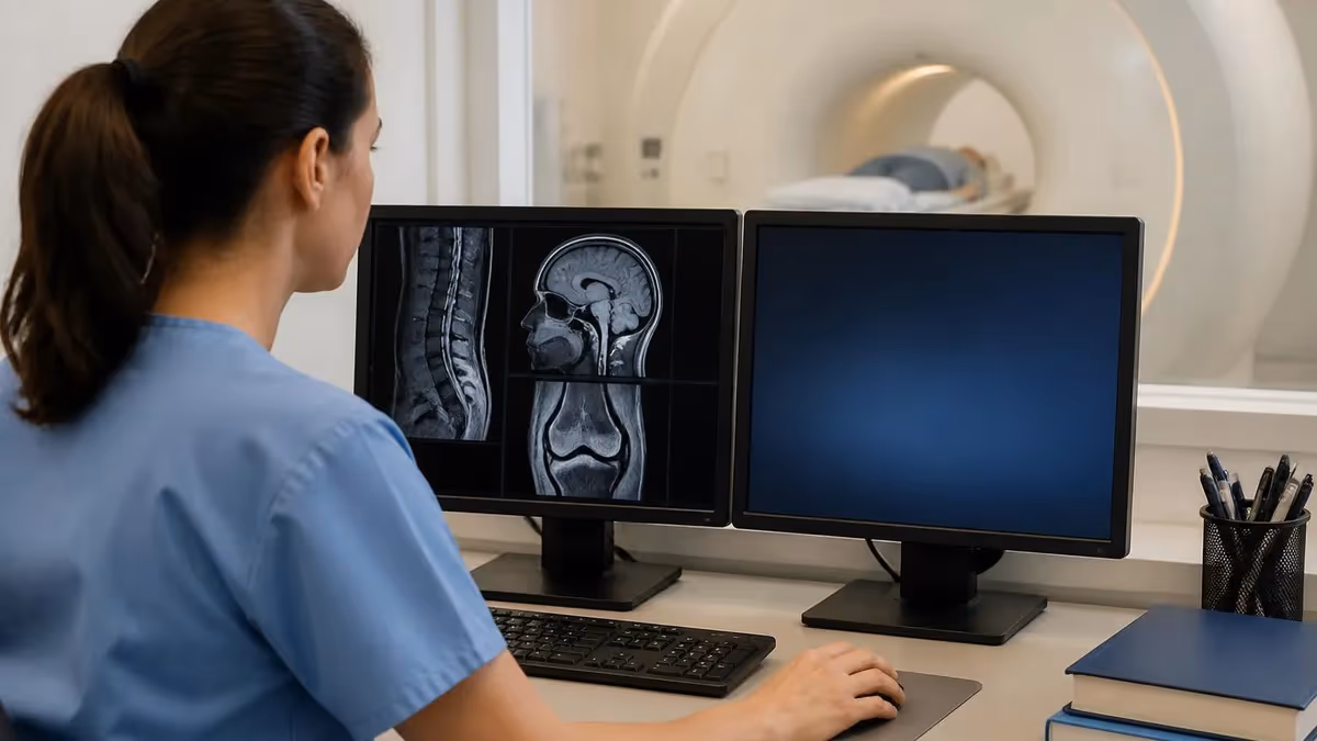
From the patient perspective, the open bore MRI experience starts before you ever see the scanner. A good imaging center will ask during scheduling whether you have a history of claustrophobia, prior incomplete scans, or anxiety around enclosed spaces. If you do, they should automatically route you to a wide-bore system rather than a conventional closed one. Tell the scheduler if you have ever had to stop an MRI partway through, because that history strongly predicts how the next scan will go without intervention.
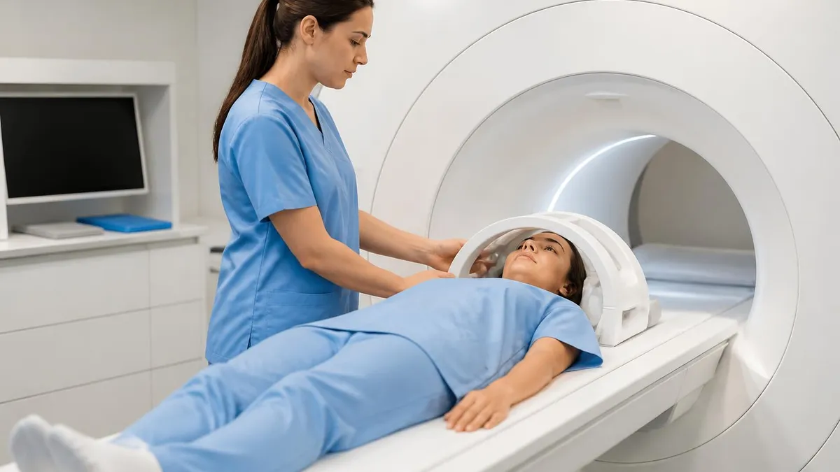
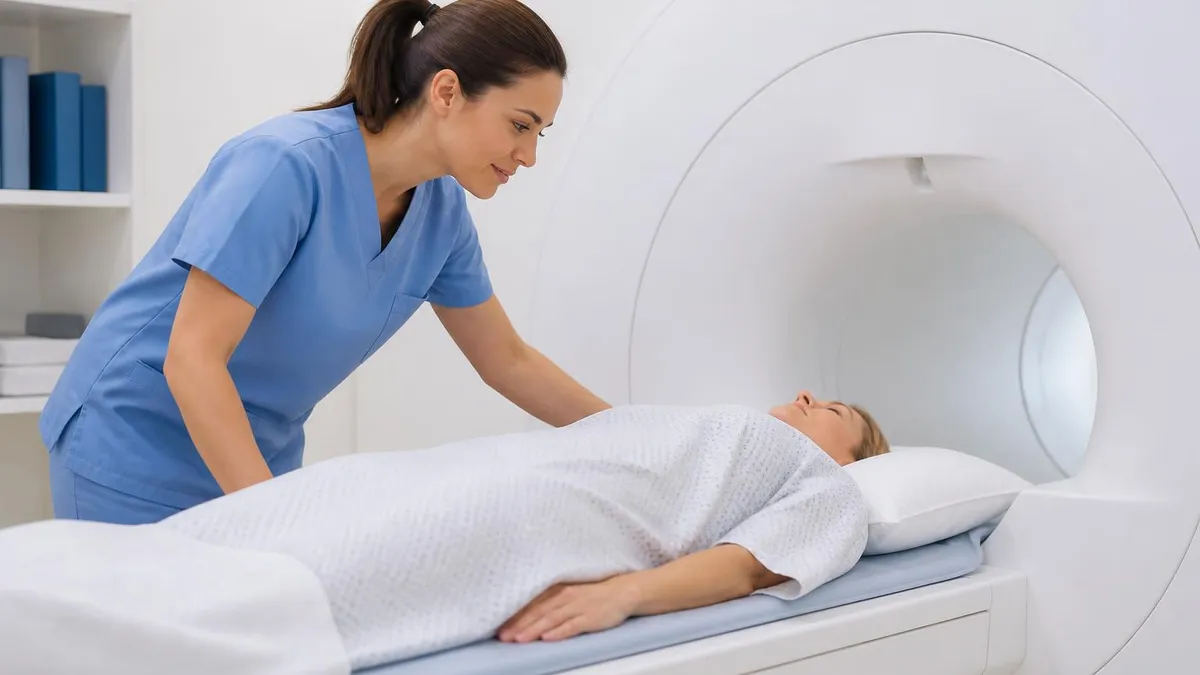
On the day of the scan, you will change into a gown or metal-free clothing, complete a safety screening that asks about implants and prior surgeries, and meet your MRI technologist. The technologist positions you on the patient table, places the appropriate receive coils on or around the body part being imaged, and provides hearing protection in the form of earplugs, headphones, or both. Many wide-bore systems offer music streaming, which substantially reduces perceived noise and time.
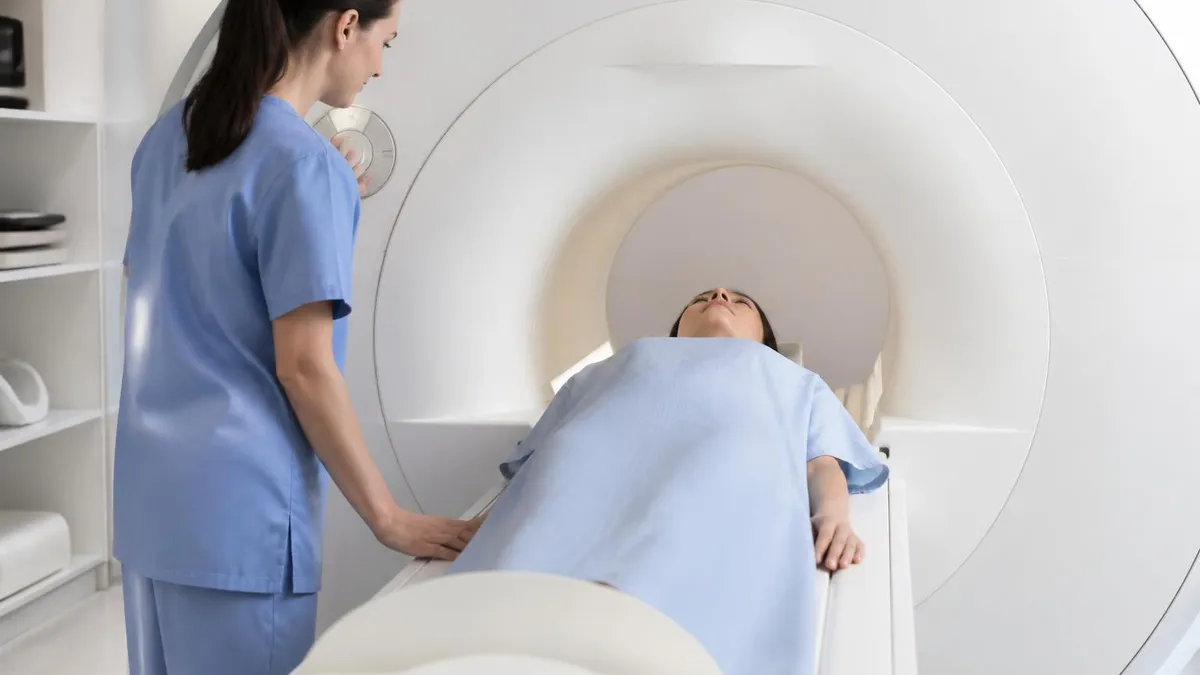
Once the table slides you into the open bore MRI, you will notice immediately that there is more space around your face, chest, and shoulders than older systems offered. A mirror mounted above the eyes lets you see out of the bore toward the technologist console, which helps many patients feel less enclosed. In-bore lighting and a small fan circulating air at face level further reduce the closed-in sensation. You will hold a squeeze ball connected to an alarm in case you need to stop.
The scan itself consists of multiple sequences, each lasting two to seven minutes, separated by short pauses while the technologist confirms image quality and adjusts parameters. You will hear loud knocking, buzzing, and chirping sounds caused by the rapid switching of gradient coils. This noise is intrinsic to MRI and unrelated to bore size, although many modern wide-bore systems include quiet pulse technology that reduces acoustic output by 70 to 80 percent. Patients curious about the sounds often appreciate reading about the mri machine noise to set expectations.
If contrast is needed, the technologist will pause the scan partway through to inject a gadolinium-based agent through an IV in your arm. Contrast enhances vascular structures and certain pathology, particularly tumors, infections, and active inflammation. The injection is brief and painless apart from the IV stick. After contrast, you return to the bore for the post-contrast sequences, which typically add ten to fifteen minutes to total scan time.
When the scan finishes, the table slides out, the technologist removes the coils, and you are free to leave unless you received sedation, in which case you will need monitoring and a driver. Most patients report that an open bore MRI felt significantly easier than a previous closed-bore experience. The combination of physical space, in-bore lighting, mirror viewing, music, and quiet sequences transforms what used to be a dreaded procedure into a tolerable one for the vast majority of people.
Total time from arrival to departure is typically 60 to 90 minutes for a single body part, longer if contrast or multiple regions are involved. Plan accordingly and bring a book or a companion for the waiting room. Knowing what to expect removes most of the uncertainty that drives pre-scan anxiety in the first place.
Even on an open bore MRI, the magnetic field is always on at full strength. Pacemakers, cochlear implants, certain aneurysm clips, retained metallic foreign bodies, and some neurostimulators can be dangerous or fatal inside the bore. Complete the safety screening honestly and disclose every implant, surgery, and prior occupational exposure to metal fragments before entering Zone IV.
Cost is a frequent question when patients learn their facility uses an open bore MRI. The good news is that wide-bore scans are billed identically to conventional closed-bore scans under US insurance contracts. Medicare, Medicaid, and commercial payers use the same CPT codes regardless of bore size, so your out-of-pocket cost will not increase because you chose the more comfortable scanner. List prices for a single-body-part MRI in the US typically range from 400 to 3,500 dollars before insurance, with hospital outpatient settings on the higher end and freestanding imaging centers on the lower end.
With commercial insurance, expected patient responsibility usually falls between 100 and 800 dollars depending on your deductible, copay structure, and whether the facility is in-network. Medicare patients typically pay 20 percent of the Medicare-allowed amount after the Part B deductible, which usually works out to 80 to 250 dollars per scan. Some imaging centers offer cash-pay discounts of 30 to 60 percent off the list price for uninsured patients, often making a wide-bore MRI affordable even without coverage.
Availability of open bore MRI scanners has grown rapidly. As of the mid-2020s, more than 70 percent of new MRI installations in the United States are wide-bore systems, and the majority of outpatient imaging chains advertise wide-bore capability as a competitive differentiator. Major manufacturers including Siemens, GE, Philips, Canon, and United Imaging all offer 1.5T and 3.0T wide-bore models. Smaller community hospitals may still rely on older closed-bore equipment, so it is worth asking before you schedule, especially if comfort is a priority.
For patients who genuinely cannot tolerate even a wide-bore MRI, alternatives exist. Truly open low-field systems remain available in many markets. Sedation, anti-anxiety medication, and in some cases general anesthesia can also make a closed or wide-bore scan possible. In situations where MRI is not feasible at all, your physician can discuss whether CT, ultrasound, or PET imaging would answer the clinical question. For deeper context, the article on MRI alternatives explores when other modalities are the smarter choice.
Image quality differences between facilities are mostly driven by magnet strength, coil technology, and protocol design rather than bore size. A 3.0T wide-bore system at an academic center will generally produce more detailed images than a 1.5T scanner at a community facility, regardless of bore geometry. For most routine indications, however, 1.5T wide-bore imaging is fully diagnostic, and the comfort advantage often outweighs any marginal benefit from a higher field strength scanner that the patient cannot tolerate.
When choosing a facility, the most important questions are accreditation, radiologist subspecialty expertise, and turnaround time for the report. ACR accreditation ensures that the scanner is maintained to industry standards and that protocols are appropriately designed. Subspecialty radiologists, particularly for neurological, musculoskeletal, and breast imaging, produce more accurate and clinically useful reports than general radiologists working outside their primary expertise. Turnaround time matters because a beautifully comfortable scan is wasted if the report takes two weeks to reach your referring doctor.
Finally, consider geography and convenience. An open bore MRI close to home that you can actually complete is far more valuable than a higher-field scanner two hours away that you might cancel. The wide-bore revolution has made comfortable, diagnostic-quality MRI available in nearly every US metropolitan area, and that accessibility may be its most important contribution to modern radiology.
Practical tips can turn an open bore MRI from tolerable to genuinely easy. First, communicate aggressively with the scheduler and technologist about any history of anxiety, claustrophobia, or prior failed scans. Imaging staff are trained to help, and they have an arsenal of strategies including warm blankets, weighted pads, in-bore music, prism glasses that let you look out of the bore lengthwise, and longer breaks between sequences. The more they know about your concerns in advance, the better they can tailor the experience to you specifically.
Second, practice slow breathing in the days abdominal mri prep. Box breathing, in which you inhale for four seconds, hold for four, exhale for four, and hold for four, activates the parasympathetic nervous system and reduces the heart rate response to enclosed spaces. Patients who practice for a week before the scan consistently report less anxiety during the procedure. This costs nothing, requires no medication, and works regardless of which scanner you use, but it pairs especially well with the additional space of an open bore MRI.
Third, ask whether you can be positioned feet-first. For knee, ankle, foot, lower spine, pelvis, and even some abdominal exams, the patient enters the bore feet first with the head outside or near the opening of the magnet. This single change eliminates the visual trigger that causes most claustrophobic episodes. Combined with a short-bore wide system, feet-first positioning means many patients never feel enclosed at all. Not every exam can be done this way, but most extremity and lower-body imaging absolutely can.
Fourth, consider bringing a trusted friend or family member to the appointment. Many facilities allow a companion to stand in the scan room during the exam, provided they pass the same safety screening. Having a familiar person within sight or audible distance provides reassurance that no medication can match. Even a hand resting on your shin can ground you during the longer sequences. Ask the facility about their companion policy when scheduling, because not all sites permit this.
Fifth, talk to your physician about anti-anxiety medication if behavioral strategies are not enough. A single low dose of lorazepam, alprazolam, or a similar short-acting benzodiazepine taken 30 to 60 minutes before the scan can dramatically reduce anxiety without affecting image quality. You will need a driver and should plan to rest the remainder of the day. For most patients with significant claustrophobia, the combination of a wide-bore scanner and a single dose of pre-medication transforms the experience entirely.
Sixth, focus on the sounds rather than fighting them. Many patients find that paying attention to the rhythm and pattern of the gradient noise, almost like listening to industrial music, makes the time pass faster than trying to block it out. The brain habituates quickly to repetitive sounds, and within a few minutes the knocking and buzzing fade into the background. Reviewing what to expect from knee MRI images or other specific scans you might be having also gives your mind something concrete to focus on.
Finally, remind yourself that the scan will end. The longest single sequence on a typical MRI lasts about seven minutes, and most run two to five minutes. Between sequences the technologist will check in with you, often offering a brief break or position adjustment. Knowing that each individual segment is short, that you have a squeeze ball to stop the scan if needed, and that the technologist is just a few feet away makes the entire experience manageable for the vast majority of patients, especially in the spacious environment of a modern open bore MRI.
MRI Questions and Answers

MRI Medical Abbreviation: What MRI Stands For and Why It Matters

The History of MRI: From Discovery to Modern Medicine

Knee MRI Images: A Complete Guide to Reading, Understanding, and Interpreting Knee Scans

Noise of MRI Machine: Why MRI Scanners Are So Loud and What to Expect
MRI Alternatives: When CT, Ultrasound, X-Ray, and PET Make More Sense Than an MRI
About the Author
Medical Laboratory Scientist & Clinical Certification Expert
Johns Hopkins UniversityDr. Sandra Kim holds a PhD in Clinical Laboratory Science from Johns Hopkins University and is certified as a Medical Technologist (MT) and Medical Laboratory Scientist (MLS) through ASCP. With 16 years of clinical laboratory experience spanning hematology, microbiology, and molecular diagnostics, she prepares candidates for ASCP board exams, MLT, MLS, and specialist certification tests.
Join the Discussion
Connect with other students preparing for this exam. Share tips, ask questions, and get advice from people who have been there.
View discussion (6 replies)