When Are MRI Scans Used? A Complete Guide to MRI Indications, Conditions, and Clinical Applications
When are MRI scans used? Complete guide to MRI indications, conditions diagnosed, body parts scanned, and when doctors choose MRI over CT or X-ray. 📚
Understanding when are MRI scans used helps patients, students, and clinicians make informed decisions about diagnostic imaging. Magnetic resonance imaging is a powerful, radiation-free technology that uses strong magnetic fields and radiofrequency pulses to produce highly detailed images of soft tissues, organs, blood vessels, and the nervous system. Unlike X-rays or CT scans, MRI excels at distinguishing between different types of soft tissue, making it the gold standard for diagnosing conditions ranging from torn ligaments to brain tumors and multiple sclerosis lesions.
Physicians order MRI scans when they need exceptional anatomical detail without exposing patients to ionizing radiation. Common scenarios include unexplained neurological symptoms such as persistent headaches, seizures, numbness, or weakness; suspected joint injuries that physical examination cannot fully characterize; staging of cancers; evaluation of the spine for herniated discs or nerve compression; and assessment of cardiovascular abnormalities. MRI also plays a critical role in pregnancy when avoiding radiation is paramount, particularly during the second and third trimesters.
The decision to use MRI typically follows a stepwise diagnostic process. A physician first performs a clinical examination and may order initial imaging such as ultrasound or X-ray. When those modalities cannot provide enough detail, or when soft tissue contrast is essential, MRI becomes the next logical step. Radiologists collaborate with referring physicians to choose the appropriate MRI protocol, including whether contrast agents like gadolinium are needed to highlight tumors, inflammation, or vascular abnormalities.
MRI is particularly valuable in neurology, orthopedics, oncology, cardiology, and gastroenterology. In neurology, it visualizes the brain and spinal cord with unmatched clarity. In orthopedics, it shows cartilage, tendons, ligaments, and bone marrow edema invisible on X-rays. Oncologists rely on MRI for tumor characterization, surgical planning, and treatment monitoring. Cardiologists use cardiac MRI to assess heart function, scarring after a heart attack, and congenital defects. Each clinical scenario has specific protocols that radiologists tailor to the diagnostic question.
Despite its power, MRI is not always the first imaging choice. It is more expensive, slower, and less accessible than CT or ultrasound, and certain patients with implanted devices, claustrophobia, or unstable conditions may not be candidates. Understanding when MRI is the right tool, and when alternatives may serve the patient better, helps avoid unnecessary delays, costs, and discomfort while ensuring accurate diagnoses.
This comprehensive guide explains when MRI scans are used, what conditions they detect best, how doctors decide between MRI and other modalities, and mri center locations can expect when referred for an MRI study. Whether you are preparing for an upcoming scan, studying for an imaging exam, or simply curious about how this remarkable technology supports modern medicine, this resource covers the indications, anatomy, contrast use, and clinical workflows that define MRI practice today. For background on the technology itself, see our overview of the history of MRI and how it became a diagnostic cornerstone.
By the end of this article, you will recognize the most common MRI indications, understand why physicians sometimes prefer MRI over CT or X-ray, learn how contrast and specialized sequences extend diagnostic capability, and identify the situations where MRI may not be appropriate. This knowledge empowers better conversations with your healthcare team and a clearer understanding of one of medicine's most sophisticated imaging tools.
MRI Usage by the Numbers
Primary Indications for MRI Scans
Persistent headaches, seizures, vision changes, weakness, numbness, memory loss, or suspected stroke. MRI evaluates the brain and spinal cord with superior soft-tissue contrast.
Torn ligaments, meniscal tears, rotator cuff damage, cartilage defects, and stress fractures. MRI shows tendons, ligaments, and bone marrow that X-ray cannot reveal.
Tumor characterization, staging, treatment planning, and follow-up surveillance for cancers of the brain, breast, prostate, liver, pancreas, and pelvis.
Cardiac MRI evaluates heart muscle function, infarct scarring, congenital defects, cardiomyopathies, and aortic aneurysms without radiation exposure.
Liver lesions, biliary obstructions, gynecologic conditions, prostate cancer, and inflammatory bowel disease are well visualized with MRI.
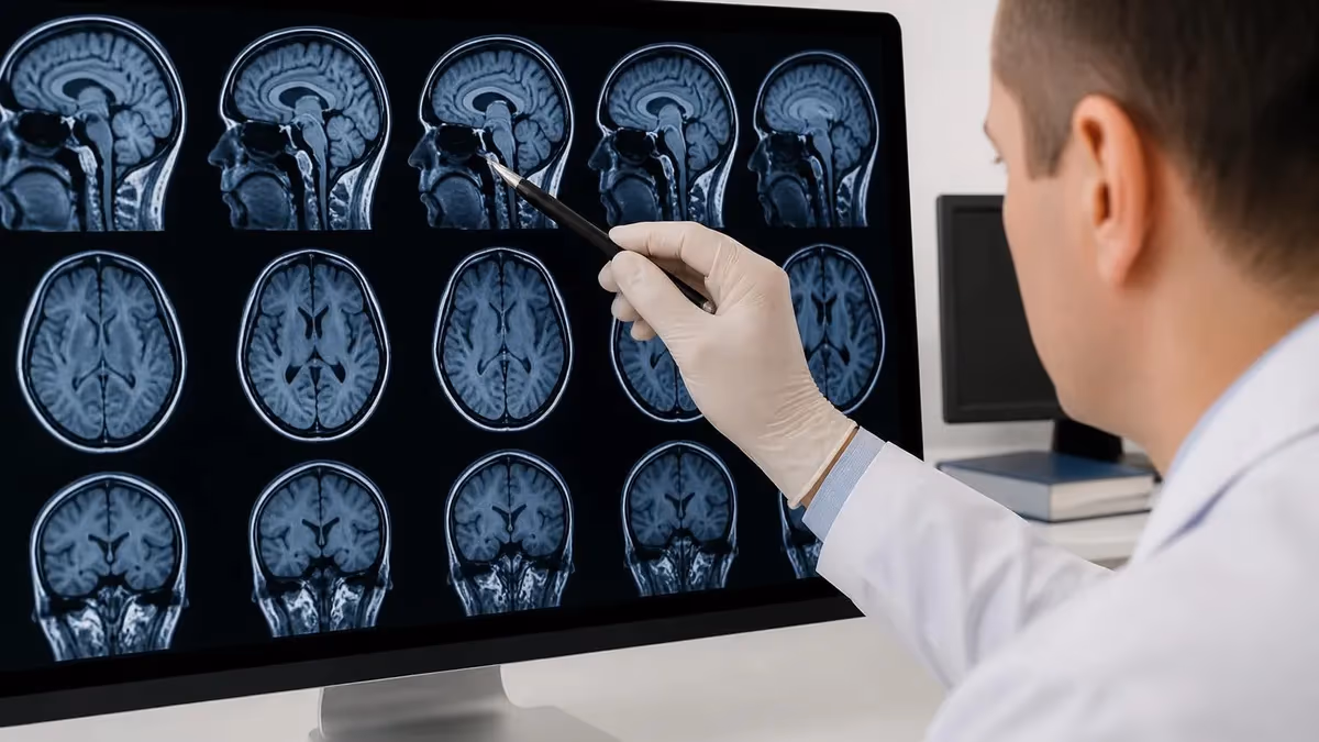
MRI scans are used across nearly every body system because of their unmatched ability to differentiate soft tissues. In the brain, MRI is the imaging modality of choice for diagnosing strokes (particularly in the early hours), demyelinating disorders such as multiple sclerosis, brain tumors, infections like meningitis or encephalitis, and degenerative diseases such as Alzheimer's. Functional MRI extends these capabilities further by mapping brain activity, which is invaluable for pre-surgical planning when tumors lie near eloquent areas controlling speech or movement.
The spine is another area where MRI dominates. Patients with back pain radiating into the legs, neck pain with arm symptoms, or suspected spinal cord compression typically undergo MRI to evaluate intervertebral discs, nerve roots, the spinal cord itself, and surrounding ligaments. Conditions like herniated discs, spinal stenosis, cauda equina syndrome, and spinal tumors are diagnosed and characterized through dedicated spine MRI protocols that include sagittal and axial T1- and T2-weighted sequences with optional contrast.
Orthopedic MRI is the workhorse of sports medicine. A knee MRI can reveal anterior cruciate ligament tears, meniscal injuries, cartilage damage, and bone bruises within a 30-minute scan. Shoulder MRIs evaluate rotator cuff tears, labral injuries, and impingement syndromes. Hip MRIs detect labral tears, avascular necrosis, and stress fractures. Ankle, wrist, and elbow MRIs investigate similar soft-tissue pathologies. To learn more about reading these specialized images, our guide on knee MRI images walks through the most important sequences and findings.
In abdominal and pelvic imaging, MRI is preferred when ultrasound or CT cannot fully characterize a lesion. MRI of the liver detects and classifies tumors with exquisite detail, especially when using hepatocyte-specific contrast agents. Magnetic resonance cholangiopancreatography (MRCP) visualizes the biliary and pancreatic ducts non-invasively, replacing many diagnostic endoscopic procedures. Pelvic MRI assesses uterine fibroids, adenomyosis, endometriosis, ovarian masses, and prostate cancer, often guiding surgical or oncologic decisions.
Cardiac MRI has emerged as a powerful tool for cardiologists. It quantifies ejection fraction, identifies myocardial scarring after a heart attack, characterizes cardiomyopathies, evaluates congenital heart defects, and detects cardiac masses or pericardial disease. Unlike echocardiography, cardiac MRI provides reproducible, high-resolution images regardless of body habitus, making it especially useful in patients where ultrasound windows are limited.
Vascular MRI, known as magnetic resonance angiography (MRA), images arteries and veins throughout the body without iodinated contrast. MRA is commonly used to evaluate the carotid arteries, intracranial vessels (for aneurysms or arteriovenous malformations), renal arteries, and peripheral arteries. For patients who cannot receive iodinated contrast due to kidney disease or allergies, MRA offers a safer alternative for vascular assessment.
Finally, MRI plays an important role in pediatric and obstetric imaging because it avoids ionizing radiation. Fetal MRI is increasingly used to evaluate congenital anomalies detected on prenatal ultrasound. In children, MRI is preferred for evaluating brain tumors, epilepsy, developmental disorders, and complex musculoskeletal problems where radiation exposure must be minimized over a lifetime.
MRI Practice Test Questions
Prepare for the MRI - Magnetic Resonance Imaging exam with our free practice test modules. Each quiz covers key topics to help you pass on your first try.
MRI Knowledge
MRI Exam Questions covering Knowledge. Master MRI Test concepts for certification prep.
MRI Physics
Free MRI Practice Test featuring Physics. Improve your MRI Exam score with mock test prep.
MRI Anatomy and Pathology
MRI Test Prep for MRI Anatomy and Pathology. Practice MRI Quiz questions and boost your score.
MRI Anatomy and Positioning
MRI Questions and Answers on MRI Anatomy and Positioning. Free MRI practice for exam readiness.
MRI Contrast Agents
Free MRI Quiz on MRI Contrast Agents. MRI Exam prep questions with detailed explanations.
MRI Patient Care and Positioning
MRI Practice Questions for MRI Patient Care and Positioning. Build confidence for your MRI certification exam.
When Is MRI Used Compared to Other Imaging Modalities
MRI and CT both produce cross-sectional images, but they answer different questions. CT excels in emergency settings because it scans rapidly, visualizes bone fractures, acute hemorrhage, and pulmonary conditions, and is more accessible. However, CT uses ionizing radiation, which raises concerns with repeated use, pregnancy, and pediatric imaging.
MRI is preferred for soft-tissue evaluation: brain disorders beyond acute bleeds, spinal cord pathology, musculoskeletal injuries, abdominal organ characterization, and pelvic disease. When detail of cartilage, ligaments, white matter, or tumor extent matters more than speed, MRI delivers superior contrast resolution. Choosing between them depends on urgency, the tissue in question, and patient safety considerations.
Advantages and Limitations of MRI
- +No ionizing radiation exposure, making it safe for repeat imaging and pregnancy
- +Superior soft-tissue contrast resolution compared to CT or X-ray
- +Multi-planar imaging in any orientation without repositioning the patient
- +Functional imaging including diffusion, perfusion, and spectroscopy capabilities
- +Excellent visualization of the brain, spinal cord, joints, and pelvic organs
- +Effective use of non-iodinated contrast agents for patients with iodine allergies
- +Highly detailed vascular imaging without arterial puncture (MRA/MRV)
- −Significantly longer scan times (30 to 90 minutes) than CT or X-ray
- −Higher cost and reduced availability in rural or under-resourced areas
- −Contraindicated for many implanted metallic devices and certain pacemakers
- −Loud acoustic noise during scanning can cause patient discomfort
- −Claustrophobia in closed-bore scanners limits patient tolerance
- −Motion artifact can degrade images, especially in pediatric or anxious patients
- −Gadolinium contrast carries small risk in patients with severe renal impairment
Preparation Checklist Before Your MRI Scan
- ✓Inform the technologist about any implanted devices, surgical clips, or metal in the body
- ✓Remove all jewelry, watches, hearing aids, hairpins, and removable dental work
- ✓Wear loose, comfortable clothing without zippers, snaps, or metal embellishments
- ✓Tell staff if you have claustrophobia so they can offer sedation or open-bore options
- ✓Disclose pregnancy or possible pregnancy before the scan begins
- ✓Mention any kidney problems if gadolinium contrast may be used
- ✓Avoid wearing makeup with metallic particles or cosmetic glitter
- ✓Eat and drink normally unless instructed otherwise for specific abdominal protocols
- ✓Bring prior imaging studies on CD if the radiologist requested comparison
- ✓Plan to lie still for the entire scan duration to ensure diagnostic image quality
MRI Is Often the Final Diagnostic Step
In most clinical pathways, MRI is ordered after initial imaging or physical examination raises concern for a condition that requires detailed soft-tissue evaluation. Because it confirms diagnoses and guides treatment planning, MRI often determines whether a patient undergoes surgery, chemotherapy, physical therapy, or watchful waiting.
Contrast-enhanced MRI uses gadolinium-based contrast agents to highlight specific tissues, primarily areas of increased vascular permeability or inflammation. When a physician orders MRI with contrast, they typically want to better characterize a tumor, identify active inflammation in multiple sclerosis, evaluate infections such as abscesses, or assess vascular structures. Gadolinium shortens the T1 relaxation time of nearby water molecules, causing those tissues to appear bright on T1-weighted images and enabling subtle pathology to stand out clearly from normal background tissue.
Not every MRI requires contrast. Many musculoskeletal scans, routine brain MRIs for headache evaluation, and lumbar spine studies for disc herniation are performed without contrast because the underlying T1- and T2-weighted sequences provide sufficient diagnostic information. Avoiding unnecessary contrast spares patients an IV stick, reduces scan time, lowers costs, and eliminates the small but real risk of contrast reactions. The decision to use contrast is made by the radiologist based on the clinical question and patient history.
Specialized MRI sequences extend diagnostic capability far beyond standard anatomical imaging. Diffusion-weighted imaging (DWI) detects restricted water movement, making it indispensable for identifying acute strokes within minutes of onset and characterizing abscesses or highly cellular tumors. Susceptibility-weighted imaging (SWI) shows microbleeds and venous structures with exceptional sensitivity. Magnetic resonance spectroscopy (MRS) analyzes biochemical composition non-invasively, helping distinguish tumor types or detect metabolic disorders.
Functional MRI (fMRI) measures changes in blood oxygenation that correlate with neural activity, allowing neurosurgeons to map critical areas of the brain before tumor resection or epilepsy surgery. Diffusion tensor imaging (DTI) maps the orientation of white matter tracts, providing a roadmap of neural connectivity that helps avoid damage during surgery. These advanced techniques transform MRI from a static anatomical tool into a dynamic window on brain function and connectivity.
Cardiac MRI sequences include cine imaging to assess wall motion, late gadolinium enhancement to identify scar tissue, T1 and T2 mapping to quantify diffuse myocardial disease, and phase-contrast imaging to measure blood flow. Together, these techniques deliver a comprehensive evaluation of cardiac structure and function that often outperforms any single alternative modality, making cardiac MRI the reference standard for many cardiomyopathies and post-infarct assessments.
MR enterography is a specialized abdominal MRI protocol for evaluating the small bowel in patients with Crohn's disease and other inflammatory bowel conditions. It avoids radiation while providing detailed information about bowel wall thickness, inflammation, strictures, and fistulas. Patients drink oral contrast to distend the bowel, and IV contrast highlights areas of active disease. Repeat imaging is safer over a lifetime of disease monitoring compared with CT enterography.
Whole-body MRI is emerging as a screening and surveillance tool for cancer patients, individuals with genetic predispositions such as Li-Fraumeni syndrome, and those seeking comprehensive health checks. While not yet standard for the general population, its ability to scan from head to toe without radiation makes it valuable when systemic disease must be excluded or monitored efficiently in a single visit.
Always disclose every metallic implant, surgical clip, cochlear device, pacemaker, defibrillator, or shrapnel injury before entering the MRI room. The powerful magnetic field can heat, dislodge, or malfunction these objects, causing serious injury or device failure. MRI technologists screen every patient carefully, but you must provide accurate information about your medical history.
The MRI workflow begins with a clinical referral. A physician evaluates the patient, formulates a differential diagnosis, and submits an imaging request specifying the body part, suspected condition, and whether contrast is needed. A radiologist reviews the request to confirm appropriateness, select the optimal protocol, and approve scheduling. This protocoling step is critical because MRI sequences vary substantially based on the diagnostic question, and choosing the right combination ensures the scan answers what the referring physician needs to know.
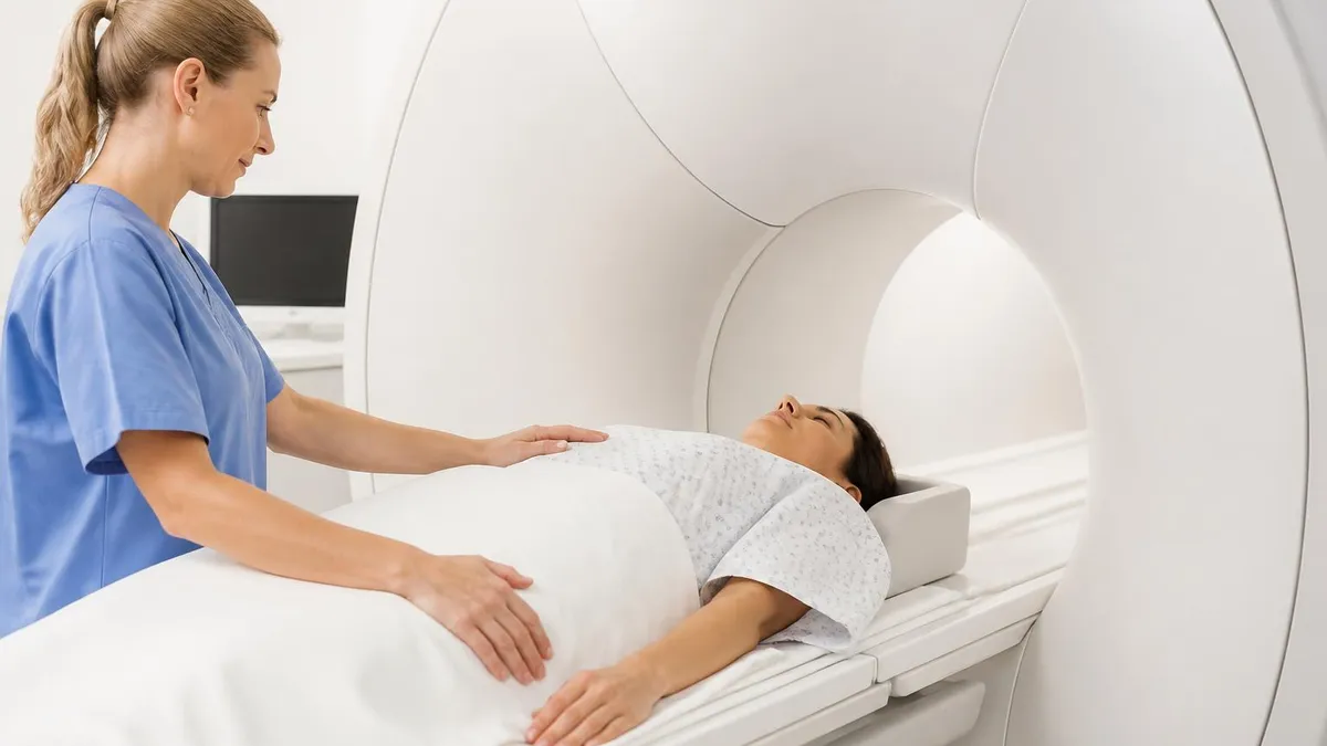
Once the patient arrives at the imaging center, a technologist performs a comprehensive safety screening. The patient completes a detailed questionnaire about implants, prior surgeries, allergies, kidney function, and pregnancy status. Anyone entering Zone IV, the scanner room, must be metal-free. Even patients are asked to change into gowns to eliminate potential hazards from undergarments, belts, or jewelry. This rigorous screening protects against catastrophic incidents that have occurred when ferromagnetic objects became projectiles in the magnet's field.
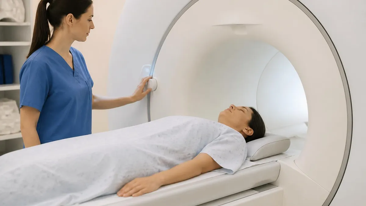
The scan itself proceeds in a series of sequences, each producing a different type of image contrast. Patients lie on a movable table that slides into the scanner. Coils are positioned over the body part being imaged. Through a microphone, the technologist communicates instructions, asks about comfort, and monitors patient well-being. The pronounced knocking and buzzing sounds are caused by gradient coils rapidly switching on and off, and patients wear earplugs or headphones to reduce noise. To understand more about this signature acoustic experience, our article on mri machine noise explains the physics behind these loud bangs.
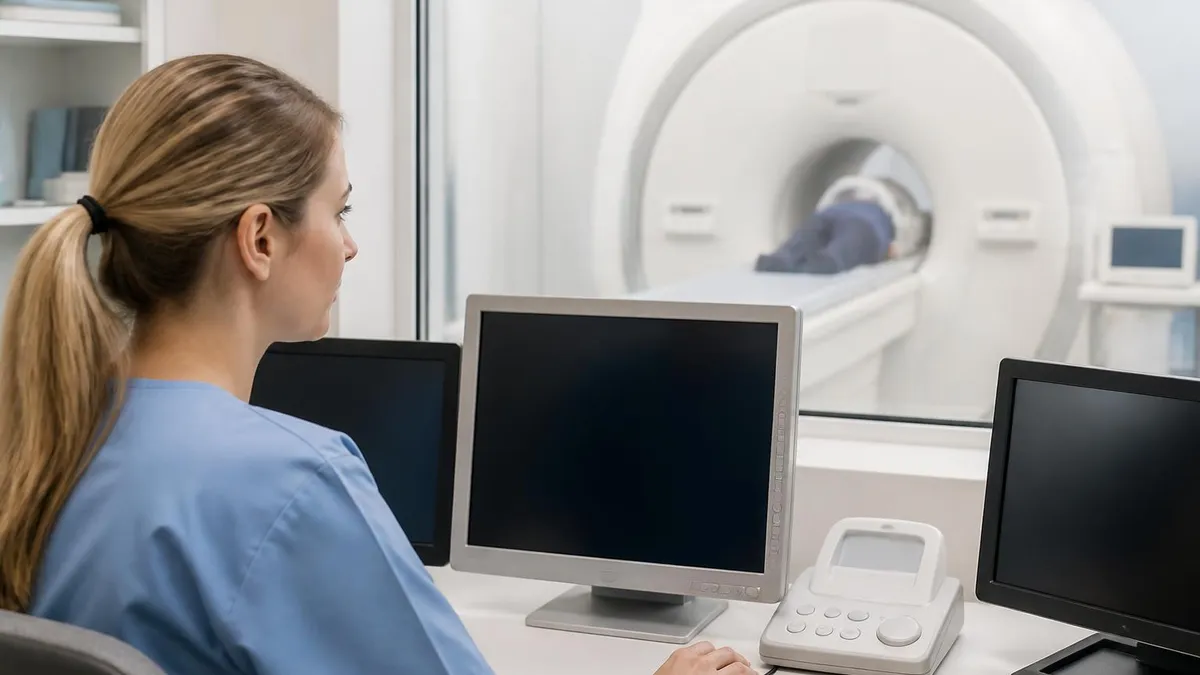
After scanning concludes, the radiologist reviews the images on dedicated workstations. They evaluate every sequence in multiple planes, compare with prior studies when available, and dictate a report that describes findings, addresses the clinical question, and offers an impression with recommendations. The report is sent to the referring physician, usually within hours to a day, and the patient typically learns the results during a follow-up appointment or via patient portal messaging.
Safety considerations extend beyond metal implants. Pregnant patients are screened carefully, with most centers avoiding MRI in the first trimester unless absolutely necessary. Gadolinium contrast is generally avoided during pregnancy unless the benefit clearly outweighs potential fetal risk. Patients with severe kidney disease (eGFR below 30) require careful consideration of contrast, given the rare but serious risk of nephrogenic systemic fibrosis associated with older gadolinium agents.
Claustrophobia affects up to 10% of MRI patients. Strategies to manage it include open-bore scanners, prone positioning, eye masks, calming music, breathing exercises, and oral or IV anxiolytics. Some patients benefit from a brief tour of the scanner room before the scan and from having a family member nearby. When sedation is needed, monitoring requirements add complexity but allow patients to complete diagnostic studies that would otherwise be impossible.
Pediatric MRI presents additional challenges. Younger children may require general anesthesia to remain still, and dedicated child-friendly facilities use distraction techniques, video goggles, and parental presence to minimize sedation use. Advances in faster pulse sequences and motion correction algorithms are gradually reducing the need for anesthesia, making MRI safer and more accessible for the youngest patients.
For patients scheduled for an MRI, a few practical tips can make the experience smoother and more diagnostically successful. Arrive 15 to 30 minutes early to allow time for paperwork, changing, and safety screening. Bring a list of current medications, prior imaging studies if requested, and contact information for your referring physician. If you typically take anxiety medication, ask your doctor whether to take it before the scan. Eat normally unless told otherwise, and use the restroom before your appointment because the scan can take an hour or more.
Wear simple, comfortable clothing without metal. Many facilities will provide a gown, but layered clothing without zippers, snaps, underwire bras, or metallic threads can sometimes be worn. Leave jewelry, watches, and accessories at home or in a secure locker. Remove transdermal medication patches, as some contain metallic backing that can heat during scanning. Tell the technologist about every patch you wear, even nicotine or birth control patches, before stepping into the magnet room.
During the scan, focus on staying as still as possible. Even small movements blur images and may require sequences to be repeated, lengthening the appointment. Many patients find it helpful to close their eyes, listen to provided music, or practice slow breathing to relax. Communicate with the technologist through the call button if you feel uncomfortable. Brief breaks between sequences are usually possible, although some sequences must run uninterrupted for several minutes.
If contrast is given, you may feel a cool sensation as it enters the IV line, and a brief metallic taste is possible. Most patients experience no adverse effects, though minor reactions such as nausea, headache, or itching occur in a small percentage. Severe reactions are rare but should be reported immediately. After the scan, drink plenty of water to help flush the contrast from your system, particularly if you have normal kidney function.
Once the scan is complete, you can usually resume normal activities immediately. If you received sedation, arrange for someone to drive you home and avoid operating machinery for the rest of the day. Results typically follow within one to two business days, although urgent cases are read sooner. Schedule a follow-up appointment with your referring physician to discuss the findings, ask questions, and plan next steps. Never assume no news is good news; always confirm that your provider has reviewed the report.
If you are studying MRI for professional reasons, such as preparing for the ARRT MRI registry exam or training as a radiologic technologist, understanding clinical indications is just as important as mastering physics and safety. Memorize the conditions most commonly imaged with MRI, the sequences that best demonstrate each pathology, and the contraindications that require alternative imaging. Practice exams help reinforce this knowledge, and reading actual radiology reports trains your eye to recognize how findings translate into clinical decisions.
Finally, remember that MRI is one tool among many. Although it is remarkably powerful, it is not always the best choice for a given clinical scenario. If MRI is contraindicated, alternatives include CT, ultrasound, nuclear medicine, and traditional X-ray. To learn when these alternatives may serve a patient better, see our companion guide on MRI alternatives covering CT, ultrasound, X-ray, and PET. Empowered with this knowledge, you can navigate diagnostic imaging decisions with confidence and clarity.
MRI Questions and Answers

MRI Medical Abbreviation: What MRI Stands For and Why It Matters

The History of MRI: From Discovery to Modern Medicine

Knee MRI Images: A Complete Guide to Reading, Understanding, and Interpreting Knee Scans

Noise of MRI Machine: Why MRI Scanners Are So Loud and What to Expect
MRI Alternatives: When CT, Ultrasound, X-Ray, and PET Make More Sense Than an MRI
About the Author
Medical Laboratory Scientist & Clinical Certification Expert
Johns Hopkins UniversityDr. Sandra Kim holds a PhD in Clinical Laboratory Science from Johns Hopkins University and is certified as a Medical Technologist (MT) and Medical Laboratory Scientist (MLS) through ASCP. With 16 years of clinical laboratory experience spanning hematology, microbiology, and molecular diagnostics, she prepares candidates for ASCP board exams, MLT, MLS, and specialist certification tests.
Join the Discussion
Connect with other students preparing for this exam. Share tips, ask questions, and get advice from people who have been there.
View discussion (6 replies)